Functional analysis of syp1, a novel substrate of the serine threonine kinase prk1
Bạn đang xem bản rút gọn của tài liệu. Xem và tải ngay bản đầy đủ của tài liệu tại đây (5.33 MB, 152 trang )
FUNCTIONAL ANALYSIS OF SYP1, A NOVEL SUBSTRATE OF
THE SERINE/THREONINE KINASE PRK1
QIU WENJIE
NATIONAL UNIVERSITY OF SINGAPORE
2007
FUNCTIONAL ANALYSIS OF SYP1, A NOVEL SUBSTRATE OF
THE SERINE/THREONINE KINASE PRK1
QIU WENJIE
A THESIS SUBMITTED
FOR THE DEGREE OF DOCTOR OF PHILOSOPHY
INSTITUTE OF MOLECULAR AND CELL BIOLOGY
NATIONAL UNIVERSITY OF SINGAPORE
2007
i
Acknowledgements
Foremost, I would like to express my sincere gratitude to my supervisor A/P Mingjie
Cai, for providing me the opportunity to continue my Ph.D. research work in his laboratory
after my former supervisor left IMCB. I am deeply grateful to A/P Cai for his guidance,
tolerance, encouragement and support throughout my graduate studies. Heartfelt
appreciation also goes to my graduate supervisory committee members, A/P Thomas
Leung and A/P Uttam SURANA, for their invaluable advice and encouragement during the
course of this study. I am also grateful to A/P Uttam Surana and A/P Alan Munn for
sharing some strains used in this study.
I would like to thank the past and present members in CMJ laboratory, for their
helpful discussion, technique assistance, cooperation and friendship. Special thanks go to
Dr. Guisheng Zeng, Dr. Yu Xianwen and Miss Suat Peng Neo for their help, advice, and
sharing of experience. Desmond Dorairajoo and Jun Wang are thanked for the work with
microscopy and other general technical assistance. Thanks also go to Dr. Guisheng Zeng,
Dr. Chee Wai Fong and Miss Suat Peng Neo for their critical reading of my thesis.
Many thanks also to the past and present members in US laboratory, to Dr. Hong
Hwa Lim, Miss Karen Crasta, Mr. Tao Zhang, Mr. Jenn Hui Khong and Mr. Saurabh
Nirantar, for interesting discussions and help with the project.
I am also indebted to my former supervisor Dr. Sheng-Cai Lin for his help to begin
my PH.D study and for the training of molecular biology techniques in his laboratory.
I cannot express in words my gratitude to my family: my wife Liqin Hu and my
lovely little daughter Elim. Thanks for their love and encouragement over these years. I
would like to thank my parents and parents-in-law too. Without their support and help, it
would be impossible to finish my Ph.D. work.
ii
Table of contents
Acknowledgements i
Table of Contents ii
List of Figures vii
List of Tables ix
Abbreviations x
Summary
xv
Chapter 1 Introduction
1
1.1 Cell polarity and its mechanism in yeast 2
1.1.1 Bud site selection for polarized growth 5
1.1.2 Establishment of polarized growth by Cdc42p 6
1.2 Yeast actin cytoskeleton 8
1.2.1 The roles of yeast actin cytoskeleton in polarized growth 8
1.2.1.1 Bipolar bud site selection 8
1.2.1.2 Maintenance of the polarity of Cdc42p 9
1.2.1.3 Actin cytoskeleton in polarized growth 10
1.2.2 Actin assembly and actin turn over 10
1.2.3 Cortical actin patches 11
1.2.3.1 Dynamic localization of cortical actin patches 11
iii
1.2.3.2 Assembly of actin filaments by the Arp2/3 complex and its NPFs 12
1.2.3.3 Actin patches and endocytosis 16
1.2.3.4 Role of Pan1p and Sla1p in patch development 18
1.2.3.5 Regulation of actin cytoskeleton and endocytosis by Prk1p 19
1.2.4 Actin cables 21
1.2.4.1 Actin cable formation by formins 21
1.2.4.2 Profilin promotes actin filament elongation 22
1.2.4.3 Regulation of actin cable assembly by polarisome 23
1.2.4.4 Regulation of actin cable assembly by Rho GTPase 24
1.2.5 Actin ring formation and cytokinesis 25
1.3 Septin cytoskeleton 26
1.3.1 Roles of septins in cell division and polarized growth 26
1.3.1.1 Roles of septins in cytokinesis 26
1.3.1.2 Axial bud site selection 27
1.3.1.3 Septins and cell wall in polarized growth 27
1.3.1.4 Morphogenesis checkpoint 28
1.3.2 Organization and dynamic localization of septins 31
1.3.3 Regulation of septin organization 33
1.4 Objectives and significances of the study 35
Chapter 2 Materials and Methods 36
2.1
Materials 37
2.1.1 Reagents and antibodies 37
2.1.2 Strains 37
iv
2.1.3 Constructs 40
2.2
Methods 45
2.2.1 Strains and culture conditions 45
2.2.2 Recombinant DNA methods 46
2.2.2.1 DNA transformation of E.coli cells 46
2.2.2.2 Plasmid DNA preparation 47
2.2.2.3 Site-directed mutagenesis 48
2.2.2.4 Plasmid constructions 48
2.2.3 Yeast manipulations 48
2.2.3.1 Yeast transformation 48
2.2.3.2 Two-hybrid assays 49
2.2.3.3 Uracil uptake assay 49
2.2.3.4 Lucifer yellow uptake 50
2.2.4 Fluorescence microscopy studies 50
2.2.4.1 Staining of F-actin and chitin 50
2.2.4.2 Real time imaging of proteins with fluorescent tags 51
2.3
Protein Analysis 52
2.3.1 Preparation of crude protein extracts using
acid-washed glass beads 52
2.3.2 Preparation of total protein extracts using
TCA precipitation
53
2.3.3 in vitro kinase assay and GST- fusion protein binding assay 53
2.3.4 Immunoprecipitation and Western blot 55
v
Chapter 3 Syp1p, a new phosphorylation target of Prk1p 57
3.1
Introduction 58
3.2
Results 58
3.2.1 Phosphorylation of Syp1p by Prk1p in vitro and in vivo 58
3.2.2 Effect of Prk1p phosphorylation on Syp1p 61
3.3 Discussion 63
3.3.1 Syp1p is a new regulatory target of Prk1p 63
Chapter 4 Relationship between Syp1p and actin cytoskeleton 65
4.1 Introduction 66
4.2 Results 67
4.2.1 Functional relationship between Syp1p and Pfy1/Bni1p 67
4.2.1.1 Syp1p overexpression partially suppressed the phenotypes of
profilin deletion mutant 69
4.2.1.2 Syp1p overexpression suppressed the phenotypes
of bni1∆ mutant 69
4.2.1.3 Polarized localization and function of Syp1p depend on
profilin and Bni1p 71
4.2.2 Localization interdependency between Syp1p and actin cytoskeleton 73
4.2.2.1 Dependency of Syp1p polarized localization on actin cytoskeleton 7
3
4.2.2.2 Polarity defect of actin patches in cells overexpressing Syp1p 75
4.2.3 Association of Syp1p with Sla1p 77
4.2.3.1 Interaction between Syp1p and Sla1p in vitro and in vivo 77
4.2.3.2 Mapping binding regions on Syp1p for Sla1p 83
vi
4.2.3.3 No endocytosis defect in syp1Δ cells
or cells overexpressing Syp1p 85
4.3 Discussion 87
4.3.1 Evidence for Syp1p functioning in actin cytoskeleton organization 87
4.3.2 The role of Syp1p in the function of profilin and Bni1p 89
4.3.3 Functional relationship between Syp1p and Sla1p 90
Chapter 5 Relationship between Syp1p and the septin cytoskeleton 92
5.1
Introduction 93
5.2
Results 93
5.2.1 Syp1p overexpression causes septin disorganization 93
5.2.2 Abnormal septin structures in HU-arrested syp1∆ cells 98
5.2.3 Association of Syp1p with septins 100
5.2.4 Dynamic localization of Syp1p in live cells 103
5.2.5 The effects of SYP1 deletion on septin dynamics 105
5.2.6 Effects of SYP1 deletion on budding site selection 108
5.3 Discussion 110
5.3.1 Evidence for Syp1p functioning in septin organization 110
5.3.2 Interaction between Syp1p and septins 111
5.3.3 Regulation of septin dynamics by Syp1p 112
5.3.4 The possible links between actin cytoskeleton and septins
through Syp1p 113
References
116
vii
List of Figures
Figure:
1.1
Three forms of polarized cell growth
in the Saccharomyces cerevisiae life cycle
2
1.2
Different stages of budding during the cell cycle 4
1.3
Axial and bipolar budding patterns in yeast cells 6
1.4
Summary of signaling pathways that lead to the polarity establishment
during bud formation
7
1.5
Schematics of yeast NPFs 13
1.6
Model for actin patch development 17
1.7
Domain organization of budding yeast formins Bni1p and Bnr1p 22
1.8
Swe1p localization and degradation in yeast 29
1.9
Primary structure and organization of S. cerevisiae mitotic septins 32
3.1
Identification of Syp1p as a new phosphorylation target of PRK1p 59
3.2
Effect of Syp1p phosphorylation by Prk1p on pfy1Δ suppression (A) and bud
morphogenesis (B)
62
4.1
Syp1p overexpression partially suppressed phenotypes of pfy1
Δ
mutant
70
4.2
Syp1p overexpression partially suppressed phenotypes of bni1Δ mutant
70
4.3
Depolarization of Syp1p localization in pfy1 and bni1 mutants 72
4.4
BNI1 deletion abolished the elongated bud induced by Syp1p overexpression-
-
73
4.5
Colocalization between Syp1p and actin cytoskeleton 74
4.6
The dependence of Syp1p polarized localization on actin cytoskeleton 76
4.7
Syp1p overexpression depolarized actin cytoskeleton and chitin deposition 78
4.8
Sla1p is required for the polarized localization of Syp1p 80
viii
4.9
Physical interaction between Syp1p and Sla1p 82
4.10
Co-immunoprecipitation between Syp1p and Sla1p 82
4.11
The regions of Syp1p required for Sla1p interaction 84
4.12
SYP1 deletion and overexpression did not cause endocytosis defects
-
86
4.13
The conserved domains in Syp1p through searching the proteins databases 88
5.1
Septin disorganization caused by Syp1p overexpression 94
5.2
Cytokinesis defect and septin disorganization in α-factor treated cells
caused by Syp1p overexpression 96
5.3
Synthetic lethality between cdc10 and Syp1p overexpression 98
5.4
Septin abnormality of the syp1∆ cells upon HU treatment 99
5.5
Co-localization of Syp1p and septins 101
5.6
Physical interaction between Syp1p and septins 102
5.7
Dynamic localization of Syp1-GFP during the cell cycle 104
5.8
Abnormal septin dynamics in the syp1∆ cells
and the cells overexpressing Syp1p 106
5.9
Effect of the syp1∆ mutation on bud site selection 109
ix
List of Tables
Table:
1
Yeast strains used in this study 37
2
Plasmids used in this study 40
3
The homologous domains with Syp1p through searching against database 89
x
Abbreviations
a.a. or aa amino acid
AAK1 adaptor-associated
kinase 1
ADF actin depolymerizing factor
ADFH actin depolymerizing factor homologous region
ADP adenosine 5’-diphosphate
AP2 adaptor protein 2
ATP adenosine 5'-triphosphate
BAR BIN-amphiphysin-RVS domain
bp base pair
BSA bovine serum albumin
°C
degree Celsius
CAP adenylyl cyclase-associated protein
CC coiled coil
CDC Cell Division Cycle
CDK Cyclin-dependent kinase
CFP cyan fluorescent protein
CFW Calcofluor White
CIP calf intestinal phosphatase
Cln cyclin
C-terminal carboxy-terminal
COPII coated vesicle complex II
DAD Diaphanous autoregulatory domain
DAPI 4',6-diamidino-2-phenylindole
DID Diaphanous inhibitory domain
DMSO Dimethyl Sulfoxide
DNA deoxyribonucleic acid
DPW Asp-Pro-Trp motifs
xi
DTT dithiothreitol
ECL enhanced chemiluminescence
E. coli
Escherichia coli
EDTA ethylenediamine tetraacetic acid
EGFP enhanced green fluorescent protein
EH Eps15 homology
ENTH Epsin amino-terminal homology
EVH Ena/VASP homology
F-actin filamentous actin
FCH Fes/CIP4 homology domain
FH formin homology domain
FRAP fluorescence recovery after photo-bleaching
FUR Fluoro Uracil Resistance
G-actin actin monomer
GAP GTPase-activating protein
GBD GTPase binding domain
GDP guanosine diphosphate
GED GTPase effector domain
GEF guanine-nucleotide exchange factor
GFP green fluorescent protein
GST glutathione S-transferase
GTP guanosine triphosphate
GTPase guanosine triphosphatase
hrs hours
HA haemagglutinin
HEPES hydroxyethylpiperazine ethanesulfonic acid
HIP1 Hungtingtin interacting protein-1
HRP horseradish peroxidase
HU hydroxyurea
xii
IgG immunoglobulin G
IP immunoprecipitation
IPTG
isopropyl-β-D-thiogalactoside
kb kilobase(s)
kDa kilodalton
Lat-A Latrunculin-A
LB Luria-Bertani medium
LY Lucifer Yellow
M molar
MAT mating locus
min minute
ml milliliter
mM millimolar
μg
microgram
μm
micrometer
nm nanometer
N-terminal amino-terminal
NPF Asp-Pro-Phe motifs
NPFs nucleation-promoting factors
N-WASP neuronal Wiskott-Aldrich syndrome protein
OD optical density
ORF open reading frame
PAGE polyacrylamide gel electrophoresis
PBS phosphate-buffered saline
PCR polymerase chain reaction
PEG polyethylene glycol
PH pleckstrin homology
PIP
2
phosphatidylinositol-4,5-bisphosphate
PMSF phenylmethylsulfonyl fluoride
xiii
PNPP p-nitrophenylphosphate
poly-P proline-rich region
PP2A Protein Phosphatase 2A
PVDF polyvinylidene difluoride
RBD Rho-binding domain
RFP red fluorescent protein
rpm revolutions per minute
SBD Spa2-binding domain
sec second
SC synthetic complete (medium)
SDS Sodium dodecyl sulphate
SH2 Src homology 2
SH3 Src homology 3
SHD Sla1 homology domain
SR the C-terminal QxTG repeats of Sla1p
SYP Suppressor of Yeast Profilin deletion
TAP tandem-affinity purification
TCA Trichloroacetic Acid
TE Tris-EDTA buffer
TEMED N,N,N',N'-tetramethylethylenediamine
Tris Tris(hydroxymethyl)aminomethane
VASP
vacuolar protein sorting
VPS
vacuolar protein sorting
WA WH2 domain and acidic motif
WASP Wiskott-Aldrich syndrome protein
WAVE WASP-family verprolin homologous protein
WH WASP homology region
WIP WASP interacting protein
WT wild type
xiv
YEPD yeast extract-peptone-dextrose (rich medium)
YFP yellow fluorescent protein
xv
Summary
Cell polarity is a fundamental property of cells. The budding yeast
Saccharomyces cerevisiae is a model system for the study of cell polarity. Yeast cells
first select a proper site to establish cell polarity. In this site, actin and septin
cytoskeletons are organized to achieve polarized cell growth. Actin patches and actin
cables are two essential organizations of actin cytoskeleton which are involved in the
establishment and maintenance of polarized cell growth. Actin patches are required for
endocytosis while actin cables are essential for the polarized vesicle transport. Upon
internal and external signals, actin cytoskeleton undergoes a dramatic reorganization
regulated by a large number of cytoskeleton-associated proteins, such as Pan1p, Sla1p
and Bni1p. The functions of Pan1p and Sla1p are regulated by an important
serine/throrine kinase Prk1p.
Septin cytoskeleton is required for cell morphogenesis and division in budding
yeast. Septins form a heterooligomeric complex which localizes at the mother-daughter
junction. Septin filaments also undergo assembly and disassembly in accordance with the
progression of the cell cycle.
Syp1p was first identified as a multi-copy suppressor of profilin deletion mutant
and its overexpression was found to cause an elongated bud phenotype. The functions of
Syp1p in actin and septin cytoskeletons were investigated in depth in this study. Firstly,
Syp1p is shown to be a novel substrate of Prk1p and its phosphorylation by Prk1p
negatively regulates Syp1p’s functions. Secondly, Syp1p overexpression suppresses the
bni1Δ mutants at non-permissive temperature. Syp1p overexpression also partially
rescues the depolarized localization of actin of the bni1Δ mutant. Thirdly, Syp1p is
xvi
found to colocalize with the actin cytoskeleton. The localization of Syp1p is dependent
on the intact actin cytoskeleton. Fourthly, Syp1p is discovered to physically interact with
the actin patch-associated protein Sla1p. These results indicate that Syp1p has functional
relationships with both actin cables and actin patches.
In addition to its roles in the actin cytoskeleton, Syp1p is also discovered to be a
new regulator of septin dynamics. Firstly, Syp1p is found to colocalize with septin
throughout the cell cycle. Secondly, Syp1p is able to interact directly with the septin
subunit Cdc10p. Thirdly, Syp1p overexpression disorganizes the septin structure and
induces the Swe1p-dependent elongated bud phenotype. Fourthly, in the syp1
Δ
mutant,
the formation of a complete septin ring at the incipient bud site and the disassembly of
the septin ring at the end of cell division were both significantly delayed. These results
suggest that Syp1p is involved in the regulation of cell cycle-dependent dynamics of the
septin cytoskeleton in yeast.
In summary, Syp1p is a novel regulator of cell polarity through its regulation of
both actin and septin cytoskeleton organization.
Chapter 1 Introduction
Chapter 1 Introduction
1
Chapter 1 Introduction
The cytoskeleton filaments are fundamental structures to achieve polarized cell
growth and directional cell division. The cytoskeleton is involved in the positioning of
organelles or protein complexes, vesicle trafficking, cell shape maintenance and
remodeling, and cell movement (Bretscher, 2003; Pruyne et al., 2004b). There are
basically three forms of cytoskeleton elements: actin cytoskeleton, intermediate filaments
and microtubules. Recently, septin filament has been known as another type of
cytoskeleton critical for cell polarity (Douglas et al., 2005; Kinoshita, 2006; Spiliotis and
Nelson, 2006). A central feature of the cytoskeleton is its ability to reorganize rapidly in
response to internal and external stimuli to allow a cell to perform its function and to
survive in a harsh environment (Moseley and Goode, 2006). This dynamic organization
of the cytoskeleton has to be properly regulated. Therefore, it is critical to understand
how different associated proteins regulate the reorganization of the cytoskeleton.
The yeast Saccharomyces cerevisiae is a powerful system to study the
mechanisms of cell polarity and regulation of cytoskeleton. Many findings from yeast
have been shown to be conserved in higher organisms such as vertebrates (Pruyne et al.,
2004b). In the following literature reviews, the cellular polarization and the role of
actin/septin cytoskeletons in polarized cell growth in yeast will be discussed in detail.
1.1 Cell polarity and its mechanism in yeast
S. cerevisiae undergoes polarized growth during several stages of its life cycle
(Fig.1.1) (Roemer et al., 1996b). In the presence of rich nutrients, yeast grows by
budding, and the position of bud growth is known as the cell division plane (Fig. 1.1 and
Fig. 1.2).
2
Chapter 1 Introduction
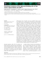
Figure 1.1 Three forms of polarized
cell growth in the Saccharomyces
cerevisiae life cycle.
Cells grown in a
rich medium are round or oval and
have defined budding patterns. When
exposed to a low-nutrient medium,
cells elongate and bud from the distal
end to form pseudohyphae. Haploid
cells exposed to pheromone from cells
of the opposite mating type arrest in
G1 and extend a projection toward
their mating partner. (Reproduced with
permission from Trends Cell Biol.)
(Roemer et al., 1996b)
A second form of polarized growth in yeast is called pseudohyphal growth, which
occurs when there is shortage of nutrients. Under these conditions, yeast cells elongate to
form chains of cells (Fig. 1.1)(Gimeno et al., 1992; Roberts and Fink, 1994).
A third form of polarized growth in yeast occurs during the mating response.
Haploid yeast has two cell types, MATa and MATα. Upon exposure to pheromone from
cells of the opposite mating type, the cells are arrested in late G1 and form an elongated
mating projection (shmoo) (Fig. 1.1) (Cross et al., 1988; Marsh et al., 1991).
Although bud growth, pseudohyphal growth, and the mating response are
different cellular processes, each process undergoes similar steps to achieve polarized
growth (Madden and Snyder, 1998; Casamayor and Snyder, 2002). First, a proper site for
polarized growth is selected and established upon internal or external signals. Next,
cytoskeletons are organized and polarized to the chosen sites. The cytoskeleton then
targets polarized secretion to that site. During these different stages of polarized growth,
both actin and septin cytoskeletons play critical roles in establishment and maintenance
of cell polarity.
3
Chapter 1 Introduction
Figure 1.2 Different stages of budding during the cell cycle. 1) The cell first selects a
defined site according to its ploidy for bud emergence during the late G1 stage of the cell
cycle. 2) The established site then organizes a cytoskeleton network, which is required
for targeting secretion to that site for bud emergence. After bud emergence, cell growth is
restricted first at the bud tip (apical growth) (3) and then throughout the bud (isotropic
growth) (4). When the bud reaches certain sites, the cell undergoes mitosis (5) and
cytokinesis (6), and secretion is directed to the bud neck for the synthesis of septum and
actomyosin ring that separates the mother and daughter. (Modified with permission from
Annu Rev Cell Dev Biol.) (Pruyne et al., 2004b)
4
Chapter 1 Introduction
1.1.1 Bud site selection for polarized growth
Yeast cells choose a bud site according to its ploidy, with diploid cell budding from
the poles of cells (bipolar pattern), and haploid budding from sites adjacent to their
previous bud site (axial pattern) (Fig. 1.3) (Chant and Pringle, 1995; Roemer et al.,
1996b; Casamayor and Snyder, 2002). These budding patterns suggest that the
polarization machinery recognizes the cortical cues that persist from the previous cell
cycle. Initial insights into how this occurs came from a screen for mutants that altered the
axial and bipolar budding patterns (Chant et al., 1991; Chant and Herskowitz, 1991).
Three classes of proteins have been identified to be important for bud site selection. One
class is required for axial budding, but does not affect the bipolar pattern (Fig. 1.4, Gene
set I) (Chant and Herskowitz, 1991). These proteins include Bud3p (Chant et al., 1995),
Bud4p (Sanders and Herskowitz, 1996), Axl2p/Bud10p (Halme et al., 1996; Roemer et
al., 1996a) and Axl1p (Fujita et al., 1994). Mutations of these genes result in bipolar
budding in haploid cells. Another class is important for the bipolar budding pattern of
diploid cells and not required for haploid axial budding (Fig. 1.4, Gene set II) (Zahner et
al., 1996), including Bud8p, Bud9p (Taheri et al., 2000; Harkins et al., 2001) and Rax2p
(Chen et al., 2000). The third class is required for both axial and bipolar budding which
includes the Ras-related GTPase, Rsr1p/Bud1p, and its regulatory GTPase-activating
protein (GAP) Bud2p and guanine-nucleotide exchange factor (GEF) Bud5p (Fig. 1.4,
Gene set III) (Chant et al., 1991; Bender, 1993; Park et al., 1993). The Bud1p GTPase
signaling module is thought to recruit bud formation components, such as Cdc42p,
Cdc24p, and Bem1p (Fig. 1.4, Gene set IV), to the cortical region at the presumptive bud
sites (Zheng et al., 1995; Park et al., 1997; Kozminski et al., 2003).
5
Chapter 1 Introduction
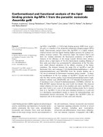
Figure 1.3 Axial and bipolar budding patterns in yeast cells. Staining with the
calcofluor dye permits visualization of two types of scars on any yeast cell surface. The
scar marking the place where the cell was initially attached to its mother (M) cell is called
the birth scar, whereas smaller scars that originated by cytokinesis of the daughter (D)
cells are named bud scars. Examination of the pattern of bud and/or birth scars reveals
different budding patterns. The axial budding pattern is typically found in haploid MATa
and MATα cells, and is characterized by adjacent budding to the birth scar in both mother
and daughter cells. Diploid MATa/MATα cells follow a bipolar budding pattern in which
daughter cells usually bud distally (that is, at the opposite pole to the birth scar), and the
mother cell buds at either pole. The birth scar is represented by a curved black line, and
subsequent bud scars are represented by curved white lines. (Reproduced with permission
from Curr Opin Microbiol.) (Casamayor and Snyder, 2002).
1.1.2 Establishment of polarized growth by Cdc42p
Deletion of any one of the bud site selection genes is not lethal. However, some
genes that are involved in bud formation are essential. Factors required for bud formation
were identified in screens for temperature-sensitive mutants that were arrested as
enlarged, round unbudded cells at the restrictive temperature. Two essential factors
identified in this way are the Rho-family GTPase Cdc42p (Adams et al., 1990; Johnson
and Pringle, 1990) and its Rho-GEF Cdc24p (Sloat and Pringle, 1978; Zheng et al.,
1994). The third component Bem1p was identified as a scaffold protein that binds
Cdc24p and Cdc42p (Zheng et al., 1995; Bose et al., 2001).
6
Chapter 1 Introduction
Figure 1.4 Summary of signaling pathways that lead to the polarity establishment
during bud formation. Proteins belonging to the same functional group are framed in
the same color (Gene sets I–VI). The dotted arrow represents hypothetic regulation of
gene set II by the specific bud-site selection signals present in diploid cells. (Modified
with permission from Curr Opin Microbiol.) (Casamayor and Snyder, 2002)
The
polarity-establishing proteins are thought to promote the assembly of
cytoskeleton components such as actins and septins to target the secretory vesicles to the
bud site for bud formation (Fig. 1.4). The earliest events of polarized growth are the
7