Regulation of DNA (cytosine 5) methyltransferase 1 in the cell cycle and its role(s) in doxorubicin mediated micronuclei formation
Bạn đang xem bản rút gọn của tài liệu. Xem và tải ngay bản đầy đủ của tài liệu tại đây (4.34 MB, 208 trang )
REGULATION OF DNA (CYTOSINE-5)
METHYLTRANSFERASE 1 IN THE CELL CYCLE AND
ITS ROLE(S) IN DOXORUBICIN-MEDIATED
MICRONUCLEI FORMATION
TAN HWEE HONG
NATIONAL UNIVERSITY OF SINGAPORE
2008
REGULATION OF DNA (CYTOSINE-5)
METHYLTRANSFERASE 1 IN THE CELL CYCLE AND
ITS ROLE(S) IN DOXORUBICIN-MEDIATED
MICRONUCLEI FORMATION
TAN HWEE HONG
(B.Sc, Murdoch University)
A THESIS SUBMITTED FOR THE DEGREE OF
DOCTORATE OF PHILOSOPHY
INSTITUTE OF MOLECULAR AND CELL BIOLOGY
AND DEPARTMENT OF PHYSIOLOGY
NATIONAL UNIVERSITY OF SINGAPORE
2008
ACKNOWLEDGEMENTS
I would like to thank my supervisor, Associate Professor Benjamin F.L.Li, for having
faith in my ability to undertake graduate studies and for his guidance and
encouragement throughout the years.
I would also like to thank Professor Alan Porter, Associate Professor Cai Mingjie and
Dr Linda Chuang, members of my supervisory committee, for their advice and
discussion.
I would like to extend my sincere appreciation to;
Prof Alan Porter for his willingness to guide me through the last year of my PhD. I am
most grateful to him for giving me constructive suggestions for my project, and for
reading and editing my thesis. I really appreciate his constant encouragement.
A/Prof Manoor Prakash Hande for his kindness in helping me cross the final hurdle.
Dr Linda Chuang for teaching me many useful techniques and for sharing with me her
invaluable knowledge on DNA methylation. I am grateful for her guidance and her
friendship all these years.
To Miss Yeo Wanlin and Dr Vinay Badal for their friendship and encouragement.
To Dr Oh Hue Kian and Miss Swa Li Foon for their collaborations.
Members of Prof Porter’s laboratory for sharing their knowledge in cell death.
Special thanks to my husband for his support and understanding throughout the years.
Finally, I would like to dedicate this thesis with great love to my parents and my
husband, without whom none of this would have been possible.
i
SUMMARY
DNA methyltransferase 1 (DNMT1) is the major methyltransferase involved in
maintenance methylation of newly synthesized DNA. The regulation of DNMT1
expression is critical in coordinating DNMT1 activity with biological processes and
therefore must be tightly regulated in the cell cycle. Interestingly, DNMT1 expression
is inversely correlated with the cell cycle inhibitor p21
WAF1
protein in mammalian
cells, which is independent of the tumor suppresser p53 as shown here. Using a
combination of experimental protocols including cell synchronization studies,
transient over-expression, siRNA-mediated depletion and luciferase reporter assays,
the roles of the transcription factors E2F1 and Sp1, and the transcriptional co-
activator p300 in the regulation of DNMT1 expression were investigated.
Transcription from the human DNMT1 promoter was shown to be dependent on E2F1
and Sp1. In addition, this study has identified p300 as a crucial transcriptional co-
activator for E2F1 and Sp1 in regulating DNMT1 expression. Most importantly, this
report demonstrates for the first time that p21
WAF1
negatively regulates DNMT1 at the
transcriptional level. The up-regulation of p21
WAF1
by in-vitro over-expression or by
treatment with Trichostatin A led to the corresponding down-regulation of DNMT1,
which consistently coincided with the reduction of p300. Although p21
WAF1
is known
to inhibit the transcriptional activity of E2F1, my data show that p21
WAF1
may
potentially inhibit p300, either directly or indirectly. Surprisingly, DNMT1 was
down-regulated by TSA treatment in the absence of p300, which may be due to the
selective depletion of Sp1 that only occurs when p300 is absent. Nevertheless, my
findings provide the outline of a mechanistic explanation for the inverse relationship
between DNMT1 and p21
WAF1
in mammalian cells. This novel p21
WAF1
-E2F1/p300-
ii
iii
DNMT1 pathway may play a pivotal role to ensure regulated DNMT1 expression and
DNA methylation in mammalian cell division.
De-regulated DNMT1 expression is often associated with tumorigenesis, and there are
reports suggesting that DNMT1 acts as a potential target for cancer therapy. I
attempted to compare the genotoxic effects of doxorubicin, a Topoisomerase II poison
commonly used in chemotherapeutic treatments, on normal and cancer human cell
lines, and the potential involvement of DNMT1 in this doxorubicin-induced
cytotoxicity. The physiological significance of the relationship between p300 and
DNMT1 was further highlighted in the doxorubicin-mediated DNA damage response,
which consistently depleted p300, and consequently DNMT1, in non-tumorigenic but
not tumorigenic cells. My data further show that doxorubicin selectively induces
micronucleation preceded by senescence-like morphological changes in transformed
or tumorigenic cells only, and this consistently correlates with a decrease in cell
viability and an increase in cell death. Importantly, I discovered for the first time a
positive link between DNMT1 protein expression and micronuclei formation.
However, although I provide strong evidence that DNMT1 plays a significant role in
doxorubicin-induced micronucleation, which may lead to cellular demise; it remains
unclear to what extent DNMT1 contributes to the ultimate cell death. Nevertheless,
my findings strongly support DNMT1 as one molecular target for doxorubicin-
induced cytotoxicity in mammalian cancer cells. I proposed that the expression levels
of DNMT1 in tumor cells may potentially determine the effectiveness of doxorubicin
in chemotherapy. This novel observation enhances the understanding of drug response
during doxorubicin administration in cancer therapy.
TABLE OF CONTENTS
Acknowledgements
Summary
Table of Contents
List of Tables
List of Diagrams
List of Figures
Abbreviations
Chapter 1: INTRODUCTION
1.1 Historical Overview of DNA Methylation
1.1.1 DNA methylation in prokaryotic cells
1.1.2 DNA methylation in eukaryotic cells
Mammalian DNA methylation
1.1.3 Demethylation of 5MeC
1.1.4 Host defense versus DNA methylation
1.2 Mammalian DNA Methyltransferases
1.2.1 DNMT1
DNMT1 variants
1.2.2 DNMT2
1.2.3 DNMT3a and DNMT3b
1.2.4 Cooperation among the DNMTs
1.3 Roles of DNA Methylation
1.3.1 Genomic imprinting
1.3.2 X-chromosome inactivation
1.3.3 Epigenetic regulation of genes
1.3.3a DNMT1 interacts with G9a histone methylase
1.3.3b DNMT1 interacts with Polycomb Group (PcG) proteins
1.3.3c DNMT1 interacts with UHRF1
1.3.4 Transcriptional suppression
i
ii
iv
ix
ix
x
xiv
1
2
3
4
5
7
9
10
11
12
13
15
16
17
17
18
19
iv
1.4 DNMT1 in the Cell Cycle
1.4.1 Regulation of DNMT1 expression
1.4.2 E2F1/RB pathway
1.4.3 p300/CBP co-activator
1.4.4 DNMT1 and DNA replication
1.4.5 DNMT1 and PCNA
1.4.6 DNMT1 and p21
WAF1
1.5 Regulation of p21
WAF1
Expression
1.5.1 p53-dependent trancriptional regulation of p21
WAF1
1.5.2 p53-independent transcriptional regulation of p21
WAF1
1.5.3 Translational regulation of p21
WAF1
1.6 DNA Damage Response
1.6.1 ATM/ATR signaling pathways
1.6.2 Cell cycle checkpoints
1.6.3 DNMT1 and p53
1.6.4 DNMT1 in DNA damage and repair
1.6.5 The nucleolus – a DNA damage response center
1.7 Micronuclei
1.7.1 Micronuclei - What are they?
1.7.2 DNMT1 and micronuclei
1.7.3 Possible DNA content in the micronuclei
1.7.4 Doxorubicin
1.8 Research Objectives
Chapter 2: MATERIAL AND METHODS
2.1 Mammalian cell cultures
2.2 Antibodies
2.3 Bacterial strain
2.4 Drugs and Chemicals
2.5 Drug treatments
2.6 Harvesting of mammalian cells
2.7 Flow cytometry analysis
2.8 Cell viability and cell proliferation assays
21
23
25
27
27
29
31
31
32
33
34
36
37
38
40
41
42
43
45
47
47
48
48
48
48
49
49
v
2.9 Lactate Dehydrogenase (LDH) cell death assay
2.10 Purification of total RNA
2.11 Reverse transcription and PCR
2.12 Purification of genomic DNA
2.13 DNA agarose gel electrophoresis
2.14 Methylation-sensitive McrBc restriction digestion
2.15 Cell lysis
2.16 Western blot analysis
2.17 DNA manipulations
2.18 Mammalian expression plasmids
2.19 Cloning of full length DNMT1 into pXJ40-Flag vector
2.20 Cloning of DNMT1 promoter construct into pGL3-Basic vector
2.21 Preparation of competent cells
2.22 Transfection
2.23 Luciferase reporter assay
2.24 siRNA experiments
2.25 Cell staining
2.26 Cell count for micronuclei frequency
2.27 Comet assay
Chapter 3: RESULTS AND DISCUSSION
3.1 Regulation of DNMT1 expression in the Cell Cycle
3.1.1 Inverse relationship between DNMT1 and p21
WAF1
in the
cell cycle
3.1.1.1 DNMT1 expression in the cell cycle
3.1.1.2 Inverse relationship between DNMT1 and p21
WAF1
in DNA
damage
3.1.1.3 Inverse relationship between DNMT1 and p21
WAF1
expression is independent of p53
3.1.1.4 Transient over-expression of DNMT1 does not inhibit
p21
WAF1
3.1.1.5 siRNA-mediated depletion of DNMT1 does not induce
p21
WAF1
49
50
50
51
52
52
52
53
54
54
56
56
57
58
58
59
60
60
61
63
63
67
68
71
72
vi
3.1.1.6 Transient over-expression of p21
WAF1
inhibits DNMT1
3.1.1.7 TSA-mediated induction of p21
WAF1
results in inhibition of
DNMT1
3.1.1.8 TSA-mediated induction of p21
WAF1
is independent of p53
3.1.2 Transcriptional regulation of human DNMT1 promoter
3.1.2.1 siRNA-mediated depletion of E2F1 results in down-
regulation of DNMT1
3.1.2.2 siRNA-mediated depletion of p300 results in down-
regulation of DNMT1
3.1.2.3 Transcriptional regulation of DNMT1 promoter by E2F1,
Sp1 and p300
3.1.3 Discussion
3.2 The Role(s) of DNMT1 in Doxorubicin-mediated micronuclei
formation
3.2.1 Doxorubicin mediates selective depletion of DNMT1 in non-
transformed cell lines
3.2.2 Doxorubicin induces micronuclei formation in transformed
cell lines
3.2.3 Doxorubicin-mediated micronuclei formation is a general
phenomenon in transformed cell lines
3.2.4 Doxorubicin retards proliferation in both transformed and
non-transformed cell lines
3.2.5 Doxorubicin induces micronuclei formation in transformed
cell lines in a dose and time-course dependent manners
3.2.6 Induction of micronuclei correlates with decrease in cell
viability and increase in cell death in the transformed cell
lines
3.2.7 Doxorubicin-mediated micronuclei structures are sites of
DNA damage
3.2.8 Depletion of DNMT1 by 5AzadC treatment attenuates Dox-
mediated micronuclei formation
73
76
85
89
89
92
97
101
108
112
118
120
124
125
129
132
vii
viii
3.2.9 Knockout of DNMT1 attenuates Dox-mediated micronuclei
formation
3.2.10 Knockdown of DNMT1 by siRNA-mediated transfection
attenuates Dox-mediated micronuclei formation
3.2.11 Discussion
Chapter 4: CONCLUSIONS, IMPLICATIONS AND FUTURE
DIRECTIONS
Conclusions
Implications and Future Directions
Chapter 5: REFERENCES
Chapter 6: APPENDIX
List of Publications
136
141
144
151
154
164
190
LIST OF TABLES
1 Forward and reverse primers used for RT-PCR of the cDNA
preparation.
2 Primers used for amplification of human DNMT1 proximal promoter
sequence for cloning into pGL3-Basic vector.
3 Description of the different combinations of treatments with 5μM
Doxorubicin and 2.5μM 5AzadC for 24 hr in MCF7 and MCF-10A
cell lines.
LIST OF DIAGRAMS
1 Schematic diagram of the 1616 amino acid human DNA
Methyltransferase 1 (DNMT1) protein showing the binding sites for
proteins.
2 Epigenetic reprogramming cycle (adapted from Morgan et al, 2005).
3 Circular map of pXJ41-neo vector.
4 Circular map of pGL3-Basic vector.
5 Schematic diagram of DNMT1 promoter spanning 340bp upstream of
the ATG site.
6 Schematic diagram of human DNMT1 proximal promoter region
spanning 340bp upstream of ATG site cloned into pGL3-Basic vector.
7 Speculative model illustrating the mechanism(s) involved for the
inverse relationship observed between DNMT1 and p21
WAF1
in
mammalian cells.
8 Speculative model illustrating the response of non-tumorigenic
(normal) and tumorigenic (cancer) cell lines to the treatment with the
Topoisomerase II poison (doxorubicin).
51
57
133
7
14
55
55
56
97
152
153
ix
LIST OF FIGURES
1 Cell cycle synchronization in MRC5 lung fibroblast non-transformed
(MRC5.NT) cell line shows an inverse relationship between the
expression of DNMT1 and p21
WAF1
.
2 Protein profiles of MRC5.NT cell line treated with 5Gy of Gamma
radiation for different time intervals shows the inverse relationship
between DNMT1 and p21
WAF1
protein expression
3 Protein profiles of MCF7 cell line treated with Gamma radiation
(10gy), methylmethane sulfonate (MMS) (500μM) or H
2
O
2
(0.2mM)
for different time intervals shows the inverse relationship between
DNMT1 and p21
WAF1
protein expression.
4 Cell cycle synchronization in HCT116 parental (WT) and p53-
knockout (p53
-/-
) cell lines shows lack of p53-dependence in the
inverse relationship between DNMT1 and p21
WAF1
expression.
5 MCF7 cells transfected with different concentrations of pXJ.Flag-
DNMT1 plasmids shows DNMT1 over-expression does not increase
p21
WAF1
protein levels.
6 MCF7 and H1299 cell lines transfected with si-RNA to DNMT1 (si-
DNMT1) for different times shows no changes in p21
WAF1
protein
levels.
7 MCF7 cell line transfected with different concentrations of pXJ-
p21
WAF1
plasmids shows over-expression of p21
WAF1
inhibits DNMT1
and p300 proteins.
8 MCF7 and MCF-10A cell lines treated with 100, 200 or 300nM
Trichostatin A (TSA) for 24 hr shows TSA-mediated p21
WAF1
induction leads to down-regulation of DNMT1 expression (Continued
in Figure 9).
9 MCF7 and MCF-10A cell lines treated with 100, 200 or 300nM
Trichostatin A (TSA) for 24 hr shows TSA-mediated p21
WAF1
induction leads to down-regulation of DNMT1 expression.
10 MCF7 and MCF-10A cell lines treated with 200nM Trichostatin A
(TSA) for different times shows TSA-mediated p21
WAF1
induction
leads to down-regulation of DNMT1 expression (Continued in Figure
11).
11 MCF7 and MCF-10A cell lines treated with 200nM Trichostatin A
(TSA) for different times shows TSA-mediated p21
WAF1
induction
leads to down-regulation of DNMT1 expression.
64
68
68
70
71
73
75
78
79
81
82
x
xi
12 Protein profiles of MCF7 cells co-treated with TSA and Mg132 shows
that the reduction of DNMT1 protein upon TSA treatment cannot be
rescued by proteosomal inhibitor
13 HCT116 parental (WT) and p53-knockout (p53
-/-
) cell lines treated
with 200nM Trichostatin A (TSA) for different times shows the lack
of p53-dependence for the down-regulation of DNMT1 by TSA-
mediated p21
WAF1
induction (Continued in Figure 14).
14 HCT116 parental (WT) and p53-knockout (p53
-/-
) cell lines treated
with 200nM Trichostatin A (TSA) for different times shows the lack
of p53-dependence for the down-regulation of DNMT1 by TSA-
mediated p21
WAF1
induction.
15 U2OS cells transfected with si-RNA to E2F1 (si-E2F1) for 8, 32 and
48 hr shows a decrease in DNMT1 expression by E2F1 knockdown.
16 MCF7 and H1299 cell lines transfected with si-RNA to p300 (si-p300)
and si-RNA to DNMT1 (si-DNMT1) for 48 hr shows p300
knockdown depletes DNMT1 expression.
17 Methylation-sensitive McrBc digestion of total genomic DNA upon
si-RNA transfection in MCF7 and H1299 cell lines shows that the
genomic methylation status of the cells was not altered by si-RNA-
mediated p300 and DNMT1 knockdown
18 MCF7 and H1299 cell lines transfected with si-RNA to p300 (si-p300)
for 16, 32 and 48 hr or 72 hr shows decline of DNMT1 protein
associated with reduction in p300 (Continued in Figure 19).
19 MCF7 and H1299 cell lines transfected with si-RNA to p300 (si-p300)
for 16, 32 and 48 hr or 72 hr shows decline of DNMT1 protein
associated with reduction in p300.
20 Protein profiles of MCF7 cells transfected with the respective
plasmids for 48 hr.
21 Luciferase reporter assays of human DNMT1 promoter (DNMT1-Pro)
in MCF7 cells transfected for 48 hr in the absence of exogenous
p21
WAF1
or co-transfection with exogenous p21
WAF1
.
22 MCF-10A and MCF7 cell lines treated with 5μM Dox for different
times shows the preferential depletion of DNMT1 and p300 protein
expression in the non-transformed MCF-10A cells (Continued in
Figure 23).
23 MCF-10A and MCF7 cell lines treated with 5μM Dox for different
times shows the preferential depletion of DNMT1 and p300 protein
expression in the non-transformed MCF-10A cells.
84
86
87
91
93
94
96
97
98
99
109
110
24 MRC5.NT and MRC5.SV40 cell lines treated with 5μM Dox for
different times shows the preferential depletion of DNMT1 and p300
protein expression in the MRC5.NT cells (Continued in Figure 25).
25 MRC5.NT and MRC5.SV40 cell lines treated with 5μM Dox for
different times shows the preferential depletion of DNMT1 and p300
protein expression in the non-transformed MRC5.NT cells.
26 MCF-10A and MCF7 cell lines treated with 5μM Dox for different
times shows the selective induction of micronuclei structures in MCF7
cells upon Dox-mediated DNA damage.
27 Formation of micronuclei in MCF7 cells upon Dox treatment
coincides with an increase in cell size.
28 Comet assay on MCF-10A and MCF7 cell lines with no treatment
(24C) or treated with 5μM Dox for 24 hr (Dox) shows a higher level
of DNA damage, as determined by the longer average tail moment
randomly scored in 200 cells, in MCF7 cells.
29 MRC5.NT and MRC5.SV40 cell lines treated with 5μM Dox for
different times shows the selective induction of micronuclei structures
in MRC5.SV40 cells upon Dox-mediated DNA damage.
30 Formation of micronuclei in MRC5.SV40 cells upon Dox treatment
coincides with an increase in cell size.
31 Mammalian cell lines treated with 5μM Dox for 24 hr shows the
preferential depletion of DNMT1, which coincides with the absence of
micronuclei, in the non-tumorigenic cell lines (IMR90, MCF-10A and
MRC5.NT).
32 MCF-10A and MCF7 cells treated with 5μM Dox shows that Dox
inhibits proliferation in both non-transformed and transformed cell
lines.
33 MRC5.NT and MRC5.SV40 cells treated with 5μM Dox shows that
Dox inhibits proliferation in both non-transformed and transformed
cell lines.
34 MCF7 cells treated with different doses of Dox or with 5μM Dox for
different times shows that Dox induces micronuclei structures in
transformed cell lines in a dose and time-course dependent manners.
35 Induction of micronuclei correlates with a decrease in cell viability
and increase in cell death in MCF7 cells.
36 Induction of micronuclei correlates with a decrease in cell viability
and increase in cell death in MRC5.SV40 cells.
111
112
114
115
116
117
118
119
121
122
125
127
128
xii
37 Immunostaining of MCF7 cells treated with 5μM Doxorubicin for 24
hr and viewed under the fluorescent microscope or confocal imaging
shows the specific localization of H2AX (A), Nucleophosmin/B23
(B), DNMT1 (C) and MRE11 (D) in the micronuclei structures.
38 MCF7 and MCF-10A cell lines treated with 5μM Doxorubicin and
2.5μM 5AzadC for 24 hr shows that Dox can selectively block the
5AzadC-mediated DNMT1 degradation in MCF7 cells (Continued in
Figure 39).
39 MCF7 and MCF-10A cell lines treated with 5μM Doxorubicin and
2.5μM 5AzadC for 24 hr shows the depletion of DNMT1 by 5AzadC
attenuates Dox-mediated micronuclei formation in MCF7 cells.
40 HCT116 DNMT1
+/+
and DNMT1
-/-
cell lines treated with 5μM Dox
for different times shows fewer micronuclei formation in the absence
of DNMT1 (Continued in Figure 41).
41 HCT116 DNMT1
+/+
and DNMT1
-/-
cell lines treated with 5μM Dox
for 24 hr shows that loss of DNMT1 attenuates Dox-mediated
micronuclei formation but was unable to ‘rescue’ cell viability.
42 Si-RNA mediated DNMT1 knockdown leads to inhibition of
micronuclei formation in MCF7 cells (Continued in Figure 43).
43 Si-RNA mediated DNMT1 knockdown leads to inhibition of
micronuclei formation in MCF7 cells.
130
134
135
138
139
142
143
xiii
ABBREVIATIONS
5MeC 5-methyl-cytosine
6meA 6-methyl-adenine
5AzaC 5-Aza-cytidine
5AzadC 5-Aza-deoxycytidine
A.thaliana Arabidopsis thaliana
aa amino acids
mAb monoclonal antibody
pAb polyclonal antibody
ATCC American Tissue Culture Collection
avg average
bp basepairs
BRCA1 breast cancer susceptibility gene 1
BSA Bovine Serum Albumin
Caf Caffeine
ChIP Chromatin Immunoprecipitation Assay
Conc concentration
CpA cytosine-adenine dinucleotide
CpG cytosine-guanine dinucleotide
CpT cytosine-thymine dinucleotide
D. melanogaster Drosophila melanogaster
DAM DNA adenine methylase
DMEM Dulbecco’s Minimum Essential Medium
DNA deoxyribonucleotide acid
DNMT DNA (cytosine-5) methyltransferase
dNTPs deoxy-nucleotide triphosphates
Dox Doxorubicin
DTT Dithiothreitol
E. coli Escherichia coli
ECL enhanced chemiluminescence
EDTA Ethylenediamine tetraacetic acid
EGF epidermal growth factor
FBS fetal bovine serum
xiv
FITC fluorescein isothiocyanate
G, A, T, C guanine, adenine, thymine and cytosine
GAPDH glyceraldehyde-3-phosphate dehydrogenase
GFP green fluorescent protein
GST Glutathione S-transferase
H
2
O
2
hydrogen peroxide
HDAC histone deacetylase
HEPES N-(2-hydroxyethyl)piperazine-N’-2-ethanesulfonic acid
HPV Human papilloma virus
hr hour
ICF Immunodeficiency-Centromeric instability-Facial anomalies
IGF imprinted growth factor
IgG Immunoglobin type G
IP Immunoprecipitation
IPTG Isopropylthio-β-D-galactoside
Kb kilobase
kD kilodalton
LB Luria-Bertani broth
LDH Lactate Dehydrogenase
LOI loss of imprinting
Luc Luciferase
Mab monoclonal antibody
MEM Eagle’s Minimum Essential Medium
min minute
ml mililiter
mM milimolar
MMS methylmethanesulphonate
MN micronucleus/micronuclei
NEB New England Biolabs
NaF sodium fluoride
NLS Nuclear Localization Signal
NP-40 nonindet-p-40
NT non-transformed cells
xv
xvi
pAb polyclonal antibody
PAGE polyacrylamide gel electrophoresis
PBS phosphate buffered solution
PCNA proliferating cell nuclear antigen
PCR polymerase chain reaction
Pf Paraformaldehyde
PI Protease Inhibitor
PMSF phenylmethylsulfonyl fluoride
PVDF polyvinyllidene difluoride
rpm revolutions per minute
RPMI Roswell Park Memorial Institute
RT-PCR reverse transcription-polymerase chain reaction
SAM S-adenosyl-L-methionine
sec second
siRNA small interference RNA
SDS sodium dodecyl sulphate
SLP senescence-like phenotype
SV40 simian 40 virus
TEMED NNN’N-tetramethylethylenediamine
Tris Tris[hydroxymethyl]amino-methane
TSA trichostatin A
μg microgram
μl microliter
μM micromolar
WAF1 wild-type p53 activated factor
WT wild-type
Zn Zinc
Chapter 1: INTRODUCTION
1.1 Historical Overview of DNA Methylation
The genome is encoded by four fundamental base residues, Adenine (A), Guanine
(G), Cytosine (C) and Thymine (T). In addition, minor base residues in the form of
hydroxylated and methylated bases are also present in nucleic acids. Of all DNA
modifications, two methylated base residues, namely 5-methyl-cytosine (5MeC)
(Hotchkiss, 1948) and N
6
-methyladenine (6MeA) (Dunn and Smith, 1955), are
extensively studied as both serve important functions in both prokaryotic and
eukaryotic cells. They are formed by the enzymatic transfer of a methyl group from S-
Adenosyl-L-Methionine (SAM) onto the DNA by specific DNA methyltransferases.
1.1.1 DNA methylation in prokaryotic cells
Both 5-methyl-cytosine (5MeC) (1%) and N
6
-methyladenine (6MeA) (2%) are found
in the prokaryote Escherichia coli (E. coli) genome (Doskocil and Sormo'Va, 1965;
Hattman et al., 1978). DNA cytosine methylase (dcm) methylates the internal
cytosine residue in the palindromic CCA/TGG sequence, and DNA adenine
methylase (dam) methylates the adenine residue in the palindromic GATC sequences
(Razin et al., 1980).
While the function of dcm methylation still remains to be established, dam serves
vital roles related to DNA replication, mismatch repair as well as the host-defense
restriction endonuclease system in E. coli. A time lag between DNA replication and
methylation by dam results in hemi-methylation of the DNA at the replication fork of
the genome (Szyf et al., 1982). Since E. coli OriC has 11 GATC sites that can be
1
methylated by dam, and hemi-methylated OriC demonstrated a higher affinity for
binding to cell membranes (Ogden et al., 1988), this allows preferential binding of the
SeqA protein to GATC sequences, hence delaying premature methylation and re-
initiation from the OriC until DNA replication is completed (Bakker and Smith, 1989;
Kang et al., 1999).
Apart from DNA replication, several lines of evidence point to the pivotal roles of
Dam methylation in post-replication mismatch repair and genetic recombination in
bacteria. It has been shown that the DNA-binding activity of the MutL mismatch
protein is needed for dam methylation-directed mismatch repair in E. coli (Robertson
et al., 2006). Importantly, deficiency of methylation at CCA/TGG by dam is
associated with increased genetic recombination events (Korba and Hays, 1982).
Another important role of dam methylation in the bacterial genome is the restriction
endonuclease system that cleaves foreign DNA to confer protection to the host DNA
from infection. Interestingly, while the mismatch repair system is well conserved in
living organisms, dam methylation system mainly functions in prokaryotes but not in
eukaryotes (Wilson and Murray, 1991).
1.1.2 DNA methylation in eukaryotic cells
The most striking difference between prokaryotes and eukaryotes is the negligible
level of 6MeA in eukaryotic DNA, suggesting the non-essential role of adenine
methylation in eukaryotes. In contrast, 5MeC, mainly located in CG and CNG
sequences, is non-randomly distributed in higher eukaryotic genomes (Gruenbaum et
al., 1981; Ramsahoye et al., 2000) . These DNA methylation patterns play important
roles in gene regulation in diverse organisms including sea urchins (Bird et al., 1979),
2
fungi, plants, protozoa and mammals, with the exception of Saccharomyces cerevisiae
and Caenorhabditis elegans which do not display methyltransferase activities despite
the existence of putative DNA methyltransferase genes, as reviewed by Bestor (Goll
and Bestor, 2005).
Mammalian DNA methylation
The mammalian genome contains about 3 X 10
7
residues of 5MeC which represent
1% to 3% of the total cytosine content of mammalian DNA (Bestor and Tycko, 1996).
Even though 5-methylcytosine has been observed in CpA, CpT and CpC
dinucleotides, CpG is the major methylated dinucleotides that accounts for 40% to
80% of the cytosine methylation in different mammalian genomes (Woodcock et al.,
1987).
While the functional roles of CpA, CpT and CpC methylation remain unresolved,
methylation of CpG in mammalian cells has been implicated in epigenetic
modifications of chromatin such as genomic imprinting, X-chromosome inactivation,
aging and mammalian development (Bird, 2002). However, imprinted genes and
genes subjected to X-chromosome inactivation only account for <10% of the 5-
methylcytosine content in the genome, with the majority residing in transposons
(Walsh and Bestor, 1999; Yoder et al., 1997). This is in agreement with the fact that
transposable elements are generally abundant in the genome (>40%) and are relatively
rich in CpG dinculeotides (Goll and Bestor, 2005; Smit and Riggs, 1996). Genomic
imprinting has been proposed to involve allele and tissue-specific methylation
patterns which are heritable in somatic cells (Wigler et al., 1981). These parental-
specific methylation patterns are erased in primordial germ cells, but are re-
3
established during gametogenesis or early embryogenesis (Chaillet et al., 1991).
Additionally, DNA methylation patterns may also alter during development and
differentiation. Disruption of DNA methylation patterns leads to abnormal
development and recessive embryonic lethality, as demonstrated in the mouse system
by gene targeting mutation of the murine DNA methyltransferase gene (Li et al.,
1992). Knockout of the murine oocyte-specific DNMT1o gene was also shown to
result in the disruption of genomic imprinting in homozygous embryos, further
demonstrating the importance of DNA methylation in mammalian development
(Howell et al., 2001).
1.1.3 Demethylation of 5MeC
DNA methylation in mammals was also shown to be a reversible biochemical process
and the mechanism for DNA demethylation involves DNA demethylases that encode
a methyl-CpG-binding domain (Patra et al., 2008). One such protein identified earlier
in the chick embryo is the 5MeC DNA glycosylase (5-MCDG) which requires RNA
for its demethylating function (Jost et al., 1997). Interestingly, methylcytosine binding
protein MBD4 has also been shown to act as a demethylase with similar 5-MCDG
activity (Hendrich et al., 1999; Zhu et al., 2000). Similarly, MBD2 was shown to
possess dual functions as a transcription repressor and as a DNA demethylase that
target demethylation and activation of specific gene promoters (Detich et al., 2002).
This novel finding was however heavily disputed by others (Ng et al., 1999; Hendrich
et al., 2001).
Demethylation of DNA can also occur by the treatment of cells with demethylating
agents such as 5-azacytidine (5AzaC) and 5-aza-deoxycytidine (5AzadC) (Wolffe et
4
al., 1999). Their incorporation into the DNA causes DNMT1 to become irreversibly
bound to these cytosine structural analogues. This leads to rapid loss of DNMT1 upon
repeated replications, resulting in significant demethylation (Ghoshal et al., 2005).
Similarly, procainamide, a local anesthetic, has been shown to demethylate genes by
inhibiting the maintenance activity of DNMT1 (Lee et al., 2005), and constrained
analogues of procaine targeting DNMT1 have recently been synthesized (Castellano
et al., 2008). In addition, an anti-sense oligodeoxynucleotide to DNMT1, identified as
MG98, has been discovered to effectively deplete DNMT1 expression in mammalian
cells. Though MG98 was shown to have therapeutic potential, clinical studies are still
extensively carried out in patients with myelodysplastic syndrome (MDS) and acute
myeloid leukemia (AML) to determine the efficacy of this drug in targeting aberrant
epigenetics in MDS/AML (Klisovic et al., 2008).
1.1.4 Host defense versus DNA methylation
Cytosine methylation is part of a genome defense system involved in the inactivation
of parasitic sequences such as transposable elements and proviral DNA, and most of
these elements are found to be methylated and transcriptionally inert as
heterochromatin in mammalian, plants and fungi genomes (Bestor and Tycko, 1996).
Indeed, the majority of cytosine methylation in plants and mammals, and all cytosine
methylation in the ascomycete fungus Neurospara crassa, reside in repetitive
elements such as transposons (Goll and Bestor, 2005; Smit and Riggs, 1996).
Strikingly, Drosophila which is deficient in 5meC suffers significantly larger numbers
of insertion mutations (50% to 85%) than mammals, suggesting that cytosine
methylation may have a primary role in regulating the activities of transposons (Yoder
et al., 1997).
5
RNA interference (RNAi) which occurs in plants, mammals and some fungi, is also
capable of directing DNA methylation. Several plant RNA viruses have been shown
to trigger de novo methylation and co-suppression of homologous DNA sequences
during the course of infection (Jones et al., 1998a). The viral double-stranded RNAs
(dsRNAs) are cleaved by the dicer class of ribonuclease into small 21-26 nucleotides
RNAs termed RNAi, which are then degraded by the argonaute protein of the RISC
complex (Liu et al., 2004). Methylation of homologous DNA sequences in the host
results in the formation of a transcriptionally silent heterochromatin, and therefore
confers protection against cleavage and resistance to further infection as a means of
host defense (Mathieu and Bender, 2004). RNAi was also shown to induce DNA
methylation and histone H3 methylation in human cells, as demonstrated by the
silencing of the E-cadherin gene using synthetic RNAi targeted to the CpG islands of
the E-cadherin promoter (Kawasaki and Taira, 2004). However, the importance of this
pathway in mammals is unknown.
Other than host defense, the ability of DNA methyltransferases to recognize
secondary structures may also lead to genomic defects. During DNA replication,
slippage of replication intermediates can result in the extrusion of a segment of
GGC/GCC triplet sequences, which form stable three-way junctions associated with
DNA instability; and this can lead to chromosome fragility (Usdin and Woodford,
1995). Furthermore, this abnormal DNA structure can be preferentially recognized by
DNA methyltransferases for de novo methylation. In such a scenario, the host defense
system attacks an innocuous alteration of an endogenous gene because of its similarity
to a parasitic sequence element. As a result, the GGC/GCC repeats expansions may
lead to diseases related to auto-immune disorders (Bestor and Tycko, 1996).
6
1.2 Mammalian DNA Methyltransferases
DNA (cytosine-5) methyltransferases (DNMT) catalyze the enzymatic transfer of
methyl groups from S-adenosyl-methionine (SAM) to the 5-carbon position of
cytosine residues, predominantly in CpG dinucleotides, to form 5MeC bases
(Robertson, 2001). DNMTs bear ten characteristic sequence motifs, Motifs I to X.
Most of these motifs are discernable in the DNA cytosine-methyltransferases from
bacteria, fungi and plants to mammals (Bestor, 2000). In mammals, three families of
DNA (cytosine-5) methyltransferases have been identified, namely DNMT1,
DNMT2, DNMT3a and DNMT3b.
1.2.1 DNMT1
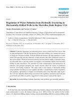
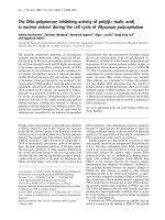
Human DNA Methyltransferase 1 (DNMT1)
Regulatory Domain Catalytic Domain
DMAP1
PCNA
NLS
Replication
Foci Targeting
Cys-rich
Zn binding
(KG)n
HDAC1
DNA
binding
I IV VI IX X
Conserved methyltransferase motifs
BAH
p53 pRb
Catalysis
Diagram 1: Schematic diagram of the 1616 amino acid human DNA methyltransferase 1
(DNMT1) protein showing the binding sites for proteins (Robertson, 2001).
DNA methyltransferase 1 (DNMT1) was the first eukaryotic DNA methyltransferase
to be purified and cloned from the mouse (Bestor et al., 1988). The human homologue
was identified in 1992 (Yen et al., 1992), and the full-length DNMT1 gene encoding
1616 amino acids was mapped to chromosome 19p13.2 (Yoder et al., 1996). The
7