Regulation of subcellular localization and functions of RGK proteins by 14 3 3 and calmodulin
Bạn đang xem bản rút gọn của tài liệu. Xem và tải ngay bản đầy đủ của tài liệu tại đây (6.32 MB, 164 trang )
REGULATION OF SUBCELLULAR LOCALIZATION AND
FUNCTIONS OF RGK PROTEINS BY
14-3-3 AND CALMODULIN
RAMASUBBU NARAYANAN MAHALAKSHMI
DEPARTMENT OF PHYSIOLOGY
NATIONAL UNIVERSITY OF SINGAPORE
INSTITUTE OF MOLECULAR AND CELL BIOLOGY
2006
REGULATION OF SUBCELLULAR LOCALIZATION AND
FUNCTIONS OF RGK PROTEINS BY
14-3-3 AND CALMODULIN
RAMASUBBU NARAYANAN MAHALAKSHMI
(B.Pharm. (Hons.), MSc. (Hons.))
A THESIS SUBMITTED
FOR THE DEGREE OF DOCTOR OF PHILOSOPHY
DEPARTMENT OF PHYSIOLOGY
NATIONAL UNIVERSITY OF SINGAPORE
INSTITUTE OF MOLECULAR AND CELL BIOLOGY
2006
- I -
Acknowledgements
I would like to thank my supervisor Dr. Walter Hunziker for giving me an
opportunity to work in his lab, and also for his patience, kindness and continuous support
throughout the work.
I am grateful to Dr. Pascal Beguin for the collaborations, and for being my mentor
and imparting immense knowledge.
I am thankful to my committee members, Prof. Hong Wanjin and Dr. Edward
Manser for their suggestions and guidance during the annual committee meetings.
I thank my lab mates for their support and help and in particular, Damien and Mei
Yong, for their technical assistance and Carola for her support. I would also take the
opportunity to thank my friend Sumana, for being with me in all ups and downs over the
past five years, for sharing cell lines and providing insights during various discussions.
My sincere thanks to all IMCBites, who have been a part of my work, including
staff of ComIT, administration and support facilities.
I am extremely grateful to my family and friends for all the love, understanding
and patience, especially my husband Rajesh for having enormous trust in me and
providing courage and support during hardship.
Finally, I dedicate this thesis to my dear and late father, without whose blessings,
I could not have been successful.
- II -
Table of contents
Acknowledgements……………………………………………………………………….I
Table of contents…………………………………………………………………… II
Summary of work ……………………………………………………………………V
List of publications… VII
List of figures…………………………………………………………………… VIII
Abbreviations… ……………….…………………………………………………… X
Chapter1: Introduction…………………………………………………………… 1
1.1 Ras superfamily of small GTPases 1
1.2 RGK subfamily of Ras related GTPases 9
1.2.1 Kir/Gem
1.2.2 Rad
1.2.3 Rem1
1.2.4 Rem2
1.3 Regulators and effector of RGK proteins 18
1.3.1 Calmodulin
1.3.2 14-3-3 proteins
1.3.3 β
3
subunit of VDCCs
1.4 Biological functions of small GTPases 26
Chapter 2: Materials and Methods……………………………………………………
38
2.1 Cloning techniques
38
2.1.1 ESTs
2.1.2 Polymerase chain reaction
2.1.3 Restriction digestion and gel electrophoresis
2.1.4 Ligation
2.1.5 Preparation of competent cells
2.1.6 Transformation
- III -
2.1.7 Miniprep and Midiprep
2.1.8 Sequencing
2.2 Cell culture and transfection 40
2.2.1 Propagation of cells
2.2.2 Freezing of cells
2.2.3 Thawing of cells
2.2.4 Transfection
2.3 Protein analysis
43
2.3.1 Cell lysis and homogenate preparation
2.3.2 Preparation of GST fusion proteins
2.3.3 Immunofluorescence
2.3.4 Co-immunoprecipitation
2.3.5 GST pull down
2.3.6 Western blot
2.4 Electrophysiology 47
Chapter 3: Regulation of RGK proteins by CaM and 14-3-3 … 49
3.1 Identification of 14-3-3 binding sites in RGK proteins 49
3.2 Characterization of 14-3-3 binding to RGK proteins 51
3.3 14-3-3 regulates the subcellular distribution of RGK proteins 57
3.4 Modulation of subcellular localization of RGK proteins by CaM
63
3.5 Modulation of localization of RGK proteins by 14-3-3 in the absence of
CaM binding 65
Chapter 4: Roles of 14-3-3 and CaM in cell shape remodeling and down regulation
of calcium channel activity by RGK proteins………………………… .77
4.1 14-3-3 and CaM modulate RGK mediated cell shape changes 77
4.2 Introduction to voltage dependent calcium channels 84
4.3 CaM, but not 14-3-3 plays a role in RGK-mediated down regulation of
calcium channel activity 86
4.4 RGK proteins block cell surface expression of α subunit of VDCCs 88
- IV -
Chapter 5: Identification and characterization of nuclear localization signals in
RGK family of proteins………………………………………………… 94
5.1 Introduction 94
5.2 Identification of NLSs in Kir/Gem 98
5.3 Importin α5 interacts with Kir/Gem and is required for its nuclear localization 104
5.4 The NLSs in Kir/Gem can mediate nuclear localization independently
106
5.5 CaM associated to Kir/Gem interferes with importin α5 binding 108
5.6 Rad, Rem and Rem2 share conserved NLSs 110
Chapter 6: C-terminal phosphorylation modulates the subcellular localization
of RGK proteins……………………………………………………… 116
6.1 Nuclear accumulation of Kir/Gem is regulated by C-terminal
phosphorylation 116
6.2 Regulation of subcellular localization of Rad, Rem and Rem2 by serine
phosphorylation differs as compared to Kir/Gem 122
6.3 Serine (S286) phosphorylation modulates 14-3-3 mediated nuclear exclusion of
Kir/Gem 123
6.4 Serine phosphorylation of Rad, Rem and Rem2 regulates 14-3-3
binding and function 126
Chapter 7: Discussion………………………………………………………………….
132
Chapter 8: Conclusion…………………………………………………………………142
References…………………………………………………………………………… 144
- V -
Summary of work
Kir/Gem, Rad, Rem and Rem2 (RGK) are members of a distinct family of Ras
GTPases. Two important functions of RGK proteins are the regulation of voltage gated
calcium channels (VDCCs) and cell shape remodeling.
In the current study, I did a comprehensive analysis of the interaction of RGK
proteins with 14-3-3 and calmodulin (CaM). The two proteins alter the subcellular
localization of RGK proteins through regulation of nucleocytoplasmic transport. While
14-3-3 binding sequesters the RGK proteins in the cytosol, abolition of CaM binding
allows them to translocate to the nucleus. In addition to the effect on cellular
localization, 14-3-3 and CaM also modulate the cell shape changes induced by RGK
proteins.
The mechanism of regulation of calcium channel activity by RGK proteins was
also studied. Current results show that RGK proteins interact with the β
3
subunit of
calcium channel and this association prevents the interaction of the β
3
subunit with the α
subunit, thereby affecting cell surface expression of the α subunit, which in turn
downregulates calcium channel activity. Further, any possible roles for CaM or 14-3-3 in
the regulation of VDCCs by RGK proteins was investigated and found that CaM but not
14-3-3 affects the modulation of calcium channel activity by RGK proteins.
Since nucleocytoplasmic transport was found to play a significant role in
regulating the functions of RGK proteins, I analyzed if RGK proteins possess any nuclear
localization signals. Indeed, three NLSs were identified in Kir/Gem, which were
conserved in the other RGK members. While NLS1 and NLS2 are non-canonical signals,
- VI -
NLS3 is a typical bi-partite motif consisting of basic amino acid clusters. The study also
revealed that RGK proteins associate with specific importins, which are essential for
nuclear transport of RGK proteins. Furthermore, phosphorylation regulates the
subcellular localization of RGK proteins and 14-3-3 binding to RGK proteins.
Thus our investigations reveal that RGK family of Ras related small GTPases are
subjected to multiple regulatory mechanisms, which may be critical for the selective
control of their effects on the dynamics of cytoskeleton and calcium channel activity.
- VII -
List of publications
1. *Béguin, P., *Mahalakshmi, R.N., Nagashima, K., Cher, D.H., Takahashi, A.,
Yamada, Y., Seino, Y. and Hunziker, W. (2005a). 14-3-3 and calmodulin control
subcellular distribution of Kir/Gem and its regulation of cell shape and calcium
channel activity. J. Cell Sci. 118, 1923-1934
2. *Béguin, P., *Mahalakshmi, R.N., Nagashima, K., Cher, D.H., Kuwamura, N.,
Yamada, Y., Seino, Y. and Hunziker, W. (2005b). Roles of 14-3-3 and calmodulin
binding in subcellular localization and function of the small G-protein Rem2.
Biochem. J. 390, 67-75
3. *Béguin, P., *Mahalakshmi, R.N., Nagashima, K., Cher, D.H., Ikeda, H., Yamada,
Y., Seino, Y. and Hunziker, W. (2006). Nuclear sequestration of beta-subunits by Rad
and Rem is controlled by 14-3-3 and calmodulin and reveals a novel mechanism for
Ca2+ channel regulation. J. Mol. Biol. 355, 34-46.
4. Mahalakshmi, R.N., Nagashima, K., Ng, M.Y., Inagaki, N., Hunziker, W. and
Beguin, P. (2007). Nuclear transport of Kir/Gem requires specific signals, importin
α5 and is regulated by calmodulin and serine phosphorylation. Traffic.
5. Mahalakshmi, R.N., Ng, M.Y. Beguin, P and Hunziker, W. (2007). Nuclear transport
blocks cell shape remodeling and serine phosphorylation regulates 14-3-3 binding and
subcellular distribution of RGK proteins. Traffic.
6. Béguin, P., Kruse, C., Ng, A., Mahalakshmi, R.N., Ng, M.Y. and Hunziker, W.
(2006). RGK small G protein interaction with the nucleotide kinase domain of Ca
2+
channel beta-subunit using an uncommon effector binding domain. J. Biol. Chem.
* First co-authors
- VIII -
List of figures
1-1 Mechanism of action of small GTPases
1-2 Classification of Ras superfamily
1-3 Clustal alignment between Ras and RGK proteins
1-4 Binding of CaM and β
3
subunit to Kir/Gem
1-5 Properties of a 14-3-3 dimer
3-1 Sequence analysis of Rem2 and critical binding sites in RGK proteins
3-2A Binding of 14-3-3 to RGK proteins
3-2B Association of RGK proteins with 14-3-3 dimers
3-3 Cytoplasmic relocalization of RGK proteins by 14-3-3
3-3A Regulation of localization of Kir/Gem by 14-3-3
3-3B Regulation of localization of Rad by 14-3-3
3-3C Regulation of localization of Rem1 by 14-3-3
3-3D Regulation of localization of Rem2 by 14-3-3
3-4 RGK proteins deficient in CaM binding localize to nucleus
3-5 Cytoplasmic relocalization of RGK mutants lacking CaM binding
3-5A Regulation of localization of Kir/Gem W269G and mutants by 14-3-3
3-5B Binding of 14-3-3 to Kir/Gem mutants lacking CaM binding
3-5C Regulation of localization of Rad L281G and mutants by 14-3-3
3-5D Regulation of localization of Rem1 L271G and mutants by 14-3-3
3-5E Regulation of localization of Rem2 L317G and mutants by 14-3-3
3-5F Binding of 14-3-3 to RGK mutants lacking CaM binding
3-6 Quantification of cytoplasmic redistribution and dendritic extensions in
RGK proteins
4-1A Nuclear localization of Rad and Rem reduced RGK induced cell shape
changes
- IX -
4-1B Quantification of induction of dendritic extensions
4-1C Comparison of localization of Rem2 WT and mutants in different cell
lines
4-2 Schematic diagram of the subunits of VDCCs
4-3 Electrophysiology to study the regulation of calcium channel activity by
RGK proteins
4-4 RGK proteins block cell surface expression of α subunit in PC12 cells
4-5 Working model for the regulatory role of 14-3-3 and CaM on Kir/Gem
localization and function
5-1 Mechanism of cargo import by the importin α/β pathway
5-2A List of mutants used in the identification of NLSs in Kir/Gem
5-2(B-E) Identification of NLSs in Kir/Gem
5-2F Localization of mutants used in the study of NLSs in Kir/Gem
5-3 Association of importins with Kir/Gem
5-4 Nuclear translocation of isolated NLSs
5-5 Binding of importin α5 and CaM to Kir/Gem is mutually exclusive
5-6 NLSs in RGK proteins are conserved
5-7 Mutants used in the identification of NLSs in Rad, Rem and Rem2
5-8 Association of importins with Rad and Rem
6-1 Serine phosphorylation regulates subcellular distribution of Kir/Gem
6-2 Phosphorylation state of the serine residue located within the NLS3
determines subcellular localization of Rem but not Rad and Rem2
6-3 Regulation of 14-3-3 binding by C-terminal phosphorylation in Kir/Gem
6-4 Serine phosphorylation modulates 14-3-3 mediated subcellular
redistribution of Rad, Rem and Rem2
6-5 Phosphorylation of a serine upstream from the 14-3-3 binding site
regulates 14-3-3 binding to RGK proteins
6-6 Working model for the regulation of the nucleocytoplasmic shuttling of
RGK proteins
- X -
Abbreviations
aa or a.a amino acids
ADP adenosine 5’-diphosphate
AMP adenosine 5’-monophosphate
ATP adenosine 5'-triphosphate
bp base pair
BSA bovine serum albumin
°C degree Celsius
Ca
2+
Calcium
cAMP cyclic AMP
CC coiled coiled
CACN Calcium channel
CaM Calmodulin
COOH-terminus carboxy-terminus
DMSO Dimethyl Sulfoxide
DNA deoxyribonucleic acid
DTT dithiothreitol
ECL enhanced chemiluminescence
E. coli Escherichia coli
EDTA Ethylenediamine tetraacetic acid
ER Endoplasmic reticulum
GAP GTPase-activating protein
GDP Guanosine diphosphate
GEF Guanine exchange factor
GFP Green fluorescent protein
GK Guanylate Kinase
GST Glutathione S-transferase
GTP Guanosine triphosphate
GTPase Guanosine triphosphatase
HA Haemagglutinin
HEPES Hydroxyethylpiperazine ethanesulfonic acid
HRP Horseradish peroxidase
IgG Immunoglobulin G
IP immunoprecipitation
Kb kilobase(s)
kDa kilodalton
L Liter
LB Luria-Bertani
M Molar
MAGUK Membrane Associated Guanylate Kinase
- XI -
Min minute
ml milliliter
miniprep Small scale plasmid isolation
midiprep Medium scale plasmid isolation
NLS Nuclear localization signal
NES Nuclear export signal
NPC Nuclear pore complex
mM millimolar
µg microgram
µm micrometer
nm nanometer
NH
2
-terminal amino terminal
OD optical density
ORF open reading frame
PAGE polyacrylamide gel electrophoresis
PBS phosphate-buffered saline
PCR polymerase chain reaction
PEG polyethylene glycol
PKA protein kinase A
PKC protein kinase C
PLCγ phospholipase Cγ
PM Plasma membrane
PMSF phenylmethylsulfonyl fluoride
PVDF polyvinylidene difluoride
rpm revolutions per minute
sec second
SDS Sodium dodecyl sulphate
SH3 Src homology 3
SV40 Simian virus 40
TE Tris-EDTA buffer
TEMED N,N,N',N'-tetramethylethylenediamine
Tris Tris(hydroxymethyl)aminomethane
WT wild type
1
CHAPTER 1
Introduction
1.1 Ras Superfamily of small GTPases: GTPases, along with their regulators
and effectors, function as central elements in signal transduction pathways that control
virtually every aspect of cell biology. These GTPases fall within a superfamily named
RAS. The Ras superfamily of GTPases are a large group of proteins comprising over 150
members with evolutionarily conserved proteins in D.melanogaster, C.elegans,
S.cerevisiae, S.pombe, D.discoideum and plants (Colicelli, 2004). GTPases of the Ras
superfamily regulate a wide variety of cellular functions, acting as biotimers that initiate
and terminate specific cell functions and thus control the duration of different processes.
They play key roles not only in the temporal but also in the spatial determination of
cellular functions. Generally small GTPases act as molecular switches alternating
between an inactive GDP bound “OFF” state and an active GTP bound “ON” state. They
exhibit high affinity binding for GDP and GTP and possess low intrinsic GTP hydrolysis
rates. The key regulators of the switch process are GTPase activating proteins (GAPs)
and Guanine exchange factors (GEFs), which determine the equilibrium between active
and inactive states. Following an upstream signal, GEFs exchange the GDP to GTP, thus
promoting the formation of active GTP bound protein. This leads to a conformational
change in the effector binding region of the GTPases, which allows interaction with
downstream effectors, thereby triggering a number of cellular functions. GAPs
accelerate the GTP hydrolysis rate in the active protein and help in the conversion to the
inactive GDP bound state, leading to the release of the bound effectors. Thus, the GEFs
function as positive regulators and GAPs function as negative regulators of Ras signalling
2
and together, they alternate cycles of activation and inactivation, which transduces an
upstream signal to a downstream effector. In addition to the switch process, the spatial
and temporal distribution of the small GTPases , as well as of their regulators, are equally
important determinants of Ras signaling. These include variations in structure, post-
translational modifications that may specify defnite subcellular localizations and the
regulators and effectors, which allow the small GTPases to function as sophisticated
modulators of cellular processes.
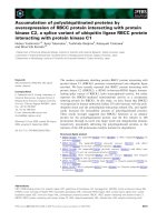
Fig. 1-1 Mechanism of action of small GTPases. Guanine exchange factors or
GEFS bind to inactive small GTPases and convert them to the active GTP bound
form. The proteins in the active state can interact with effectors and trigger a
number of signaling events. The active GTPase is inactivated by GTPase
activating proteins or GAPS.
Effector
GDP bound-
Inactive-
Off state
GTP bound-
active-
On state
Guanine Exchange Factors
(GEFs)
(GAPs)
GTPase Activating Proteins
3
The Ras superfamily can be grouped into five subfamilies based on their sequence
and functional similarities: Ras, Rho, Rab, Arf and Ran (Takai
et al., 2001). In general,
the subfamilies have distinct functions: the Ras subfamily mainly regulates gene
expression, the Rho subfamily regulates cytoskeletal reorganization and gene expression,
the Rab/Arf members control intracellular vesicle trafficking and the Ran members are
involved in nucleocytoplasmic transport.
Members of the Ras superfamily share a conserved core region composed of a set
of so called G domains namely G1, G2, G3, G4 and G5. These domains play specific
roles in phosphate binding, guanine binding and effector binding. Key amino acids and
motifs in the various domains of the core region are conserved among the different
members of the Ras family: G1-GXXXXGKS/T; G2-T; G3-DXXGQ/H/T; G4-T/NKXD
and G5-C/SAK/L/T (Colicelli, 2004 and Wennerberg et al., 2005). The structural
changes during the conversion between inactive and active states are confined to two
loop regions called switch I (Ras residues 30-38) and switch II (Ras residues 59-67).
These regions exhibit pronounced differences in conformations during the switch process
and it is mainly through these conformations that the regulators and effectors sense the
nucleotide status of the small GTPases. A second important biochemical feature of Ras
proteins is their post translational modification by lipids. Majority of the Ras GTPases
contain a C-terminal CAAX (C-Cysteine, A-aliphatic and X-any amino acid) sequence.
The CAAX motif is the recognition sequence for farnesyl transferase and geranylgeranyl
transferase, which catalyze the covalent addition of a farnesyl or geranylgeranyl group to
the cysteine residue. This modification is normally required for the specific subcellular
location of the GTPases.
4
Rho and Rab GTPases are alos regulated by a third class of molecules called
Guanine Dissociation Inhibitors (GDIs), which mask the prenyl modification and
promote cytosolic sequestration of these GTPases (Seabra et al., 2004). It is interesting
to note that some Ras members do not appear to be modified by lipids, but still associate
with membranes (e.g. Rit, Miro, sar1) while some others are not lipid modified and are
not bound to membrane (e.g. Ran, Rerg) (Wennerberg et al., 2005).
While the branching of the Ras superfamily into 5 subfamilies is largely based on
functional and structural similarities, grouping into the various subfamilies is often
arbitrary (Wennerberg et al., 2005).
5
Fig. 1-2 Classification of Ras superfamily
(Reproduced from Wennerberg, K. et al. J Cell Sci 2005;118:843-846, with permission of the company of Biologists)
`
6
Subfamilies of the Ras Superfamily
The Ras subfamily: The RAS (Rat Sarcoma) oncoproteins have been of
immense interest due to their critical role as human oncogenes. Mutations in three Ras
genes have been detected in ~ 30% of human cancers. Ras proteins were first identified
in the Harvey and Kersten strains of acutely transforming reteroviruses. There are about
35 members in this subfamily and they show high conservation in the core domain.
Most Ras proteins are localized in the membrane due to prenylation of the Cys in
a C-terminal CAAX motif. While some Ras proteins possess other lipid modifications
like geranylgeranylation, farnesylation, acetylation and palmytoylation, others lack lipid
modifications. N-terminal lipidation like myristoylation and palmitoylation are also
found in some Ras proteins. Well characterized Ras effectors include RAF1, PI3K,
RIN1, RAL GEFs etc. The best characterized Ras signaling pathway is the activation of
Ras by the epidermal growth factor receptor tyrosine kinase through the Ras GEF SOS
(Repasky et al., 2004). Activated Ras binds to Raf and translocates it to the plasma
membrane, where it undergoes additional phosphorylation for complete kinase activation.
Raf phosphorylates and activates MEK, which further phosphorylates and activates ERK,
a MAP kinase. Once activated, ERK translocates into the nucleus, where it triggers
activation of downstream promoters. Thus a number of Ras family proteins regulate
different signaling networks. They not only regulate cell proliferation, but also control
differentiation, cellular morphology and apoptosis. Interestingly, some Ras proteins such
as Rerg, Noey2 and D-Ras, seem to act as tumor suppressors, rather than as oncogenes
(Colicelli, 2004). This exemplifies the diversity and complexity of the functional roles of
the different members of the Ras subfamily.
7
The Rho subfamily: Rho (Ras homolog) proteins are key regulators of signaling
networks that mediate actin organization, cell cycle progression and gene expression
(Etienne-Manneville and Hall, 2002). RhoA, Rac and Cdc42 are the best studied among
the 23 members that have been categorized into this subfamily. RhoA is involved in
actin stress fibre formation and focal adhesion assembly, Rac1 promotes lamellipodia
formation and membrane ruffling and Cdc42 promotes actin micropikes and filopodia
formation (Wennerberg et al., 2005). Rho GTPases have been implicated in multiple
aspects of cell polarity, cell motility, cell shape remodeling, cell-cell interaction and
regulation of endocytosis and exocytosis (Ridley, 2001). Rho GTPases are regulated by a
large diversity of GAPs and GEFs and possess a varied range of downstream effectors.
Some of the Rho family effectors include ROCK1, mDia, PLCB etc. Like the Ras
proteins, this subfamily is also subject to a number of post translational modifications.
Although the G boxes are highly conserved between the Rho and Ras family members,
the former has an additional insert sequence that is absent in Ras subfamily members.
Significantly distinct proteins of the Rho subfamily are the Miro or RHOT proteins which
show a notable sequence divergence from the other members, lack the insert sequence as
well as lipid modifications. Miro proteins possess two EF-hand domains that may confer
calcium binding, a function that is unique to them. They also possess a C-terminal
GTPase like domain, the significance of which is not known yet. Miro proteins, being
localized to mitochondria, regulate the integrity of the compartment (Krister et al., 2005).
The Rab subfamily: The Rab proteins were originally identified as Ras proteins
in
brain. They are the largest subfamily of the Ras superfamily with around 61 members
(Pereira-Leal and Seabra, 2001). Rab proteins function in protein trafficking pathways
8
by regulating vesicle formation, budding of vesicles from donor compartments, transport
to acceptor compartments and vesicle fusion. Effectors of Rabs are rabphilin, RILP,
M6PRBP1 etc. Most Rab proteins undergo C-terminal prenylation, which determines
their subcellular localization and function. Rab5, for example, localizes to early
endosomes and regulates clathrin coated vesicle mediated transport from plasma
membrane to early endosomes.
The Ran subfamily: The Ras like nuclear proteins (RAN) are very closely
related to Rab proteins by sequence homology and are the most abundant small GTPase
in cells. They are involved in the regulation of nucleocytoplasmic transport of RNA and
proteins, which is primarily dependent on the spatial gradient of the GTP-bound Ran.
The presence of Ran GAP in the cytosol and Ran GEF (RCC1) in the nucleus establishes
a gradient of Ran activity across the nuclear membrane and pore complex, determining
the directionality of nuclear import and export. Nuclear Ran-GTP binds to importins and
transports them to the cytosol where they are released and Ran GTP is converted to Ran
GDP by Ran GAP. Ran-GDP binds to exportin and diffuses back to the nucleus where
GDP is exchanged to GTP by Ran GEF. Ran is characterized by an acidic C-terminal
region and is not known to undergo prenylation.
The ARF subfamily: ADP Ribosylation Factors or ARFs are regulators of
intracellular trafficking and cytoskeletal remodeling. Arf proteins lack C-terminal lipid
modifications but are subject to N-terminal myristoylation. Conformational differences
between the GDP and GTP bound forms not only occur in the switch I and II regions, but
also in the N-terminal region, where the myristate group interacts with membranes in the
active state. Effectors mediating Arf functions include Arfaptins and Arfopilins.
9
There are six known Arf members, Arf 1-6. Arf1 is the best characterized and is
involved in the regulation of vesicle formation in endocytic and exocytic pathways. Arf6
is known to function in actin reorganization and endocytosis.
1.2 RGK subfamily of Ras related proteins: The RGK (Rad, Rem/Gem/Kir)
family belongs to the Ras-related GTPase family and consists of
Kir/Gem (Cohen et al.,
1994; Maguire
et al., 1994), Rad (Reynet and Kahn, 1993), Rem or Rem1 (Finlin and
Andres, 1997) and
Rem2 (Finlin et al., 2000).
The RGK proteins exhibit several unique structural and functional features that
differentiate them from other Ras related proteins. The basic structure of RGK proteins
consists of a ras related core domain flanked by distinct C- and N-terminal extensions.
The Ras related core domain is divided into G1, G2, G3, G4 and G5 regions which are
involved in guanine, phosphate and effector binding. The RGK proteins differ among
themselves in the effector (G2) domain, which implies that they might have different
interacting partners. The RGK family members differ from other Ras like GTPases in a
number of characteristic features. Firstly, the G3 motif, which in Ras participates in
binding and hydrolysis of GTP, is unique (DXWE instead of DXXG), implying that the
RGK proteins probably share an exclusive molecular mechanism for GTP hydrolysis or
do not hydrolyze GTP. Secondly, RGK members contain a notable N- and C-terminal
extension flanking the Ras-like core region. The C-terminal extension includes a
calmodulin binding region, linking these proteins to calcium signaling events (Fischer et
al., 1996; Moyers et al., 1997). Thirdly, they do not have classical lipid modification
motifs at the C-terminus, which in other Ras-like proteins undergo lipid modifications
that are important for membrane anchorage. Another distinctive characteristic is their
10
tissue specific expression patterns and regulation at the transcriptional level (Cohen et al.,
1994; Finlin and Andres, 1997; Maguire et al., 1994; Reynet and Kahn, 1993). Further
key amino acids in Ras like the G12 and Q61 have been substituted for other amino acids
in RGK proteins.
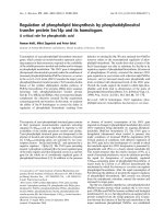
(Reproduced with permission from Finlin et al., 2000, Biochemical Journal, 347, 223-231, the Biochemical society)
Fig. 1-3 Clustal alignment between Ras and RGK proteins. Comparison of the amino
acid sequences of rat (r) Rem2, mouse (m) Rem, human (h) Gem, hRad, hKir and rK-ras-
2B proteins.
Hyphens represent gaps introduced for optimal alignment. Numbers are
residue numbers. Amino acid residues that are conserved in at least two of the five
proteins in the alignment are shaded in grey. Consensus sequences for GTP-binding
regions (G1–G5) and the conserved C7 sequence motif are labelled. The G3 consensus is
unique to the RGK family and is underlined and in italics.
11
1.2.1 Kir/Gem: Gem (Gene expressed in Mitogen stimulated T cells) is the
human orthologue of mouse Kir (
Kinase inducible ras like). Kir was identified by a
differential screen aimed at isolating genes involved in malignant transformation by the
BCR/ABL oncogene. BCR/ABL is a fusion gene resulting from the t(9;22) chromosomal
translocation, a cytogenic marker of chronic myelogenous leukemia. Kir and Gem are
about 35kDa in size and their expression is highly regulated. Gem has been shown to be
an immediate early gene in primary lymphocytes, monocytes, endothelial cells and
human embryonic fibroblasts (Maguire et al., 1994, Van hove et al., 1997). Gem mRNA
is detected in thymus, spleen, kidney, testis and lungs (Maguire et al., 1994). Gem is
constitutively expressed in neuroblastoma cell lines and ectopic Gem expression
stimulates cell flattening and neurite extensions in human (SH-SY5Y) and mouse
(N1E115) neuroblastoma cell lines (Leone et al., 2001). It could be interesting to analyze
the metastatic potential of neuroblastoma expressing ectopic Gem. Overexpression of
Kir in Saccharomyces cerevisiae induces invasive pseudohyphal growth. Kir induced
pseudohyphae formation requires a MAP kinase cascade involving ste20, ste11 and ste7
(Dorin et al., 1995). Calmodulin binds to Kir/Gem and inhibits GTP binding to the
protein. The C-terminus of Kir/Gem exhibits hallmarks of a typical calmodulin binding
domain. A point mutant W269G was identified to completely abolish calmodulin
binding to Gem (Fischer et al., 1996).
Gem interacts with a kinesin like molecule called KIF9 and has been shown to be
associated with microfilaments and microtubules. The dynamics of Gem mediated
formation of long cellular extensions were studied by time lapse video microscopy and
events such as cell body retraction, increased filopodia formation, increased nuclear
12
migration and cortical contraction were observed. It was also reported that the Gem
induced extensions need intact actin and microtubules. Both Gem and Kif9 display
identical expression patterns in different tissues and developmental stages indicating a
molecular link between Gem and the cytoskeleton. The group also showed that
nucleotide binding was required for the complete elongation activity of Gem since Gem
S89N, bearing a single point mutation in the GTP binding site was significantly less
active compared to the WT, in terms of cell morphology changes (Piddini et al., 2001).
The potential role of Gem-Kif9 interaction could be manifold. Gem might be regulating
the motor activity of KIF9, as already proposed for rab6 on rabkinesin6 or the interaction
could provide ways of recruiting Gem on microtubules, which would put it in the vicinity
of its effectors/regulators. A number of evidences state that microtubules act as signal
transduction platforms where key components are recruited through kinesins for
downstream signaling.
Kir/Gem is involved in the negative regulation of Rho pathway through its
interaction with Rho kinase β. It prevents Rok-β mediated cell rounding and neurite
retraction, thereby implying a role in cytoskeletal organization. Gem binds Rokβ
independently of RhoA in the Rokβ coiled-coil region. The interaction affects the
substrate specificity of Rokβ by inhibiting phosphorylation of myosin light chain and
myosin phosphatase, but not LIM kinase (Ward et al., 2002).
Kir/Gem also functions in the regulation of voltage dependent calcium channels.
The Ca
2+
transporting α
1
subunit of voltage-dependent Ca
2+
channels associates with
auxiliary subunits (
β, α
2
δ and γ subunits) that have regulatory functions (Catterall,
1998)). Kir/Gem interacts with the Ca
2+
channel β-subunit in a GTP dependent manner