Group 2 allergens from dust mite epitope mapping and functional characterization of der p 2, and identification of a paralogue of der f 2
Bạn đang xem bản rút gọn của tài liệu. Xem và tải ngay bản đầy đủ của tài liệu tại đây (2.2 MB, 235 trang )
GROUP 2 ALLERGENS FROM DUST MITE:
EPITOPE MAPPING AND FUNCTIONAL
CHARACTERIZATION OF DER P 2, AND
IDENTIFICATION OF A PARALOGUE OF DER F 2
KAVITA REGINALD
(B.Sc. (Hons.), UPM)
A THESIS SUMBITTED FOR THE DEGREE OF
DOCTOR OF PHILOSOPHY
DEPARTMENT OF BIOLOGICAL SCIENCE
NATIONAL UNIVERSITY OF SINGAPORE
2006
Acknowledgements
It is close to impossible to do good scientific research in graduate school without help.
As most other students who have taken the same path will agree, it is a long and
arduous journey, with short bursts of satisfactions when discoveries are made. There
have been many people who have helped me in my journey as a young scientist in
graduate school, and I wish to extend my thanks to them in this section.
My supervisor, Dr Chew for the research opportunity and guidance in the last 5 years.
My lab mates, for assistance in laboratory techniques and continually giving me
constructive suggestions. A big thanks goes out to Tan Ching for the immunological
experiment techniques, and Siew Leong for advice and assistance in the protein
studies.
The allergic patient volunteers, for your time and cooperation throughout this study.
My friends in university. Special thanks goes out to Sai Mun and Souvik for always
giving me timely assistance and advice in so many areas of research. Also to Shruthi,
who has been of great assistance for databasing, and analysis of some sections. To
Vaane, Grace, and Dr. Tan for patiently reading and editing the thesis. To Siva, for
lending technical help.
My collaborator, Dr. Markus, for being an inspirational scientist, and giving me the
opportunity to venture into the study of lipids. Also to members of his research lab,
especially Guanghou and Gek Huey whom I have worked closely with.
My friends outside university. I thank all of them for making my stay in Singapore a
fulfilling one. To Radhi, Shion, Ken, Shih Lene and Wan Yee, needless to say, I have
thoroughly enjoyed all our house parties, cook outs and chill out sessions. To Shashi,
Harveen, Jam and Punam, you have evolved from friends to my family. I have no
words to express my gratitude.
The Sahaja Yogis. How would you thank those who have helped you to connect to
your spirit? There are no words, just bliss. You have helped me discover the true
meaning of life, and made it possible for me to go through the difficult moments. A
huge hug for the ‘world collective’ especially to Geethanjali and Michalis for your
assistance.
My family. Without their blessing and continual support, none of this would be
possible.
The Divine.
i
Disclaimer
Some parts of the experiments were done together with other lab members, and are
listed here.
In chapter 3, IgE inhibitions were done together with Aaron Chen.
In chapters 3-8, screening of IgE reactions was done together with Yap Kwong Hsia.
In chapter 4, cloning of Blo t 2 was done together with Kway Kwee Theng.
Quantification of allergen concentration in dust samples was done with Kelly Goh.
Extraction of native Blo t 2 was done with Alvin. Histamine release assay was done
with Gavin. Study of isoforms was done with Jia Yi.
In chapter 5, immmunostaining and southern blot analysis was done together with Dr.
Tan Chye Ling. Quantification of allergen concentration in dust samples was done
with Chen Simin. Phylogenetic analysis was done with Shruthi and Dr. Yap Von Bing.
In chapter 6, mass spectrometry was done together with Shui Guanghou. Docking
analysis was done with Chua Gek Huey.
In chapter 8, the cytokine assay was done together with Dr. Ong Tan Ching.
ii
List of conference abstracts and book chapters
International Conference Abstracts
Reginald K, L Haroon-Rashid, YS Sew, SH Tan, FT Chew (2002). Identification of
putative Tyrophagus putrescentiae allergens with sequence homology to other known
allergens by expressed sequence tagging. In: XXIth European Academy of
Allergology and Clinical Immunology Annual Meeting (EAACI), June 2002, Naples,
Italy. Allergy 57 (Suppl. 73): 286-7.
Loo AHB, SPL Tan, AC Angus, KT Kuay, K Reginald, YF Gao, FT Chew (2003).
Genetic Relationship Between Allergy-Causing Dust Mites: Phylogenetic Inference
From Random Amplified Polymorphic DNA (RAPD) Markers, Housekeeping Gene
(18S rDNA) And Group 2 Allergens. In: The 60th American Academy of Allergy and
Immunology Annual Meeting, 7 - 12 March 2003, Denver, USA. J Allergy Clin
Immunol 111 (2): S162.
K Reginald, XZ Bi, ST Ong, FT Chew (2003). Profiling of Crude Allergen Extracts
Using SELDI Mass Spectrometry for Rapid Standardization. In: The 60th American
Academy of Allergy and Immunology Annual Meeting, 7 - 12 March 2003, Denver,
USA. J Allergy Clin Immunol 111 (2): S242.
AHB Loo, SY Goh, K Reginald, YF Gao, H Jethanand, HS Shang, FT Chew (2004).
Validation of the Purity of Acarid Mite Cultures Used for Allergen Extract
Preparation and Identification of Contaminants by Ribosomal DNA Sequencing via a
PCR-Cloning- and Sequence Homology-Based Approach. In: The 61th American
Academy of Allergy and Immunology Annual Meeting, 19 - 24 March 2004, San
Francisco, USA. J Allergy Clin Immunol 113 (2): S140.
K Reginald, YF Gao, YS Siew, HS Shang, FT Chew (2004). Cross Comparison of
the IgE Binding Profiles to Recombinant Allergens from Suidasia medanensis,
Blomia tropicalis and Dermatophagoides farinae using Sera from Blomia- and
Dermatophagoides-Predominant Environments. In: The 61th American Academy of
Allergy and Immunology Annual Meeting, 19 - 24 March 2004, San Francisco, USA.
J Allergy Clin Immunol 113 (2): S228-9.
Reginald K, Gao YF, Lim YP, Chew FT (2004). The expressed sequence tag
catalogue and allergens of dust mite, Suidasia medanensis. In: XXIIIth European
Academy of Allergology and Clinical Immunology Annual Meeting (EAACI), June
2004, Amsterdam, The Netherlands.
Tay ASL, Shang HS, Bi XZ, Reginald K, Gao YF, Angus AC, Ong ST, Wang WL,
Kuay KT, Wang DY, Mari A, Chew FT (2005). Component-Resolved Diagnosis Of
House Dust Mite Allergy With A Large Repertoire Of Purified Natural And
Recombinant Allergens From The Major Species Of Mites Worldwide. In: The 62th
American Academy of Allergy and Immunology Annual Meeting, March 2005, San
Antonio, USA. J Allergy Clin Immunology, 115 (2): S164
iii
Reginald K, Wenk MR, Chew FT (2005). The major mite allergen from
Dermatophagoides pteronyssinus, Der p 2, is a sterol binding protein. In: The 62th
American Academy of Allergy and Immunology Annual Meeting, March 2005, San
Antonio, USA. J Allergy Clin Immunology, 115 (2): S88
Tan CL, Reginald K, Chew FT (2006). Genomic organization and characterization of
group 2 allergen paralogs from Dermatophagoides farinae. In: The 63th American
Academy of Allergy and Immunology Annual Meeting, March 2006, Miami, Florida,
USA. J Allergy Clin Immunology, 117 (2): S120
Reginald K, Chew FT (2006). Epitope mapping of Der p 2 by site directed
mutagenesis: Differential IgE binding epitope profile among individuals sensitized to
only Dermatophagoides spp. and those with non-pyroglyphid mite responses. In: The
63th American Academy of Allergy and Immunology Annual Meeting, March 2006,
Miami, Florida, USA. J Allergy Clin Immunology, 117 (2): S118
Book Chapter
Reginald K, Sew YS, Haroon-Rashid L, Kulaveerasingam H, Tan SH, Chew FT.
Chapter 49. Identification of putative Tyrophagus putrescentiae allergens with
sequence homology to other known allergens by Expressed Sequence Tagging.
Progress in Clinical Immunology and Allergy in Medicine. Edited by Gianni Marone.
(invited Book Chapter)
iv
Table of contents
Page
Acknowledgements
i
Disclaimer
ii
List of conference abstracts and book chapters
iii
Table of contents
v
List of figures
xi
List of tables
xv
List of abbreviations
xvi
Summary
xx
Chapter 1: Introduction
1
1.1 Literature Review
1
1.1.1 Allergy
1
1.1.2 Dust mites
4
1.1.3 Dust mite allergens
7
1.1.4 Immunotherapy as a treatment for allergic diseases
12
1.2 Aims
14
Chapter 2: Materials and methods
16
2.1 Cloning, mutagenesis, DNA sequencing and gene characterization
16
2.1.1 Sub-cloning and site-directed mutagenesis
16
2.1.2 RT-PCR of putative Blo t 2 using degenerate primers
16
2.1.3 DNA sequencing
17
v
2.1.4 Isolation of Blo t 2 isoforms
18
2.1.5 Isolation of the genomic DNA encoding for Der f 2 and Der f 22
18
2.1.6 Genomic DNA extraction, Southern Blot analysis and hybridization
19
2.2 Protein expression, purification, CD analysis and antibody generation
20
2.2.1 Expression and purification of wild type and mutant allergens
20
2.2.2 Isolation of native Blo t 2
21
2.2.3 Circular dichroism (CD) spectropolarimetry
21
2.2.4 Gel Filtration
21
2.2.5 Generation of rabbit polyclonal antibodies
22
2.3 Serum samples
22
2.4 Immunological assays
22
2.4.1 Immuno dot blot
22
2.4.2 Specific IgE binding ELISA
23
2.4.3 Inhibition ELISA
24
2.4.4 Histamine release assay
25
2.4.5 Dust sample collection, processing and quantification.
25
2.4.6 Staining and immunoprobing
26
2.4.7 Skin prick test
26
2.4.8 Isolation of PBMC and measurement of proliferation upon stimulation with
wild type or mutant allergen
27
2.4.9 Measurement of excreted cytokines
28
2.4.10 Mouse immunization
28
2.4.11 Inhibition of human IgE binding to Der p 2 by specific mouse IgG
antibodies
2.5 Computer based characterization and analysis
28
29
vi
2.5.1 Analysis of DNA and protein sequences
29
2.5.2 Three dimensional protein structure predictions
29
2.5.3 Phylogenetic sequence analysis
30
2.5.4 Docking of cholesterol to Der p 2
31
2.6 Lipid assays
2.6.1 Liposome preparation
33
33
2.6.2 Detection of liposome binding to Der p 2 by liposome sedimentation and
SDS-PAGE
33
2.6.3 Lipid ELISA
34
2.6.4 Extraction of lipid fraction from Der p 2
34
2.6.5 HPLC/APCI/MS/MS analysis of cholesterol
35
2.7 Statistical analyses
36
2.8 Approval
36
Chapter 3: IgE reactivity and cross reactivity profiles of group 2 allergens
37
3.1 Introduction
37
3.2 Cloning and sequence analysis of Ale o 2, Sui m 2 and Blo t 2
39
3.3 IgE reactivity to group 2 allergens
50
3.3.1 IgE reactivity to group 2 allergens in the Singaporean population
50
3.3.2 IgE reactivity to group 2 allergens in the Italian population
56
3.4 Further characterization of Blo t 2
61
3.4.1 Isolation of native Blo t 2
61
3.4.2 Histamine release of Blo t 2
61
3.4.3 Blo t 2 is present in the environmental dust samples
64
3.4.4 Isoforms of Blo t 2
66
vii
3.5 Discussion
69
Chapter 4: Identification and characterization of Der f 22, a novel allergen from
Dermatophagoides farinae: a paralogue of Der f 2?
75
4.1 Introduction
75
4.2 Identification, isolation and characterization of Der f 22
76
4.3 Genomic organization of Der f 22 and Der f 2
83
4.4 Southern blot analysis
86
4.5 IgE binding capacities of Der f 22 and Der f 2
88
4.6 Localization of Der f 22 and Der f 2 on sectioned D. farinae
92
4.7 Concentration of Der f 22 and Der f 2 in the indoor environment
94
4.8 Der f 2 and Der f 22 binds to cholesterol
96
4.9 Presence of paralogues of group 2 allergens
98
4.10 Discussion
101
Chapter 5: Der p 2 is a cholesterol binding protein
105
5.1 Introduction
105
5.2 Der p 2 binding to liposomes
107
5.3 Binding of Der p 2 to purified lipids
109
5.4 Analysis of lipid extracts from nDer p 2 and rDer p 2
112
5.5 Characterization of potential cholesterol binding sites in Der p 2 via site
directed mutagenesis
114
5.6 Native Der p 2 and two other allergens of the ML domain family bind to
cholesterol
117
5.7 Docking of cholesterol on Der p 2
119
viii
5.8 Discussion
121
Chapter 6: IgE epitope mapping of Der p 2
125
6.1 Introduction
125
6.2 Design and production of Der p 2 alanine mutants with single amino acid
substitution.
127
6.3 Reaction profile of dust mite allergic patients from Singapore and Italy
133
6.4 IgE epitope mapping of Der p 2 in the Singaporean and Italian populations
based on sensitization profiles.
139
6.5 Evaluation of the changes in secondary structures of mutant E102A and
unfolded Der p 2
151
6.6 Discussion
155
Chapter 7: Evaluation of hypoallergen vaccine candidates for Der p 2
158
7.1 Introduction
158
7.2 Specific IgE binding to site directed mutants of Der p 2 in five allergic
individuals
159
7.3 Skin Prick Test
167
7.4 Mouse IgG antibodies raised against mutant E102A and unfolded Der p 2 are
able to block allergic individuals’ IgE binding to WT Der p 2
169
7.5 T cell reactivity and cytokine profile
172
7.6 Discussion
175
ix
Chapter 8: Conclusion and future direction
181
8.1 Conclusion
181
8.2 Future directions
186
References
190
Appendix I
210
Appendix II
213
x
List of figures
Figure 1.1
A simplified overview of the allergic reaction
3
Figure 1.2
Taxonomic classification of common dust mites
5
Figure 3.1
Nucleotide and translated amino acid sequence of Ale o 2
40
Figure 3.2
Nucleotide and translated amino acid sequence of Sui m 2
41
Figure 3.3
Nucleotide and translated amino acid sequence of Blo t 2
43
Figure 3.4
Predicted three dimensional structures of recombinant
allergens
45
Figure 3.5
Far UV circular dichroism spectra of recombinant Ale o 2,
Sui m 2, Blo t 2 and Der p 2
46
Figure 3.6
Multiple alignments of the mature protein sequences of group
2 allergens
48
Figure 3.7
Phylogenetic tree of group 2 allergens from nine mite species
49
Figure 3.8
IgE binding of Singaporean dust mite positive individuals’
sera to eight group 2 allergens
51
Figure 3.9
Correlation between IgE binding of dust mite allergic
individuals from Singapore
53
Figure 3.10 IgE inhibitions of selected group 2 allergens using ELISA in
three allergic individuals from the Singaporean population
55
Figure 3.11 IgE binding of Italian dust mite positive individuals sera to
eight group 2 allergens
57
Figure 3.12 Correlation between IgE binding of dust mite allergic patients
from Italy
58
Figure 3.13 IgE inhibitions of selected group 2 allergens using ELISA in
three allergic individuals from the Italian population
60
Figure 3.14 Correlation between the amount of IgE binding of 20 dust
mite allergic individuals sera to native Blo t 2 (nBlot 2) and
recombinant Blo t 2 (rBlo t 2)
62
Figure 3.15 In vitro histamine release from whole blood of two dust mite
allergic individuals
63
Figure 3.16 Concentration of Blo t 2 and Der f 2 in dust samples from 101 65
xi
homes
Figure 3.17 Location of the 8 polymorphic residues on the predicted three
dimensional structure of Blo t 2
68
Figure 4.1
Nucleotide and translated amino acid sequence of Der f 22
77
Figure 4.2
Alignment between the mature protein sequences of Der f 2
and Der f 22
79
Figure 4.3
Cystein pairing of Der f 22 and Der f 2
80
Figure 4.4
Ribbon structures of Der f 22 and Der f 2
80
Figure 4.5
CD spectra of Der f 22 (solid line) and Der f 2 (dashed line)
82
Figure 4.6
Location of intron sequences of Der f 22 and Der f 2
84-85
Figure 4.7
Genomic Southern blot analysis of Der f 2 and Der f 22
87
Figure 4.8
IgE binding capacities of Der f 22 and Der f 2
89
Figure 4.9
Correlation between IgE binding of dust mite allergic
individuals’ sera to Der 22 and Der f 2
90
Figure 4.10 Competitive IgE ELISA assay between Der f 22 and Der f 2
91
Figure 4.11 Cross reactivity of polyclonal IgG antibodies raised against
Der f 22 and Der f 2, and immunolocalization on D. farinae
sections
93
Figure 4.12 Concentration of Der f 22 and Der f 2 in dust samples
95
Figure 4.13 Binding of Der f 22 and Der f 2 to cholesterol and POPG
97
Figure 4.14 Phylogenetic relationship of known and putative group 2
allergens
99
Figure 5.1
Liposome pull down assay
108
Figure 5.2
Binding of recombinant Der p 2 to five sterol as well as nonsterol lipids
110
Figure 5.3
Binding of recombinant Der p 2 to a selection of sterols,
phospholipids and sphingiolipids
111
Figure 5.4
LC-MSMS analysis of cholesterol
113
Figure 5.5
Multiple alignments of the amino acid sequences of Der p 2,
Der f 2 and Der f 22 coded by their mature proteins
115
xii
Figure 5.6
Binding of single site directed mutants of Der p 2 to
cholesterol
116
Figure 5.7
Binding of native or recombinant Der p 2 to cholesterol and
POPG
118
Figure 5.8
Predicted binding site of cholesterol in Der p 2
120
Figure 6.1
Amount of IgE binding to Der p 2 and hNPC2
128
Figure 6.2
Alignments of the mature protein sequence of Der p 2 and
hNPC2
130
Figure 6.3
Far UV circular dichroism spectra of wild type Der p 2, the
unfolded protein (K96A) and mutants E25A, and E102A
132
Figure 6.4
Biplots of IgE reaction between Der p 2, Blo t 2 and hNPC2
among dust mite allergic individuals in the Singaporean
population
135
Figure 6.5
Biplots of IgE reaction between Der p 2, Blo t 2 and hNPC2
among dust mite allergic individuals in the Italian population
136
Figure 6.6
Venn diagram of the number of allergic individuals with IgE
reactivity to Der p 2, Blo t 2 and hNPC2
138
Figure 6.7
IgE binding of the 21 single alanine mutants of Der p 2 in
individuals of group (i) sensitization profile
140
Figure 6.8
Location of important IgE binding residues which were
conserved in the Singaporean and Italian populations on Der
p 2 in the group of patients from the group (i) sensitization
profile
142
Figure 6.9
IgE binding of the 21 single alanine mutants of Der p 2 in
individuals of group (ii) sensitization profile
144
Figure 6.10 Location of important IgE binding residues which were
conserved in the Singaporean and Italian populations on Der
p 2 in patients from the groups (ii) and (iii) sensitization
profiles
145
Figure 6.11 IgE binding of the 21 single alanine mutants of Der p 2 in
individuals of group (iii) sensitization profile
146
Figure 6.12 IgE binding of the 21 single alanine mutants of Der p 2 in the
Singapore population in the group (iv) sensitization profile,
with patients who show IgE binding to Der p 2 and Blo t 2,
but not NPC2
148
xiii
Figure 6.13 Size exclusion chromatography elution profiles and predicted
structures of WT Der p 2 and E102A mutant of Der p 2
152
Figure 6.14 Circular dichroism (CD) spectra of WT Der p 2 and alanine
mutants of Der p 2
154
Figure 7.1
Amount of IgE binding of 5 allergic individuals to Der p 2,
hNPC2, Blo t 2 and selected site directed mutants of Der p 2
161
Figure 7.2
Inhibition of IgE antibody binding from allergic patients
between WT Der p 2 and mutant E102A
165
Figure 7.3
Inhibition of IgE antibody binding from allergic patients
between WT Der p 2 and unfolded Der p 2
166
Figure 7.4
Percentage of inhibition of human serum IgE binding to WT
Der p 2 after preincubation with immunized mouse serum
170171
Figure 7.5
PBMC proliferation induced by WT Der p 2, mutant E102A
or unfolded Der p 2
173
Figure 7.6
Profile of cytokine secretion induced by WT Der p 2, mutant
E102A or unfolded Der p 2
174
xiv
List of tables
Table 1.1
House dust mite allergens
8
Table 2.1
Parameters used for Autodock
32
Table 3.1
The number of patiets showing IgE reaction to each
recombinant group 2 allergen from the Singaporean and
Italian dust mite positive patients
51
Table 3.2
Correlation of amount of IgE binding to group 2 allergens in 53
the 116 dust mite allergic patients from Singapore
Table 3.3
Correlation of amount of IgE binding to group 2 allergens in 58
the 85 dust mite allergic patients from Italy
Table 3.4
Nucleotide and amino acid changes in isoforms of Blo t 2
67
Table 6.1
List of amino acid residues selected for epitope mapping
130
Table 6.2
Summary of IgE reactions to dust mite allergic individuals
based on population and sensitization groups
150
Table 7.1
Site directed mutants with <75% IgE binding compared to
WT Der p 2 in 5 selected dust mite allergic individuals
163
Table 7.2
Inhibition of IgE binding to immobilized WT Der p 2, by
the addition of mutant E102A
165
Table 7.3
Inhibition of IgE binding to immobilized WT Der p 2, by
the addition of unfolded Der p 2
166
Table 7.4
Skin prick test for wild type Der p 2, mutant E102A or
unfolded Der p 2
168
xv
List of abbreviations
Chemical and reagents
BCIP
5-bromo-4-chloro-3-indolyl phosphate
BrdU
bromodeoxyuridine
BSA
bovine serum albumin
DIG
digitoxigenin
DTT
dithiothreitol
EDTA
ethylenediaminetetraacetic acid
FBS
fecal bovine serum
HRP
horse radish peroxidase
IPTG
isopropyl-β-thiogalactopyranoside
NBT
nitroblue tetrazolium
PBS
phosphate-buffered saline
POPC
palmitoyloleoylphosphatidylcholine
POPE
palmitoyloleoylphosphatidylethanolamine
POPG
palmitoyloleoylphosphatidylglycerol
SDS
Sodium dodecylsulphate
SDS-PAGE
Sodium dodecylsulphate polyacrylamide gel electrophoresis
Units and Measurements
bp
base pair
kb
kilo base pair
kDa
kilo Dalton
hr
hour
xvi
IU
international unit
M
Molar
min
minute
OD
optical density
pH
abbreviation of "potential of hydrogen"
rpm
revolutions per minute
sec
second
V
Volt
(w/w)
weight: weight ratio
Others
3D
three dimensional
APC
antigen presenting cell
A. ovatus
Aleuroglyphus ovatus
BLAST
Basic Local Alignment Search Tool
B. tropicalis
Blomia tropicalis
CD
circular dichroism
cDNA
complementary deoxyribonucleic acid
DNA
deoxyribonucleic acid
D. farinae
Dermatophagoides farinae
D. pteronyssinus
Dermatophagoides pteronyssinus
Ek/LIC
enterokinase/ ligation independent cloning
ELISA
enzyme linked immunosorbant assay
EST
expressed sequence tag
GM-CSF
granulocyte-macrophage colony-stimulating factor
xvii
GST
glutathione-S-transferase
G. domesticus
Glycyphagus domesticus
hNPC2
human NPC2
IFN
interferon
IgE
immunoglobulin E
IgG1
immunoglobulin G, class 1
IgG4
immunoglobulin G, class 4
IL
interleukin
L. destructor
Lepidoglyphus destructor
mRNA
messenger ribonucleic acid
MHC
major histocompatibility complex
ML
MD2- related lipid domain
MW
molecular weight
n
native
NCBI
National Center for Biotechnology Information
NMR
nuclear magnetic resonance
NPC2
Niemann Pick protein type C2
ORF
open reading frame
PBMC
peripheral blood mononuclear cells
PBS
phosphate buffered saline
PBS-T
phosphate buffered saline – Tween20
PCR
polymerase chain reaction
PDB
Protein Data Bank
pI
isoelectric point
r
recombinant
xviii
RACE
random amplification of cDNA ends
RAST
radioallergosorbent test
RNA
ribodeoxyribonucleic acid
RT-PCR
reverse transcription- polymerase chain reaction
spp.
species
S. medanensis
Suidasia medanensi
SIT
Specific immunotherapys
TCR
T cell receptor
Th
T-helper cell
TNF
tumor necrosis factor
T. putrescentiae
Tyrophagus putrescentiae
WHO/ IUIS
World Health Organization/ International Union of
Immunologic Societies Subcommittee
WT
wild type
xix
Summary
Group 2 allergens from dust mites cause allergies in >60% of dust mite
sensitized individuals. Der p 2 was the most allergenic of the eight group 2 allergens
tested in individuals from two populations (Singaporean and Italian). Following this
observation, the IgE epitopes of Der p 2 were characterized in the same populations
using single alanine site directed mutants of Der p 2. Three mutants (E25A, E102A
and K96A) showing consistent reduced IgE reactions compared to wild type Der p 2
were then evaluated for their efficacy as hypoallergen vaccine candidates. Mutants
E102A and K96A (which resulted in an unfolded protein) were potential hypoallergen
vaccine candidates as they demonstrated reduced IgE reaction, no skin prick reactivity,
the ability to elicit blocking IgG antibodies and stimulation of T cell proliferation.
The biochemical function(s) of group 2 allergens are unknown to date. To aid
the elucidation of their function, the ligand of Der p 2 was characterized. Structural
analysis of Der p 2 and close homologues indicate a hydrophobic cavity with potential
for lipid binding. Using biochemical assays, cholesterol is the preffered ligand of Der
p 2 among 11 structurally similar sterols and other lipids, including glycerolipids,
glycerophospholipids and sphingiolipids. Cholesterol was also found in the lipid
extract of native and recombinant Der p 2 using tandem mass spectrometry. Site
directed mutagenesis of selected amino acid residues lining the cavity of Der p 2 was
used to investigate their role in cholesterol binding. Alanine mutation of eleven amino
acid residues lining the cavity of Der p 2 did not show a significant change in
cholesterol binding when compared to wild type Der p 2. In silico docking studies
showed multiple binding orientations of cholesterol within the cavity of Der p 2,
suggesting promiscuity in lipid recognition.
xx
In addition, a new allergen, Der f 22 was isolated and characterized. This
allergen showed 32% amino acid sequence identity to the group 2 allergen from D.
farinae, Der f 2. The full length sequence of Der f 22 coded for 155 amino acids, with
a 20 amino acid signal peptide, and 6 cystein residues. Both Der f 2 and Der f 22
belong to the MD-2 related lipid recognition domain family, and was shown to bind to
cholesterol at equal intensities. Der f 22 and Der f 2 have similar gene organization
(one intron and two exons). Both Der f 22 and Der f 2 genes are present as single
copy genes as shown by Southern Blot analysis. Fifty percent of the dust mite allergic
individuals showed IgE reactivity to Der f 22, and these reactions were not cross
reactive with that of Der f 2. The low sequence identity, but functional similarities
(staining at gut region of D. farinae and cholesterol binding) between Der f 22 and
Der f 2 suggest that these allergens may be paralogous.
xxi
Chapter 1: Introduction
1.1 Literature Review
1.1.1 Allergy
Allergy affects more than 25% of the world population and the prevalence of
this disease increases annually (Casolaro et al., 1996; Walker and Zuany-Amorim,
2001). Allergic rhinitis, asthma, conjunctivitis, dermatitis and anaphylactic shock are
examples of immediate symptoms resulting from allergic reactions (Beaven and
Metzger, 1993; Turner and Kinet, 1999). An estimated 100 – 150 million people
suffer from allergic asthma, and 180 000 die from this disease annually (Sly, 1999). In
terms of economics, approximately US$12.7 billion is spent on medical expenditure
for treatment of asthma annually (Weiss and Sullivan, 2001).
Patients having allergies are characterized by increased IgE production upon
exposure to normally non-harmful antigens from dust mites, food, fungi, pollen grains
and animal dander (Kay, 1997). Although the causes of allergies are not known,
atopic individuals (persons with allergic heredity) are at a higher risk of sensitization
and allergic diseases (Casolaro et al., 1996). According to the classification by
Coombs and Gell allergy isa type I hypersensitivity reaction. Most allergens are
proteins (or glycoproteins), usually 5-80 kDa in size, and are highly immunogeic
(Valenta and Kraft, 2001). The immediate symptoms of allergies are caused by the
cross-linking of the IgE antibodies which are bound to mast cells upon allergen
1
exposure, resulting in the release of inflammatory mediators, such as histamine and
leukotriene (Beaven and Metzger, 1993; Turner and Kinet, 1999).
In atopic individuals, the first step in IgE mediated allergic disease is
sensitization (Figure 1.1). After initial allergen exposure, the antigen presenting cells
(APC) process and present the fragmented allergen peptides on their MHC (major
histocompatibility complex) II molecules. The allergen fragments are then recognized
by the T- cell receptor (TCR), in the context of MHC II. T-helper cells (Th) can be
classified into two populations, based on the cytokines that they produce (Romagnani,
1991). Th1 cells produce interleukin 2 (IL-2), tumor necrosis factor-β (TNF-β) and
interferon-γ (IFN-γ), while Th2 cells produce IL-4, IL-5 and IL-13 cytokines. IL-4
and IL-13 promote Ig isotype switching from IgG to IgE in B-cells (Pene et al., 1988;
Punnonen et al., 1993) and IL-5 plays an important role in the growth, differentiation
and recruitment of eosinophils to the site of allergic reaction (Romagnani, 1991). B
cells secrete IgE which then binds to the high affinity FсεRI receptor on mast cells.
Re-exposure to the allergen leads to binding to and cross-linking of allergen specific
IgE on the surface of the mast cell. This results in the degranulation of mast cells,
releasing pre-formed inflammatory mediators such as histamine, leukotrine and
cytokines (IL-4, IL-5 and IL-13) (Kinet, 1999) which initiates the early phase allergic
reaction that appears within minutes of allergen exposure. Examples of the resulting
symptoms of allergies are asthma, rhinitis and conjunctivitis (Valenta, 2002). The late
phase reaction occurs 2-24 hours after allergen exposure, and is caused by the
proliferation of allergen-activated Th2 cells, releasing pro-inflammatory cytokines
that enhance eosinophil recruitment. Mediators produced by eosinophils such as
major basic protein, eosinophil cationic protein and leukotrienes promote tissue
damage (Dombrowicz and Capron, 2001).
2
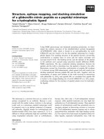
Figure 1.1 A simplified overview of the allergic reaction. Antigen presenting cell
(APC) recognizes processes and presents the allergen as on its MHC class II receptor.
The allergen fragment is then recognized by the T cell receptor (TCR) in the context
of MHC II. This drives the T cell to mature to Th2 cells, producing cytokines that
stimulate IgE production, or eosinophil recruitment. Cross linking of IgE on mast
cells causes degranulation leading to immediate hypersensitivity.
3