A systemic functional analysis of multisemiotic biology texts 5
Bạn đang xem bản rút gọn của tài liệu. Xem và tải ngay bản đầy đủ của tài liệu tại đây (590.62 KB, 49 trang )
CHAPTER FIVE
MULTIMODAL CONSTRUCTION OF BIOLOGICAL KNOWLEDGE
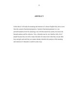
In this chapter I present the results of the analysis of the visual selections and how the
visual text relates to and interacts with the linguistic text. By visual text I refer to the
semiotic resources deployed in the print media biology textbook that depend on the
transmission and reflection of light on a treated surface (rather than the transmission of
sound in the air) to make meaning, including the typographical features of the writing
system of the English language. However, the focus of this chapter is on the visual
displays such as schematic drawings, tables, statistical graphs and micrographs. This
chapter takes as points of departure the discussion of the frameworks for the analysis
of the visual display and seeks to illustrate how meanings are made in the texts using a
variety of resources. Thus it complements in a significant measure what is presented
in Chapter Four, which is the analysis of meanings made through the linguistic
semiotic code. It will be seen that biologists do not rely on language alone to make
meaning and that visual displays are an important resource for meaning-making. In
what follows I discuss the page layout and colour schemes of the texts (Section 5.1),
present the frequencies of the categories of visual displays that occur in the texts
(Section 5.2) and then analyze one visual display of each main category (Section 5.3).
I conclude this chapter with a brief summary (Section 5.4).
190
5.1 Page Layout and Colour Schemes in the Texts
Before conducting detailed analysis of selected visual displays in the texts we need to
consider three issues that affect not only any one visual display but also almost all
meaning-making in the texts, issues that enable and constrain meaning-making in the
first place. First, how big is the page and how is it designed in a way that provides a
textual space where meanings are made? Second, how is colour deployed to make
meaning? And thirdly how is the reader expected to move his or her attention from
one semiotic mode to another in a multisemiotic text, and within one semiotic from
one part to another? Since the last issue, i.e. the reading path, has been dealt with in
Section 3.2.3 above, I discuss the first two questions below.
First, a page is a textual as well as a physical unit. At first glance, a page is a
physical object on the surface of which words and pictures are printed: we can touch it,
turn it and tear it from the book. Physically speaking, then, the pages of all three texts
adopt a size close to the standard A-4 size paper, measuring about 10.8 × 8.3 inches.
The selection of such a size can be seen as a response to the increasing number of
visual images in the texts, as well as to the size of the school bag, the desk and the
hand of a university student in his or her late teens. A bigger book makes it easy to
arrange the images and the words and a pocket size edition of these biology texts
would be hard to imagine. The page size is among the first factors affecting the page
design. At the same time, a page is also a small-scale multimodal meaning-making
unit (Baldry 2000b: 41-42): it has internal design, page layout, that organizes its
various textual elements. Kress and van Leeuwen (1996: 183) approached this issue in
terms of the systems of INFORMATION VALUE, SALIENCE and FRAMING (see
Section 3.1.2.2 above). What is equally fundamental is the column grid of the page.
191
In Texts 1 and 2, a normal page with the exception of end-of-chapter “Essential
Concepts” pages and “Questions” pages, which are in double column, and “Key
Terms”, which are listed in four-column tables in Text 1, and end-of-chapter
“Summary”, “Exercises” and text boxes, which are in double column, and “Key
words”, which are listed in three columns in Text 2, is split into main text (the body)
and text margin (see Figures 1.1 and 1.2 above for sample pages). Physically
speaking, the width of the margin is around half of the body of the text, that is, two-
thirds of the left-right page is devoted to the body of the text and one-third to the
margin (see Figure 5.1). There is a gutter of approximately 1 cm to set apart the body
and the margin.
body
Figure 5.1 Demarcation of the page in Texts 1 and 2
The demarcation of main text and text margin is intended to serve a purpose in
the texts. The major running text appears only in the body of the page, while the
caption text, visual display and in-chapter questions (in Text 1) are printed in the text
margin. That is, the text margin provides a place where questions are asked about
what has been talked about in the body of the text or explanatory texts (caption) are
provided for the visual display in the body of the text. From this we see that the page
has one part that is the chief information provider and another part that serves a
subordinate role: as a question, explanation or facilitator of the main text
1
.
The page layout of Text 3 is different. Except for the large visual displays such
as Figure 23.30 that extend from the left to the right of the page, the page contains two
192
equal columns, with a gutter of about 1cm to separate the two portions (see Figure 1.3
for a sample page).
Another aspect of the page as a textual unit is that the main text and the figures
they refer to are laid out with a page or a two-page spread as a unit. As S. Gibbs, the
managing editor of ECB (personal communication, February 8, 2001), notes,
In “laying out” book pages we always try to ensure that figures are in
close proximity to where they are discussed in the text. This means that
the reader is not distracted by having to turn pages in order to find the
relevant figure.
The second issue that affects meaning making in the texts is concerned with the
use of colour, or the colour scheme, i.e. the principled selection of a number of colours
out of “systems of colour” (Kress and van Leeuwen 2002: 366). Human vision, and
therefore visual semiosis, depends on, or is realized through colour. Without light or
colour we do not see anything. The essential issue is the colour scheme of the
textbook, black and white or full colour. Texts 1 and 2 are full colour while Text 3 is
largely black and white.
In Table 3.2 “Functions and systems in schematic drawing” above, I identify
“colour” as a semiotic resource operating across all three metafunctions. First, in a
schematic drawing, colour can be used to represent the original colour of the natural
world, for example, the green to represent the colour of the leaves. Thus colour may
function as an experiential resource. In this respect, the colour in the drawing shows
directly this aspect of the original object to a very high fidelity with or without the aid
of natural language. Colour can also be used to represent abstract entities, such as the
blue of the uniform to signal that the wearer is a policeman or policewoman. Second,
where the colour in the drawing is added, extraneous of the original object, the colour
may serve to engage the viewer. Thibault (2001: 317) notes that “[full] colour is
193
usually the unmarked choice in modern science textbooks for school pupils” and that
“the reasons are mainly interpersonal” (2001: 317). The textbook authors / publishers
engage and interact with the student readers by inviting them to experience the
colourful (though not necessarily faithful) materiality of the subject matter. Reading
textbooks thus becomes a sensory, physiological experience, as well as an intellectual
one. Thirdly, colour in the drawings may also serve to link the visual elements, “to
provide cohesion and coherence” (Kress and van Leeuwen 2001: 58). In Texts 1 and
2, for instance, green, coupled with larger font size and initial capitals, is used to signal
the headings and in Text 2 boxed essays are shaded with light green. Citing the
example of the use of colour in maps, Tufte (1990: 81; original emphasis) summarizes
“the fundamental uses of color in information design” as follows: “to label (color as
noun), to measure (color as quantity), to represent or imitate reality (color as
representation), and to enliven or decorate (color as beauty)”. Kress and van Leeuwen
(2002: 350) further argue that because colour has been culturally shaped to construct
all three metafunctions, it may be considered as “a semiotic mode in its own right,
along with language, image, music, etc”.
5.2 Categories of Visual Displays in the Texts
The types of visual displays that are selected in the texts and the frequency of each
type are summarized in Table 5.1. As seen here, the three texts show considerable
variation in the types and frequencies of visual display. For instance, structural
formulas of molecules
2
and equations of reactions occur in Text 2 only. A structural
formula describes how the various atoms in a molecule are bonded together and an
194
Text 1
Text 2
Text 3
Types
Number
%
Number
%
Number
%
Schematic
drawing
18
54.6
4
48
61.5
Photograph
13 39.4 5 24 30.8
Table
1 3.0 2 6 7.7
Statistical
graph
1
3.0
0
0
0
Equation
0 0 15 0 0
Structural
formula of
molecule
0
0
many
0
0
Total
33
100
78
100
Table 5.1 Types of visual display
equation describes what reactants participate in the reaction under certain
circumstances and what products result. They are thus well suited for the biochemistry
text. In Texts 1 and 3, on the other hand, schematic drawings are most frequent,
followed by micrographs, i.e. photographs taken through a microscope.
The three texts also differ in the extent of integration between the linguistic text
and images. Whereas in Texts 1 and 3 the visual images are separated from the
linguistic texts but are referred to in the latter, for example by “(see Figure 17-6)” (in
Text 1), in Text 2 the structural formulas and equations are very often integrated with
the linguistic text so that no separate title or caption is felt necessary for the formulas
195
and equations: they have in these cases become part of the running text. In Text 2,
only when the formulas and equations become extremely complex do they occupy a
separate section on the page (e.g. Figure 10.1 of Text 2).
In addition, the schematic drawings and the micrographs in Texts 1 and 3 occur
in two forms, either alone or in “split-screen” format, that is, one or more micrographs
are juxtaposed with their simplified schematic drawings, side by side within one figure
or occasionally in two adjacent figures. But as will be seen in the analysis of Figure
17-10 in Text 1 below, the micrograph and its schematic drawing counterpart in a split-
screen format are not equivalent. Whereas the former is believed to be a trace of
nature, the latter is a pedagogic reconstruction of the trace. Overall, it seems that the
schematic drawings are intended to contribute more to the construction of biological
knowledge in the textbooks than the micrographs, which serve merely as the
“guarantee” of the reality (Bastide 1990: 213) and “a guarantee of objectivity”
(Barthes 1977: 44). These guarantees form one of the bases for the claims made in the
linguistic and visual (i.e. schematic drawing) text. It also seems to follow that the
drawings need to be studied carefully while a glance is sufficient for the micrographs.
On the other hand, the distributional features of the visual displays described
above, like those of the verbal text that they accompany, are realizations of particular
contextual configurations; the distribution and the types of visual displays may well
differ if other texts are analyzed.
5.3 Textual Analysis of Some Figures
In this section
3
I analyze a schematic drawing (Section 5.3.1), a micrograph (Section
5.3.2), a split-screen format of a drawing and a micrograph (Section 5.3.3), a statistical
196
graph (Section 5.3.4), a scientific table (Section 5.3.5), and the structural formula of a
molecule (Section 5.3.6). The analyses are presented in decreasing detail so that only
the prominent and new features are discussed in the latter analyses.
5.3.1 A Schematic Drawing: Figure 17-3 of Text 1
Figure 17-3 (ECB: 549), together with the relevant verbal text, is reproduced in Figure
5.2.
The reader is formally introduced to Figure 17-3 when he or she reads the
following clause:
These two processes together constitute the M phase of the cell cycle
(Figure 17-3).
However, he or she may not wait until being instructed to view Figure 17-3. Since
Figure 17-3 is a full-colour drawing, a picture more attractive than the largely black
and white verbal text, a reader’s attention is more likely to be drawn to the drawing
than to the written description. Thus one plausible reading session may be that a
reader, at some point in his or her reading, turns his or her attention to the figure, and
then back to the verbal text for careful study and then back to the figure again,
following a back-and-forth type of reading path, as explained above.
The reading path within Figure 17-3 is marked in Figure 5.2 by the blue
italicised Roman letters A to G. As is clear from Figure 5.2, the reading path is not
linear, from left to right, from top to bottom, but is determined Ideationally by what is
in focus in the running text (the M phase of the cell cycle), and Interpersonally by the
visual means of directing the reader’s attention (for example, the bright yellow
197
Figure 5.2 Reading path for Figure 17-3
198
Shading and Capitalization of MITOSIS and CYTOKINESIS and light green Shading
of M phase and the large square bracket embracing MITOSIS and CYTOKINESIS).
This is, in verbal and common parlance, equivalent to saying “Hey, look at what is
highlighted first!” Indeed, in this part of the reading, Steps C and D are all an
experienced reader needs to attend to. The highlighting devices such as arrows are
equivalent to a lecturer’s cursor in an actual classroom, where he or she, while talking
to the students, points to relevant parts of the figures. Although in viewing Figure 17-3
one’s gaze, especially that of a novice, may work from Step G down to Step D due to
the Interpersonal impact of the downward-pointing arrows and the reading habit of a
normal reader, it is nonetheless arguable that the reading path suggested above is most
economical for the experienced reader, that is, one that has followed the textual
explication up to this point.
At the rank of Work, Interpersonally, this figure thus employs an array of
visual means to emphasize various parts of the cell structure and stages of cell division.
Ideationally, the figure is designed to tell a story about what happens in a cell cycle, in
particular the M phase of the cell cycle. The Ideational meanings include: (a) material
processes realized by changes in the shapes at different stages, the arrows and the
nominal groups in the linguistic text, (b) intensive identifying processes realized by the
labels, leaders and the pictorial elements, and, in the absence of leaders by the labels,
the spatial proximity between the pictorial element and the labels, and the pictorial
elements, and (c) possessive identifying relational process realized by the labels, the
square bracket, the pictorial elements and the linguistic text. The overriding
experiential content seems to be concerned with material processes, although the
intensive and possessive relational processes contribute significantly to the
construction of biological knowledge. And Textually, the drawing is not isolated from
199
the other parts of the text. It is related to the main text and the caption and is placed in
a specific position on the page, that is, in the text margin. The drawing is vertically
positioned, with the Arrows connecting one stage with another. Other resources
employed for the Textual meaning include Geometry (e.g. circles), Colour Contrast or
Similarity, Labelling (with or without leaders), and Framing. In what follows, I
analyse selected steps in terms of the Interpersonal (Modal) meaning, Ideational
(Representational) meaning and Textual (Compositional) meaning, by reference to the
functions and systems chart in Table 3.2.
Step A The Title
Distinctive typographical features, such as the boldface of the title and the greenness of
the figure’s serial number, function to attract the reader’s attention and thus attach
more importance to this linguistic message. The title is also the only explicit link to
the main text; it is the reader’s entrance to the pictorial world of the figure. It is
designed to be read first and taken as the point of departure for what is to come next.
The title is a nominal group and apparently does not select an Interpersonal
stance at the rank of clause in terms of SPEECH FUNCTIONS (Offer or Demand) and
MODALITY and MODULATION (Halliday 1994). This is a nominal group whose
function is termed by Halliday (1994: 96) as “Absolute” in that it “could be either
Subject or Complement in an agnate major clause”. Indeed all the linguistic
components except the caption in Figure 17-3 are “[u]nattached nominals” (1994: 395)
which function in this way. But such nominal groups are nonetheless far from being
free from any Interpersonal meaning. As for this title, the nominal group presents the
Process of a cell dividing as a Thing, which is objective, absolute, visible and concrete.
200
Such a high level of certainty about the state of affairs is attainable through nominal
groups or grammatical metaphor in the form of nominalisation (Halliday 1993a; 1998).
In other words, distillation of phenomena into entity or transformation of clausal
grammar to nominalised form means that the reader is not in a position to doubt the
existence of a phenomenon, but is led to believe in its absolute, timeless and
unconditional existence.
Ideationally, being a nominal group, the title serves to identify, and is thus
equivalent to an intensive identifying clause (Halliday 1994: 119-120), for example
“This is the drawing of the M phase of the cell cycle”. It is important to note that the
nominal group identifies not only through language but also by its spatial proximity to
the schematic drawing. By itself this nominal group points to a nominalised process,
the M phase of the cell cycle. Thus a sequence of dramatic events, where one cell
splits into two, has been transformed into a Thing which has consequently been
deprived of all the original vigour, liveliness and particularities.
Step C MITOSIS
This step can be broken into three sub-stages: Step C-1 the word “MITOSIS”, Step C-2
the arrow and Step C-3 the circle and the two overlapping circles which contain the
semiotic depiction of the cell.
Step C-1 the word. Typographical features such as the largest font Size,
Capitalization, and bright yellow Shading serve to attract the reader’s attention, as if
saying that MITOSIS and the drawings it refers to are what the reader needs to pay
special attention to.
201
Step C-2 the arrow. Interpersonally, the single-headed arrow is a Command; it
demands that the reader look in the direction of the arrow, in this case, from top to
bottom of the page. Here, the Command effect is strengthened by the particular
darkness and thickness of the arrow.
Ideationally, the arrow serves to signify the process and direction of movement,
change or progression, or the numerous intermediate phases between the circle above
and the circles below. In terms of Peirce’s (1985: 9-12) trichotomy of signs into an
index, an icon or a symbol
4
, the arrow is a highly stylised icon. That is, the arrow
proper does not exist in the actual world in the process of cell division; the designers
have added it to the depiction. Besides, the direction of the arrow in the physical
sense, i.e. from top to bottom, is iconic of progression in time.
Step C-3 the circles. Inside the circle (second from top), highlighting devices
such as the Colouring of the two pairs of lines and pink Shading serve to draw
attention to the essential defining features of a cell at this stage. The blank space
(Omission) between the outer ring of the circle and the pink shaded central area is, in
reality, just as occupied as other parts of the cell. This distortion functions as yet
another means of highlighting the two pairs of lines. The outermost black circle and
the adjacent blank space inward (Omission) surround the central pink shaded area,
serving as a Framing to give weight to what is highlighted in the centre. The Contrast
of colour between black, red, pink and white serves the same highlighting purpose; at
work here is the colour scheme employed: bright red and dark black against a white
and light pink background so that the former stand out.
Ideationally, the circle is drawn to represent a snapshot of a particular stage in
cell division. It focuses on the separation of the two pairs of chromosomes, omitting
the changes taking place in the cytoplasm. The Ideational meaning is realized by the
202
changes in shapes and contents of the pink-shaded area and also by the Diagonal
orientations of the two pairs of lines representing chromosomes. We need to note that
this circle is not an obvious icon (Peirce 1985). The two pairs of lines inside and the
circular shapes do somewhat resemble some types of cell components, hence they are
iconic. But the colours, the circle and the blank space testify to the symbolic nature of
the iconic sign. For instance, the colour of a particular cell component one sees in a
micrograph is the result of dyeing technique. However, what is shown in the
micrograph is not necessarily reproduced in a schematic drawing; in a drawing further
treatment is carried out to produce what appears in the final printed book. In other
words, what meets a reader’s eye in a schematic drawing is at least two steps away
from what is really there: in terms of choice of colour and diagrammatic
transformation.
Textually, several devices contribute to the organisation of the text. For
instance, Colour Cohesion and Contrast enable the viewer to recognize similarity and
difference in the Ideational meaning and Interpersonal meaning: the colours red, pink,
black and white serve as a backdrop against which the Ideational and Interpersonal
meanings are expressed. Similarly, the Shapes of the components, i.e. the lines,
circles, and the Relative Position of the components also constitute a resource to
organize the text. Below I discuss in greater detail the role of Horizontals, Verticals
and Diagonals in the Textual organization in the schematic drawing.
The two pairs of lines in the first circle in Step C are positioned diagonally
relative to the vertical-horizontal frame of the drawing. The red pair resembles the
contour of a hill or sea wave, each of which is perceived as the trace of drastic
movement or thrust resulting from the physical or geographical forces such as the
gravitational pull. The axis of the black pair is approximately 30° anticlockwise to the
203
vertical axis of the drawing. This tilt or obliqueness creates “directed tension”
(Arnheim 1974: 424-428), or “energy and dynamism” (O’Toole 1994: 23; Thibault
1997: 315-322). We may note that whereas the shape of the red pair of lines remains
roughly constant throughout the drawing, the black pair tilts most in Step C. This well
fits the Ideational theme of the step, which is concerned with drastic change in terms of
chromosomes in the nucleus. On the other hand, the Diagonal orientations of the two
pairs of lines in the step also serve to connect this step with the preceding and
following steps, thus contributing to the Textual organization or unity of the drawing.
In other words, obliqueness in orientation of the lines is echoed or shared by all the
steps in the drawing albeit to varying degrees. It is true that in the laboratory cell
biologists will know that the cells are undergoing some transformation however they
are aligned relative to the mechanical stage of the microscope. But when cells are
represented in micrographs and in particular in schematic drawings, that is, when they
are turned into lines, circles and so forth, to contribute to the Textual organization, “the
canons of classical painting” (Bastide 1990: 199-200) are often respected. One such
canon is the deployment of oblique lines to represent “energy and dynamism”
(O’Toole 1994: 23; Arnheim 1974: 424-428).
Step D CYTOKINESIS
Again, Step D can be broken into Step D-1 the word, Step D-2 the arrows and Step D-
3 the two circles (at the bottom of the figure). Below I analyze the arrows and the two
circles.
Step D-2 the arrows. Interpersonally, the fork arrows serve to draw our
attention to what the arrows point to, i.e. the two circles. What is distinct about these
204
arrows is that they point to two directions instead of one, as in previous arrows. This
means that the reader need pay attention to the two separated circles. Ideationally, the
fork arrows symbolize separation, one cell divided into two cells, or they represent the
numerous intermediate stages between the completion of mitosis and the completion of
cytokinesis. And in terms of Compositional meaning, a short line and two short
arrows are deployed to realize the Ideational and Interpersonal meanings.
Step D-3 the two circles. Interpersonally, one’s eye line is drawn to the two
circles by the two directional arrows. The two circles are quite large, designed to
attract the reader’s attention. The two smaller inner circles representing nucleuses are
highlighted by the same devices as in the above circles. The white space between the
two large circles, quite unlike the empty space inside the circles, is the background of
the image and indicates that the focus of attention now is on the fact that the two
circles have moved apart. Ideationally, if we compare the two smaller inner circles
(standing for nucleuses) at this stage with those at the end of mitosis, i.e. the contents
of the circles, we find they are identical; what has changed is that the two partially
overlapping circles have now become two separate circles. This means cytokinesis is
mostly concerned with the separation of cytoplasm (signified by the white space
between the outer and inner rings) so that each new cell gets its share of outer support
materials. The two large circles also mark the end of the M phase, hence also one
typical cell cycle. Textually, in addition to the textual resources employed earlier in
the drawing, such as Colour Shading and Framing, Symmetry of the two circles (i.e.
their identity and symmetrical horizontal alignment) and the fork arrows brought
forward from the previous phase also contribute to the Textual meaning.
205
Step E The Caption
The caption has less visual salience through smaller font size, normal type (i.e. not
boldface) and shorter leading. This suggests that the caption is to be read later in the
reading sequence. The lexicogrammatical features of the caption are discussed in the
linguistic text analysis presented in Chapter 4 and thus not repeated here. It is worth
noting, however, that Ideationally the caption presents a possessive identifying relation
and circumstantial identifying relation, realized respectively by the verbal groups
“consists of” and “followed by”. This repeats the information presented in the main
text (for example in the clause “These two processes together constitute the M phase
of the cell cycle”). The caption, however, serves in particular to specify what the
square bracket in Step B refers to, that is, it is a visual iconic expression of a
possessive identifying relation. Here we can appreciate that while the visual images
are important in biological texts they have to be given categorical meanings by
linguistic resources. The value of the visuals in this figure is that, in addition to
representing or constructing the shapes of biological entities, they are a spatialization
or icon of the temporal flow of events and also aid to construct a taxonomy of
biological terms (the relations between M phase, mitosis and cytokinesis). However,
language has to specify the relations and their visual transformation (cf. Barthes 1977:
38-41).
Step F Chromosome Replication
This step can be broken into two sub-stages: Step F-1 the words “chromosome
replication” and Step F-2 the arrow.
206
Step F-2 the arrow. Compared to the others, this arrow is short, indicating less
Prominence in the figure. This arrow also leads the reader’s attention to the next
visual representation. Ideationally, this arrow denotes the process by which one pair of
chromosomes duplicates into two pairs. One needs to note, however, that the shortness
of this arrow misrepresents the length of the time period. That is, replication in the S
phase takes much longer than the M phase. A typical eucaryotic cell spends a fraction
of its cell cycle time in the M phase, and most of it in interphase, as noted in ECB,
page 549. For example, a mammalian cell of a 24-hour cell cycle requires only about
one hour for the M phase to complete. This misrepresentation of the temporal
dimension functions to highlight the M phase of the cell cycle.
Step G The Structure of the Cell
Step G is located at the top of the figure and an uninitiated reader may begin viewing
the figure here as this step provides the background for what follows. The labels in
this step and the leaders functioning as the identifying processes disappear in the later
depictions. This means that once they have fulfilled their contextualising function,
they are discarded and are no longer made visible. Having previously established the
structure of the cell in ECB, the reader is now invited to study in detail the M phase.
As argued above, the experienced reader reads Step A first and this step last or simply
skips this step, as would perhaps a lecturer in the classroom. This step can be read in
two sub-stages: G-1 the circle and G-2 the labels.
Step G-2 the labels. Like “chromosome replication” in Step F, the words in
Step G-2 are made least prominent by means of smaller font Size, no-Shading and no-
Capitalization. The leaders are also made insignificant by means of Length and
207
Weight. Ideationally, they identify the major components of a cell, as if saying, for
example, “This is the nucleus of the cell”.
I now conclude the analysis of Figure 17-3 with a discussion of the ideational
complementarity of language and visual images. Stripped of the language, the visuals
(circles and especially arrows) seem to tell a story and speak for themselves. That is,
by viewing the visuals alone, we would get an impression of change or movement, but
would have little idea of what the visual images are about: they either say too much in
that there are too many possible interpretations one can make, or they say too little in
that they do not give the reader a clear orientation. Thus the language comes in to
capture some moments in the cell cycle, i.e. to condense and nominalise, define,
categorize, theorize about, or anchor, what the visuals show, for instance, the change
from this stage to that one is called mitosis. In the words of Barthes (1977: 38-39;
original emphasis)
all images are polysemous; they imply, under their signifiers, a ‘floating
chain’ of signifieds, … Hence in every society various techniques are
developed intended to fix the floating chain of signifieds in such a way
as to counter the terror of uncertain signs; the linguistic message is one
of these techniques.
Stripped of the visuals, on the other hand, the caption text and the relevant
main text make the relational meanings of intensive, circumstantial and possessive
types (and later in the text material relations are to dominate), but the reader would
never have any concrete idea of what a cell looks like, or how it changes its shape from
one stage to another, or what colour each cell component may be, hence the need for
the visuals to show the shape, colour, and so forth. This complementarity between
language and visual images arises from their different functionalities and limitations.
208
Language, in essence, generalizes and categorizes, discarding the non-essential
features, and is thus typologically straightforward but topologically ambiguous (Lemke
1998a: 87), while visuals are more or less effective for constructing topological
meanings, sometimes at the expense of typological meanings.
The co-deployment of both the visual and the language is absolutely essential
for biology for two reasons (cf. Section 3.2.1). First, much of what is captured under
the microscope is natural shape, colour, and process that in a sense defy a linguistic
version, or rather, natural language has not yet evolved to such a degree of accuracy as
to be of practical use to natural science, if it is the sole resource employed. Take the
example of shape. The “shape” words in English include square, circle, rectangle,
triangle, pentagon, and so on, hardly adequate for the description of the numerous
shapes we find around ourselves. If the situation requires an adequate description of
such topological meanings, language may turn out to be inadequate and other semiotic
resources may be called for. Not that natural language can never fully express the
topological meanings, but that other semiotic resources work much more efficiently
and accurately. Visual display is one such resource. It can show readily and exactly
what one sees, or perceives. Secondly, the visual devices (e.g. the square bracket, the
leaders) that serve to identify, or establish links between the linguistically coded
meaning (e.g. mitosis) and what is visible under the microscope (e.g. the change one
observes in the laboratory) are symbolic in that in real life there do not exist such
devices. Rather, they are created by the textbook authors to mean in a specific way, to
stand for and realize particular meanings. By visualizing the possessive / intensive /
circumstantial identifying meanings the readers are able to perceive the relations.
Obviously, that the possessive / intensive / circumstantial identifying meanings are
209
represented visually as a square and so forth does not mean that under the microscope
we can see them: they are the products of scientists’ conceptualization.
5.3.2 A Micrograph: Figure 17-11 of Text 1
5.3.2.1 Types of Micrographs
Cells are small, transparent and mostly colourless; in their natural state they are not
visible to human vision. So the study of cells has depended on the development of
instruments, especially microscopes, and on specimen preparation. Since Robert
Hooke (1635-1703) first observed cells with the help of a light microscope in 1665,
microscopists have been attempting to devise various types of microscopes. Based on
the form of the illuminating radiation, there are mainly two types of microscopes that
are used in the study of cells: the light microscope that exploits the characteristics of
light and makes visible objects as small as 200 nm; and the electron microscope that
makes use of a beam of electrons and has a resolving power of 0.2 nm, 1000 times
better than the resolution of a light microscope (ECB: 3). Within these two broad
types there are various subcategories. Common types of light microscopes include: (1)
the conventional light microscope, (2) the fluorescence microscope, which employs
two barrier filters and fluorescent stains (rather than ordinary stains) and produces
images of selected proteins or other molecules in bright glowing colour against a dark
background, (3) the phase-contrast microscope, differential-interference-contrast
microscope, and the dark-field microscope, which are used to visualize living cells,
and (4) the confocal scanning microscope, which constructs images of complex three-
dimensional objects. Common types of electron microscopes include: (5) the
210
transmission electron microscope (TEM), (6) the scanning electron microscope (SEM),
which gives three-dimensional images of surfaces and (7) the freeze-fracture and
freeze-etch electron microscopes that visualize the interior of cell membranes and the
exterior or interior of cells, respectively (Alberts et al 1994: 139-156).
The resulting micrographs vary accordingly in terms of both the Ideational
meaning and Interpersonal meaning. Ideationally, different types of micrographs show
different things. For instance, phase-contrast micrographs can show the structure of
the living cell, and thus suit more a text that is describing the cell division and cell
movement, and an ordinary light micrograph shows a structure of dead cells because
before viewing the cell has been chemically fixed, or killed. An electron micrograph,
on the other hand, is effective for showing fine details that light micrographs cannot
achieve. It is also obvious that there are overlaps in the functions of different
micrographs (microscopes). For example, in addition to a light micrograph, an
electron micrograph can also show the structure of the dead cell, but a possible
drawback is that an electron micrograph may show too many gratuitous details.
Interpersonally, all types of micrographs are machine inscriptions (Latour
1990: 35-36; Myers 1990: 238) and thus have the authority of presenting the true
“looks” of the cell, given the technology available at the time. Indeed, in professional
science, inscriptions are made to help to convince, win over, skeptical audience: “It
[the trend toward inscriptions] is as necessary as the race for digging trenches on the
front in 1914. He who visualizes badly loses the encounter; his fact does not hold”
(Latour 1990: 41). At the same time, some types of micrographs such as the
fluorescence micrograph have stronger interpersonal impact (“look better”) than other
types such as the bright-field unstained light micrograph, since the former are in
211
superb colour against a dark background, while the latter show vaguely in black and
white the outline of some cell components.
Given the option of the word, the schematic drawing and the various types of
micrographs, the reason why the textbook authors decide upon a particular type of
presentation at a particular point in the text lies in the Ideational development of the
co-text, the level of detail required (e.g. a labeled schematic drawing shows more
detail than a micrograph, which in turn shows more detail and specificity than the
word), the Interpersonal stance the authors intend to take, and of course the availability
of the micrographs.
5.3.2.2 Analysis of Figure 17-11 of Text 1
In Figure 17-11 (ECB: 557), we encounter a fluorescence micrograph, a micrograph
taken with the aid of a fluorescence microscope. The co-text for Figure 17-11 is the
first paragraph in the section “Chromosomes Line up at the Spindle Equator at
Metaphase”, especially the following two clauses:
thereby forming the metaphase plate.
This defines the beginning of metaphase (Figure 17-11).
The beginning of the paragraph of the main text describes a series of material
processes and then gives a name to the stage of the development (intensive identifying
process), while the figure shows a snapshot of a moment of the material processes, or
the result of the preceding changes. In freezing the moment, the dynamic nature of the
change is lost but what is captured in the moment can be viewed closely and at leisure.
Typographically, the figure is in the main text part of the page, the top left, and
its caption is on the right as the marginalia. I suggest that the reading path is as
212
follows: Step A the figure, Step B the title for the figure, and Step C the caption for the
figure. Figure 17-11, with the reading path marked with blue capitalized Roman letters
in italics, is reproduced in Figure 5.3. What follows is a selective analysis of the
figure.
B
Figure 17-11 Multiple mitotic
spindles at metaphase in a fruit fly
(Drosophila) embryo. The
microtubules are stained red, and the
chromosomes are stained green. At
this stage of Drosophila
development, there are multiple
nuclei in one large cytoplasmic
compartment, and all of the nuclei
divide synchronously. Although
metaphase spindles are usually
pictured in two dimensions, as they
are here, when viewed in three
dimensions the chromosomes are
seen to be gathered at a plate-like
region at the equator of the spindle–
the so-called metaphase plate.
(Courtesy of William Sullivan.)
C
A
Figure 5.3 Reading path for Figure 17-11
Step A The Figure
Interpersonally, this figure is very appealing. It is arguable that the selection of a
fluorescence micrograph in itself is an Interpersonal maneuver; the text, as does
science in general, attempts to reach out to the student reader and the general public.
The factors that contribute to the interpersonal appeal of the figure include the
following: (1) it deploys bright Colours: red, green, yellow and black. (2) The
Contrast in the colours is great: first, between the background black and the other
colors and second, the contrast between red and green. Of course, the outside of the
213
figure is white, which makes a greater contrast. All these serve to set up a striking
colour scheme. (3) The figure is framed by a rectangle, directing the reader’s attention
to what is inside the frame. (4) All the above features result from a fluorescence
microscope, rather than from the hands of the artists, which lends greater credibility to
the figure, compared with schematic drawings.
Within the frame, some of the mitotic spindles are chopped partially out of the
frame and some are framed inside. This Framing serves to lead the reader’s eye to the
few mitotic spindles that are placed in the centre. Within each spindle, our eye (the
Point of View) is directed toward the whole of the spindle and particularly
chromosomes because the latter are in the centre of the spindle and because they are in
different colour than the microtubules. This Prominence of chromosomes in the visual
display is in agreement with the verbal description in the main text, which is mainly
concerned with the formation of the metaphase plate by the chromosomes. While
faithful to one master, this figure betrays another – as pointed out in the caption, this is
a two-dimensional depiction: the viewer has to construct in his or her mind a three-
dimensional plate from the two-dimensional representation.
Ideationally, the figure shows a snapshot of the mitotic spindles of a fruit fly
embryo at metaphase, with chromosomes lined up in the centre of the spindles. It
shows what the spindles with chromosomes attached look like in a microscope: the
shape, colour, composition, location and orientation of each spindle, and the relative
position of one spindle to another. As such the figure complements and completes the
description given in language in the main text (the linguistic text, in its turn, also
impinges upon the visual image; see the discussion in Section 5.3.1 on the ideational
complementarity between the verbal and visual codes).
214