Cloning, characterisation and functional analysis of horseshoe crab c reactive proteins
Bạn đang xem bản rút gọn của tài liệu. Xem và tải ngay bản đầy đủ của tài liệu tại đây (2.54 MB, 129 trang )
CLONING, CHARACTERISATION AND FUNCTIONAL ANALYSIS
OF HORSESHOE CRAB C-REACTIVE PROTEINS
Tan Seok Hwee Sandra
(BSc Hons)
A thesis submitted to the
Department of Biological Sciences
The National University of Singapore
in partial fulfillment for the
Degree of Master in Science
in
Biological Sciences
2004/ 2005
Acknowledgements
I would like to express my immense gratitude and heartfelt thanks to my supervisor, Prof
Ding Jeak Ling, for her patient guidance, encouragement and endless support during my
two years in the lab.
My thanks also to Prof Ho Bow, for giving me insights into microbial work. I would also
like to thank Su Xian and Mei Ling from the Microbiology department for their many
practical pointers.
To my wonderful teacher and partner-in-crime, Patricia: you have shown me what it
truly means to be a dedicated scientist. Thank you for all that you’ve done!
To the seniors in the lab, Subha, Haifeng and Wang Jing: Thank you for all that you’ve
taught me.
I have been incredibly fortunate to work in a lab where information and ideas are
exchanged freely. My thanks goes to all my lab-mates: Siaw Eng, Lihui, Sean, Nancy,
Li Peng, Yong, Sharan, Hanh, Nicole, Belinda, Siou Ting, Geraldine and Song Yu:
you have all been such wonderful teachers and collaborators.
I also owe a note of thanks to a wonderfully supportive group of people:
My “consultant”, Cindy.
My “cheerleader”, Alphonsus.
My “classmate-of-the-year”, Derrick.
My “movie-kaki”, Nicole.
My “photographer”, John.
My “mouse supplier”, Kelvin.
Last but not least, this work is dedicated to my parents: you never understood what I was
doing, but you supported me all the same. Thank you for believing in my dream.
ii
Table of Contents
Acknowledgements
Table of Contents
List of Abbreviations
List of Figures
List of Tables
Summary
ii
iii
v
vi
viii
ix
INTRODUCTION
1.1
The horseshoe crab—a living fossil
1
1.2
1.2.1
1.2.2
1.2.3
The challenge of a pathogen-laden environment
Horseshoe crabs have a robust innate immune system
Elements of the horseshoe crab innate immunity
Plasma lectins are key components in frontline immune defense
2
2
5
10
1.3
1.3.1
1.3.2
1.3.3
1.3.4
1.3.5
The role of C-reactive proteins in frontline immune defense
Human CRP - a versatile diagnostic and prognostic marker
Gram-negative septicaemia is a widespread medical problem
LPS: ubiquitous, persistent and versatile molecules
CRP: role in bacteria neutralization?
In vivo functions of CRP remain enigmatic
11
11
12
15
18
20
1.4
Objectives and scope of the project
22
MATERIALS AND METHODS
2.1
Collection of horseshoe crab hemolymph
24
2.2
2.2.1
2.2.2
Cloning CrCRPs
Preparation of pGEX plasmid for expression in E. coli
Preparation of pYEX plasmid for expression in yeast
26
29
35
2.3
2.3.1
2.3.2
2.3.3
2.3.4
2.3.5
2.3.6
2.3.7
Expression and purification of recombinant CRPs
Large-scale expression of GST-CRPs in bacterial culture
Expression of GST-CRP-2 in yeast culture
Capturing fusion proteins by affinity column chromatography
GST tag removal by thrombin digestion
LPS removal by Triton-X 114 treatment
Recombinant Factor C (rFC) assay to monitor LPS removal
Protein quantification and determination of protein expression levels
38
41
42
43
43
44
45
45
iii
2.4
Checking the interactions of CrCRPs by GST pull-down assays
46
2.5
Antiserum production and immunoblotting of proteins
49
2.6
In-gel digestion and protein identification by mass spectrometry 50
2.7
2.7.1
2.7.2
2.7.3
Antimicrobial assays
Bacteria growth inhibition/ bactericidal assays
Bacterial agglutination assay
Neutralization of CrCRP-2 activity by LPS and its substructures
52
52
54
54
2.8
In silico analysis of DNA and protein sequences
55
RESULTS
3.1
3.1.1
3.1.2
3.1.3
3.1.4
3.1.5
3.2
3.2.1
3.2.2
3.2.3
3.2.4
3.2.5
3.2.6
3.2.7
3.2.8
3.2.9
Interactions of recombinant CRP-1 and -2
Comparison of expression efficiencies of rCRP-1 and -2
Interactions of CRPs are enhanced in the presence of calcium,
as well as during infection
CRP-1 and -2 interact preferentially with GBP and CRPs
respectively
Glycosylation primes CRP-2 for more efficient interaction
with protein partners of the hemolymph
Conclusions
The antimicrobial activity of rCRP-2
rCRP-2 exerts its antimicrobial effect on GNB
Glycosylation does not enhance the antimicrobial effects of rCRP-2
Growth inhibition was dependent on both bacterial load and rCRP
concentrations
rCRP-2 exerts exhibits potent bactericidal activity
rCRP-2 exerts its antimicrobial effects via interactions with LPS
rCRP-2 causes bacterial agglutination
The antimicrobial effect of rCRP-2 is PC- and Lipid A- but
not calcium-dependent
The C-terminal α-helix of rCRP-2 is critical for its
antimicrobial activity
Conclusions
56
56
57
65
74
75
77
77
78
83
88
88
90
91
97
99
DISCUSSIONS
4.1
The horseshoe crab as a model of innate immunity
100
4.2
Identification of CRP-interacting proteins from the plasma
101
iv
4.3
Glycosylation of CRP relieves its functional requirement for
calcium during infection
103
4.4
The antimicrobial action of CRP-2
105
4.5
CRP-2-Lipid A interactions mirrors that of other molecules in the
immune system
108
4.6
Proposed model of CRP-2 function
112
4.7
Conclusions
114
4.8
Future perspectives
117
120
REFERENCES
LIST OF ABBREVIATIONS
BPI
CDC
CFH
cfu
CL
Cr
CRP
ELISA
Fig
GBP
gly
GNB
hCRP
hpi
IPTG
kb
kDa
KDO
LAL
LALF
LBP
LPS
MALDI
MIC
MOPS
PAGE
PAMP
PC
bactericidal/ permeability-increasing protein
Centre for Disease Control and Prevention
cell-free hemolymph
colony-forming units
carsinolectin
Carsinoscorpius rotundicauda
C-reactive protein
enzyme-linked immunosorbant assay
figure
Galactose-binding protein
glycosylated
Gram-negative bacteria
human C-reactive protein
hour post-infection
isopropyl-β-D-thiogalactoside
kilobases
kilodaltons
2-keto-3-deoxyoctonate
Limulus amoebocyte lysate
Limulus anti-lippopolysaccharide factor
lippopolysaccharide binding protein
lippopolysaccharide
matrix assisted laser desorption ionization
minimum-inhibitory concentration
3-(N-morpholino) propanesulfonic acid
polyacrylamide gel electrophoresis
pathogen-associated molecular pattern
phosphorylcholine
v
PCR
PEA
PEG
PFT
r
RACE
TBS
TL
TOF
UV
polymerase chain reaction
phosphorylethanolamine
polyethyleneglycol
pore-forming toxin
recombinant
random amplification of cDNA ends
Tris-buffered saline
tachlylectin
Time-of-flight
ultraviolet
LIST OF FIGURES
Figure
Title
Page
1.1
The pathogen-laden environment of the horseshoe crab
4
1.2
The consequences of sepsis
15
1.3
LPS is a Gram-negative bacterial PAMP
17
1.4
Human CRP is an important immune defense molecule
20
2.1
Collection of horseshoe crab hemolymph
26
2.2
The bacterial and yeast expression vectors share
many similarities
28
2.3
Cloning CrCRP-1 and -2
32
2.4
Schematic diagram of the cloning process
34
2.5
The principle of automated sequencing
37
2.6
Removal of endotoxin by two-phase extraction with
Triton X-114
44
2.7
Overview of the mechanism of GST pull down
48
3.1
Interactions profiles of GST-CRP-2
62
3.2
Interactions profiles of GST-CRP-1
64
3.3
Densitometric analysis of CRP-1 and-2 interactions with
CFH proteins
65
vi
3.4
pmf profiles of CRP-2 interacting proteins
68
3.5
CRP-1 interacts preferentially with GBP
70
3.6
Glycosylation enhances CRP-2 interactions
72
3.7
Bacterial growth inhibition by rCRP-2
80
3.8
The antimicrobial activity of rCRP-2 was not
dependent on glycosylation
82
Growth inhibition effects were dependent on
both bacterial load and rCRP-2 concentrations
84
3.10
rCRP-2 exhibits bactericidal activity
86
3.11
CRP-2 exerts its antimicrobial effects via interactions
with LPS
93
3.12
CRP-2 causes agglutination of P. aeruginosa
94
3.13
Dissecting the interactions of rCRP-2 that are important for
antimicrobial activity
95
C-terminal α-helix of rCRP-2 is critical for
antimicrobial activity
98
Amphipathic profile of the C-terminal portion of
rCRP-2 α-helix
110
4.2
3-dimensional representations of functional rCRP-2s
115
4.3
Model of CRP-2 activity
116
3.9
3.14
4.1
vii
LIST OF TABLES
Table
1.1
Title
Page
Some innate immune defense molecules that may be found
in the hemocytes and hemolymph of the horseshoe crabs
8
2.1
Primers used in the cloning of CrCRP-1 and 2
31
2.2
Proteins used for calibration of MALDI TOF MS/MS
51
3.1
Assessing the expression and purification efficiencies of
recombinant CRP-1 and -2 in different host systems
60
Examples of endotoxin-binding proteins which interact with
Lipid A
109
4.1
viii
Summary
The horseshoe crab, Carcinoscorpius rotundicauda, possesses a powerful innate
immune system capable of clearing Gram-negative bacteria (GNB) infections at dosages
that would be lethal to mice. Rapid bacterial neutralization and clearance suggests the
existence of extracellular frontline defense molecules that effectively recognize
lipopolysaccharide (LPS). C-reactive protein (CRP)-1 and -2 are major extracellular
defense lectins that bind LPS. In contrast to a single CRP gene in humans, horseshoe
crabs possess numerous CRP genes, grouped into three isotypes based on sequence
homology and biochemical characterizations. The nature of CRP heterogeneity and the
roles of different isoforms remain unclear. This study aims to elucidate the roles of CRP1 and -2 during GNB infection and to verify functional differences between them.
Functional segregation of CRPs may be attributable to their different interaction
partners. Glutathione S-transferase (GST) -pull down experiments suggest that both
recombinant rCRPs interact with different plasma proteins. rCRP-1 interacts
preferentially with galactose-binding protein (GBP), while rCRP-2 oligomerise with
other CRP isoforms and interacts with carcinolectins (CLs), which are homolougous to
tachylectins (TLs) found in the Japanese horseshoe crab. The different interaction
partners of CRP-1 and -2 suggest they mediate different pathways in immune responses.
rCRP-1 and -2 interact with proteins of the naïve cell-free hemolymph (CFH).
This naïve CRP-complex represents a pool of innate immune molecules that readily
associate into a “pathogen-recognition complex” early in infection. The interactions of
both rCRP-1 and -2 are enhanced during infection and in the presence of calcium.
Calcium-dependent interaction of human CRP (hCRP) with phosphorylcholine is well
ix
known and has been the main paradigm of CRP biochemical characterization. In contrast,
the interaction of CRPs with other innate immune molecules has not been documented.
Observation of enhancement CRP interactions during infection is also novel and suggests
that CRPs mediate post-infection physiology. Recently, hCRP was reported to show
diverse glycosylation patterns upon infection (Das et al, 2003). Pull down experiments
show that glycosylation enhances the interactions between CRPs and other plasma
proteins. This suggests that glycosylation of hCRP primes it for recruitment of a similar
“pathogen-recognition complex” that is important for immune function during pathogen
challenge.
CRP-2 exhibits bacterial agglutination and bactericidal activities. Specifically, it
exerts its effects via interactions with the phosphorylethanolamine (PEA) and Lipid A
motifs of LPS. Neither glycosylation nor calcium enhanced bactericidal activity,
suggesting that these factors are not necessary for the antimicrobial properties of CRP but
are important for recruitment of the “pathogen-recognition complex”, which
consequently mediates bacterial clearance via other antimicrobial mechanisms. This is
corroborated by our observation that the “pathogen-recognition complex” mediated more
rapid bacterial clearance than just CRP-2 alone.
(429 words)
x
INTRODUCTION
1.1
The horseshoe crab—a living fossil
Horseshoe crabs are evolutionarily ancient organisms and are considered “living
fossils”. Since their first appearance in the early Paleozoic these animals have remained
largely unchanged and have, in fact, survived two mass extinction events over the past
400 million years (Stormer, 1952).
Anatomy— Horseshoe crabs derive their name from the fact that their carapace
resembles that of a horse’s hoof. They have a dorsal surface shielded by a large anterior
carapace. This extends backwards to cover the periphery of the posterior abdominal
carapace (Barnes, 1987). Under this dome-like structure, the soft body parts are
protected. The body proper consists of a pair of tri-segmented chelicerae and five pairs
of legs that border the anterior margin of the abdomen in the cephalothorax (Barnes,
1987). The posterior end of the horseshoe crab is punctuated by a spike-like telson
(Barnes, 1987). This is used by the horseshoe crab to right itself should it be flipped
over (Ng & Sivasothi, 1999).
Taxonomy and Distribution--All horseshoe crabs are members of the phylum
Arthropoda, which includes crabs, insects, scorpions and spiders. Their name is in fact
a mis-nomer, as horseshoe crabs belong to the subphylum Chelicerata and are more
closely related to the spiders. Horseshoe crabs are, literally, in a class of their own, the
Merostomata (Barnes, 1987), which describes aquatic chelicerates. All four extant
members are of the subclass Xiphosura (Barnes, 1987) and are scattered around the
globe. The American horseshoe crab, Limulus polyphemus, is distributed along the
Atlantic coast and the Gulf of Mexico. The Japanese horseshoe crab, Tachypleus
tridentatus, is scattered around the islands of Japan and the Korean peninsular (Botton,
2001). While in South-east Asia, both Tachypleus gigas and the Singapore horseshoe
1
crab, Carcinoscorpius rotundicauda, inhabit the coast and the mangroves and marshes
of the region. Both T. gigas and C. rotundicauda are found along Singapore’s northern
shoreline. In particular, C. rotundicauda is found around the Kranji estuary facing the
Johor Straits, in the north of the main island of Singapore.
1.2
The challenge of a pathogen-laden environment
In Singapore, where space is scarce and land use pressures are intense, the land-
sea margin is frequently exploited for development (Savage, 2001), leaving only tiny
pockets of natural estuarine habitats. In the marshy grounds of the Kranji estuary,
industrial development threatens to encroach upon the horseshoe crabs’ niche. During
the course of obtaining specimens, direct discharge of industrial waste into estuarine
waters has been observed. Tidal patterns have also deposited large amounts of waste
materials around the area. Much of this material is biodegradable and possibly carries
high bacterial loads.
The burrowing habits of the horseshoe crabs also cause them to come into
further contact with large numbers of microorganisms. While no attempt has been made
to quantitate the bacterial load in local mangroves, Austin (1988) has estimated that
bacterial populations in seawater range from 103 to 106 colony forming units (cfu)/ mL.
Closer to shore and in areas of highly organic sediment, counts of more than 109 cfu/ g
have been recorded (Austin, 1988). That the horseshoe crab is able to tolerate
conditions with such large numbers of microbial populations suggests it possesses a
highly sensitive and fast-acting immune system that allows it to preserve its integrity
amidst a variety of environmental insults.
1.2.1 Horseshoe crabs have a robust innate immune system
Like other invertebrates, horseshoe crabs are unable to mount adaptive immune
responses and, instead, rely on various defense systems that distinguish non-self from
2
self. These respond to common surface antigen present on pathogens and are
collectively termed the “innate immune system” (Medzhitov, 2000). This strategy has
not been detrimental; in fact, the predominant resistance mechanisms operative during
early phases of infections do not require antibody-mediated processes (Scherer &
Miller, 2001).
Innate immune responses—triggered by non-clonal cells bearing germ lineencoded recognition receptors—are operative during initial stages of infection. Acutephase pattern recognition receptors (PRRs) are produced to bind a variety of pathogenassociated molecular patterns (PAMPs). In addition, many invertebrates activate
inducible cellular and humoral defenses following stimulation with bacterial products.
For example, hemolymph coagulation, prophenoxidase-mediated melanization and host
cell agglutination (Aderen et al, 2000; Imler et al, 2000; Pieters, 2001) are induced
directly by lipopolysaccharide (LPS) of gram negative bacteria, lipotechioic acid (LTA)
of gram positive bacteria and/ or (1,3)-β-D-glucan, which is found on fungal cell walls.
The resulting activation of the complement and coagulation cascades, as well as
opsonization, phagocytosis and apoptosis by host cells (Janeway & Medzhitov, 2002;
Underhill & Ozinsky, 2002), all serve to isolate and remove the offending invader. In a
healthy host, innate immune mechanisms on their own are sufficient to combat most
pathogens and adaptive responses become unnecessary (Janeway & Medzhitov, 2002).
The fact that invertebrates are the most evolutionarily successful phylum speaks for the
success of innate immunity.
Horseshoe crabs, in particular, appear to have utilized innate immune
mechanisms most successfully. In our lab, infection studies on the Singapore horseshoe
crab, Carcinoscorpius rotundicauda, have demonstrated that106 cfu of Pseudomonas
aeruginosa was rapidly suppressed. Such a dosage would have been lethal to mice, but
3
horseshoe crabs are able to completely clear the infection within 3 days (Ng et al, 2004).
Such a robust innate immune system may be one of the key reasons for the
evolutionary persistence and success of these organisms (Iwanaga, 2002).
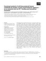
A
B
FIG 1.1: The pathogen-laden environment of the horseshoe crab. (A) The
Singapore horseshoe crab, Carcinoscorpius rotundicauda (Dorsal view) inhabits (B) a
4
pathogen-laden environment in the marshes off Kranji estuary but is able to maintain its
integrity in the midst of high pathogen loads.
1.2.2 Elements of the horseshoe crab innate immunity
Innate immune molecules are present in both the cellular and humoral systems
of the horseshoe crab. Granular hemocytes comprise of 99% of the circulating blood
cells in the horseshoe crab. The large (L)-granules of these cells selectively store more
than 20 innate immune molecules. Many of these function chiefly in hemolymph
coagulation. In contrast, the small-granular structures (S-granules) sequester only five
proteins, all of which demonstrate activities against bacteria and fungi (Toh et al, 1991).
All these cells are highly sensitive to LPS, and respond to its presence by degranulating
the hemocytes, so releasing large numbers of defense molecules. Acting together, these
form a highly complex and sophisticated innate immune system to defend the organism
from invading microbes.
Amongst the many innate immune mechanisms in invertebrates, hemolymph
coagulation has been intensively studied and is well-understood. Hemolymph
coagulation was first discovered in 1956, when Frederik Bang first described fatal
intravascular coagulation in L. polyphemus that was caused by the endotoxin of a
pathogentic Vibrio species. Levin (1968) subsequently showed that this coagulation
resulted from enzymatic conversion of a clottable protein. Proteins participating in
coagulation are derived from large granules of circulating hemocytes (Toh et al, 1991).
Specifically, Factor C, a serine protease zymogen, acts as a LPS-biosensor and induces
autocatalytic activation of itself. This in turn activates Factor B, which then converts a
proclotting enzyme to its active form for blood coagulation. The conversion of
coagulogen into coagulin results from the polymerization of noncovalently bound
coagulin in a “head-to-tail” orientation (Osaki et al, 2004). Conversion of the
preclotting enzyme is also achieved by Factor G, a sensor of β-1,3-glucan, a PAMP
5
found on fungal cell walls. Today, the Limulus amoebocyte lysate (LAL) is recognized
as a potent detector of LPS and forms the basis of the LAL assay, a test commonly
employed to check for contaminating pyrogens in a clinical environment (Iwanaga et al,
1997). The LAL used to make endotoxin detectors is traditionally taken from wild
horseshoe crab populations. As a result of intensive bio-prospecting, Limulus
populations have declined rapidly (Widener & Barlow, 1999) and the species is now
classified as a near-threatened species ( />Aside from components of coagulation cascade, another major group of proteins
present in the hemocytes are lectins. Lectins may be simply defined as proteins which
bind carbohydrates, although much about their physiological functions remain unclear.
A feature of lectin binding is its low affinity (mM range) for a single monosaccharide
residue and/ or its derivative. However, avidity for the ligand is dramatically increased
via oligomerization of the lectins to form multiple binding sites for carbohydrates,
which are themselves multivalent ligands (Loris, 2002). Multivalency is not an absolute
requirement for all lectins, but appears to be an important factor for most (Loris, 2002).
Four lectins have been found in hemocytes of the Japanese horseshoe crab, designated
tachylectin (TL)-1 to 4. These exhibit different carbohydrate specificities and are PRRs
that probably bind different moieties of conserved PAMPs. For example, TL-1 binds
KDO whilst TL-3 binds the O-antigenic region of LPS, a Gram-negative bacterial
PAMP. Unfortunately, biochemical characterizations of these lectins do not shed light
on their real-time collaborative responses following in vivo infection.
The cell-free component of the hemolymph of the horseshoe crab is also known
to contain a range of molecules highly sensitive to insults by pathogens and foreign
materials (Iwanaga et al, 1997). In particular, the circulating hemolymph consists of
three principal proteins: hemocyanin, α2-macroglobulin (α2m) and C-reactive protein
6
(CRP). Hemocyanin functions principally as an oxygen carrier, analogous to the
function of hemoglobin in mammals. α2m is conserved in mammals and is also present
in the plasma of many invertebrates. In the American horseshoe crab, Limulus
polyphemus, α2m is the only protease inhibitor present in the plasma. Melchior and coworkers (1995) have demonstrated that α2m binds active proteases, which are then
cleared from the plasma via a receptor-mediated endocytotic process. Further, α2m is
structurally related to the γ-chain of the human C8 complement factor and is thought to
be involved in the complement cascade (Iwanaga et al, 1997). Like the other TLs, CRP
is a lectin PRR and is found in the plasma of both Carsionoscorpius (Ng et al, 2004)
and Limulus (Roby & Liu, 1981). Additionally, Tachypleus also possess a plasma lectin,
TL-5. This protein exhibits broad specificity for substances containing the N-acetyl
moiety and demonstrates the strongest bacterial agglutinating activity among the all
five TLs isolated thus far (Gokukan et al, 1999).
7
Protein
Tachychitin
Tachystatins
Tachyplesins
Function/ Activity
Acts against Gram-negative and
Gram-positive bacteria, as well as
fungi
Location
SGranules
Polyphemusins
Big defensin
Factor C
Factor B
Factor G
Proclotting
enzyme
Coagulogen
Limulus anti
LPS factor
(LALF)
Tachylectin
(TL)-1
Serine proteases that participate in
the coagulation cascade
Gelling agent; final target of the
coagulation cascade
Inhibits endotoxin coagulation
Lectin. Interacts with Gramnegative bacteria probably through
2-keto-3-deoxyoctonate (KDO), one
of the constituents of LPS
TL-2
Lectin. Binds specifically to
GlcNAc, a common sugar
moietypresent on memebranes
TL-3
Lectin. Exhibits hemagglutinating
activity. High specificity for Oantigens of LPS
TL-4
Lectin. Most probable ligand is
colitose, a unique sugar in the Oantigen of Escherichia coli O111:
B4
TL-5
Lectin. Show the strongest bacterial
agglutinating activity among the
five tachylectins isolated from the
Japanese horseshoe crab. Exhibits
broad specificity for substances
containing N-acetyl groups.
Carcinoscorpius Lectin. Binds the conserved core of
CRP
LPS and is upregulated during
Gram-negative infection
Limulus CRP/ Lectins. Binds sialic acid and
limulin
phosphorylethanolamine. Isolated
Literature
Shigenaga et al,
1993
Osaki et al,
1999.
Kawano et al,
1990
Muta et al, 1990.
Kawabata et al,
1997
Wang et al, 2003
Nakamura et al,
1993
Seki et al, 1994
Muta et al, 1993
Osaki &
Kawabata, 2004
Tanaka et al,
1982
LGranules
Saito et al, 1996
Okino et al, 1995
Saito et al, 1997
Inamori et al,
2001
Inamori et al,
1999
Gokukan et al,
1999
Plasma
Ng et al, 2004
Kaplan et al,
1977
8
from Limulus polyphemus.
Tachypleus (t)
CRP-1
tCRP-2
t-CRP-3
Galactosebinding protein
(GBP)
α2macroglobulin
Hemocyanin
Lectin. Three types of CRPs have
been purified from the plasma of
Tachypleus tridentatus. These are
grouped based on their different
affinities to fetuin–agarose and
phosphory-lethanolamine–agarose.
Binds galactose residue present on
Sepharose CL-4B. Considered an
extracellular glycosylated isoform
of the hemocyte lectin TL-1, with
which it shares 67% homology.
Structurally related to the γ-chain of
the human C8 complement factor
and is involved in the complement
cascade
Oxygen transporter. Probably
invoved in pro-phenol oxidasemediated melanization.
Roby & Liu,
1981
Armstrong et al,
1996
Iwaki et al, 1999
Harum et al,
1993
Plasma
Melchior et al,
1995
Decker et al,
2001
Kawabata &
Nagai, 2000
Nellaippan &
Sugumaran,
1996
TABLE 1.1: Some innate immune defense molecules that may be found in the
hemocytes and hemolymph of the horseshoe crabs. These have been grouped
functionally according to their activities and their locations. The small (S)-granules of
the hemocytes contain about 5 proteins that act against a wide range of pathogens. In
constrast, large (L)-granules are though to contain more than 20 innate immune defense
molecules. These include components of the coagulation cascade and a range of lectins.
There are also many innate immune molecules found within the cell-free hemolymph
(CFH).
9
1.2.3 Plasma lectins are key components in frontline immune defense
Rapid bacterial clearance is dependent upon the action of fast-acting innate
immune molecules. The route of infection for many pathogens involve crossing the
blood barrier whilst systemic infection is characterized by invasion of pathogens into
the plasma. Owing to their extracellular location, plasma lectins that recognize PAMPs
represent frontline pattern recognition receptors (PRRs) that are involved in
immunosurveillance and thus play a pivotal role in halting and neutralizing pathogen
invasion.
The importance of plasma lectins is further exemplified by their evolutionary
conservation. Fibrinogen domain-containing lectins such as ficolins (Lu et al, 2002)
and TLs-5 (Gokudan et al, 1999) are found in invertebrates like the horseshoe crab,
while C-type lectins such as immunolectins (Volanakis, 2001) and collectins (Lu et al,
2002) are found in the tobacco horn worm and in mammals. C-reactive protein (CRP)
is even more prevalent, existing in many vertebrates and invertebrates (Iwaki et al,
1999).
Responses downstream to PAMP-recognition by plasma lectins such as
mannose-binding lectin (MBL), ficolin (Lu et al, 2002) and CRP (Volanakis, 2001)
include pathogen opsonization and complement cascade activation. In the horseshoe
crab, PAMP-recognition by TLs-5 (Gokudan et al, 1999) triggers agglutination of a
wide range of bacteria, leading to speculation of an opsonization function. While the
triggering of overlapping downstream responses by a range of serum lectins appears to
suggest a redundancy of function of PRR lectins, clinical manifestations of MBL
deficiency (Kilpatrick, 2002) implies that each lectin contributes differently and
significantly towards achieving the full potential of the innate immune system.
10
1.3
The role of C-reactive proteins in frontline immune defense
1.3.1 Human CRP - a versatile diagnostic and prognostic marker
One lectin thought to play an essential role in innate immunity is the C-reactive
protein (CRP). CRP was first identified in human serum in1930, as a co-precipitate of
the C-polysaccharide cell wall of Streptococcus pneumoniae. The calcium-dependent
interaction of CRP with the phosphorylcholine (PC) moiety (present in Cpolysaccharide) has been the main paradigm for CRP characterization (Kaplan et al,
1977). X-ray crystal structures indicate that CRP oligomerizes as a pentameric protein
with each subunit tipped towards the central fivefold axis. PC is bound in a shallow
pocket on the surface of each subunit and appears to interact with the two proteinbound calcium ions via the phosphate group (Thompson et al, 1998).
Like homologues found in invertebrates, human CRP (hCRP) plays a pivotal
and complex role in the immune response. hCRP is the classical acute-phase reactant
produced in response to tissue damage and inflammation (Gewurz et al, 1982; Gewurz
et al, 1995; Volanakis, 1982). CRP protein levels rise 42-684- fold above basal levels
under different pathological stresses (Black et al, 2004). Because of its predictable
behavior CRP has gained clinical utility and CRP levels have become important clinical
prognostic tools for a wide range of human diseases. CRP levels have been correlated
to insulin resistance, metabolic syndrome, atherosclerosis (Lee et al, 2004), rheumatoid
arthritis (Nielen et al, 2004) and renal failure (Ortega et al, 2004). Despite such
extensive use of CRP levels to judge disease susceptibility and progression, the actual
involvement of CRP in the pathophysiological presentation of diseases is unknown.
Overall, the evolutionary conservation of CRP across phyla suggests that CRP
possesses roles that are indispensable for survival. In addition, there are no living
11
individuals with CRP deficiency, suggesting that an apparent lethal condition can result
from the lack of just this one lectin type. All these evidences point to the fundamental
role of CRP in human innate immunity.
1.3.2 Gram-negative septicaemia is a widespread medical problem
Inflammation is a cytokine-regulated process that is both an integral part of the
normal immune reaction, as well as a detrimental bodily function. Bacterial, fungal,
viral or parasitic infestations all result in induction of the inflammatory network. When
production of pro-inflammatory molecules is pushed beyond physiologically tolerable
levels, the balance of cytokine-induced inflammatory responses is tipped and
septicaemia ensues (Oberholzer et al, 2000). The clinical pattern of this acute
inflammation is termed systemic inflammatory response syndrome (SIRS) and is
characterized by irregular haemodynamics, coagulatory malfunctions and leukocyteinduced tissue injury (Karima et al, 1999) As sepsis progresses, low perfusion of the
peripheral circulation and other organs occurs, leading to cell death by tissue anoxia,
finally resulting in organ failure, which is the main cause of mortality. (Karima et al,
1999; Brady & Otto, 2001). Clearly, the consequences of pathogen invasions are grave
when not managed properly.
Infections by Gram-negative bacteria (GNB) are the predominant cause of
clinical sepsis (Bone, 1996). Within the United States, 300,000 to 500,000 cases of
septicaemia occur annually, with mortality rates ranging from 20% to 40% (Dellinger
et al, 1997). According to the Centre for Disease Control and Prevention (CDC),
Atlanta, septicaemia and septic shock represent the thirteenth leading cause of death in
the United States and is estimated to incur up to US$10 billion worth of economic loss
(as quoted by Wenzel et al, 1995).
12
Amongst the many GNBs responsible for septicaemia, Pseudomonas
aeruginosa is a leading causative agent (Wenzel et al, 1995). P. aeruginosa is a
ubiquitous GNB noted for its environmental versatility and its resistance to a range of
antibiotics. The bacterium is capable of utilizing a wide range of organic compounds as
food source, thus giving it an exceptional ability to colonize unusual ecological niches
where nutrients are limited (Clarke, 1990). Additionally, P. aeruginosa produces a
number of proteins that cause extensive tissue damage and interfere with human
immune defense mechanisms. These range from potent toxins that kill host cells at or
near the site of colonization, to enzymes that disrupt cellular membranes and
connective tissues in various organs (Clarke, 1990). Despite its versatility, P.
aeruginosa is an opportunistic pathogen that only causes clinical manifestations of
disease in susceptible hosts (Clarke, 1990). Within a clinical environment,
immunocompromised patients, such as cancer patients and burn victims commonly
suffer serious infections by this organism, as do other individuals with immune system
deficiencies. Given the prevalence of sepsis worldwide and the pervasiveness of P.
aeruginosa in causing sepsis, this particular bacterial species is an important target for
study.
During GNB infections, detection of LPS, a pathogen-associated molecular
pattern (PAMP) anchored on the outer wall of the bacteria, triggers a series of
physiological responses. Some of these enhance the inflammatory response, while
others serve to neutralize endotoxic effects (Karima et al, 1999). The interaction of
LPS with the myeloid cell surface antigen, CD14, has been well characterized and is
known to be pivotal in mediating LPS-dependent signal transduction into macrophages.
The binding of LPS to glycosylphosphatidylinositol-anchored CD14 is facilitated by
lipopolysaccharide-binding protein (LBP), an acute phase serum component (Wright et
13
al, 1990).The binding of the LPS-LBP complex to membrane-bound CD14 triggers a
cell signaling pathway, whose mechanisms are not been fully understood (Ingalls et al,
2000; Medvedev et al, 2001). The end result is increased biosynthesis of both pro-and
anti-inflammatory cytokines such as interleukin and TNF-α (Ulevitch & Tobias, 1995).
The LBP-LPS complex has also been found to associate with soluble CD14 (sCD14).
Elevated levels of sCD14 are associated with inflammatory infectious diseases and high
mortality in Gram-negative septicaemia (LeVan et al, 2001) and LBP-LP-sCD14 can
trigger non CD14-possessing cell types such as endothelial. The presence of endotoxin
also activates the humoral arm of the immune system. Endotoxin activates the
complement cascade, which fuels the inflammatory response, and the coagulation
cascade, which results in disseminated intravascular coagulation (Glauser et al, 1991).
Live GNBs also release peptidoglycans, muramyl peptides and other as-yet unidentified
substances that induce cytokine secretions (Murphy et al, 1998). Despite attempts at
ameliorating the effects of sepsis by novel applications of cytokine antagonists (Wenzel,
1991), alternatives to antibiotics remain elusive. Further, the arsenal of antibiotics
available to treat bacterial infection becomes more limited due to the problem of
antibiotic resistance (Hall & Collis, 2001). Overall, septicaemia is a multifaceted
process involving multiple self-propagating and interconnected cascades. The
complexity of the pathogenesis of septicemia remains the greatest obstacle to its
prevention and treatment.
14
FIG 1.2: The consequence of sepsis include over production of cytokines and
uncontrolled inflammation, leading to circulatory malfunctions, shock and multiple
organ failure. Adapted from Oberholzer et al, 2000.
1.3.3 LPS: ubiquitous, persistent and versatile molecules
As discussed above, GNBs are largely responsible for the prevalence of
septicaemia in clinical settings. The key to the primacy of GNBs in causing sepsis is its
PAMP, lippopolysaccharide (LPS). LPS form a class of macromolecules unique to
GNB (FIG 1.3). These are PAMPs located on the outer membrane of GNB and are
referred to as endotoxins because of their pyrogenic (fever-causing) properties in
humans and other mammals (Opal & Gluck, 2003). Structural analysis indicates that
GNBs depend on endotoxin for protection against external assaults. Endotoxin is
arranged so that the more hydrophilic polysaccharide chains face away from the
membrane while the hydrophobic fatty acyl chains of Lipid A are anchored in the
bacterial membrane. Lipid A is tilted at an angle greater than 45˚ at the membrane
interface (Seydel et al, 2000). Such an arrangement effectively packs columns of fatty
acid tightly against one another and confers a “sealed armour” to the bacteria. An
additional layer of polysaccharides around the membrane forms an additional protective
15