Cloning, expression and characterization of oxysterol binding protein homologue 7 (OSH7) in yeast saccharomyces cerevisiae
Bạn đang xem bản rút gọn của tài liệu. Xem và tải ngay bản đầy đủ của tài liệu tại đây (1.83 MB, 123 trang )
CLONING, EXPRESSION AND CHARACTERIZATION
OF OXYSTEROL-BINDING PROTEIN HOMOLOGUE 7
(OSH7) IN YEAST Saccharomyces cerevisiae
LI HONGZHE
(Bachelor of Medicine, Capital University of Medical Sciences)
A THESIS SUBMITTED
FOR THE DEGREE OF MASTER OF SCIENCE
DEPARTMENT OF BIOCHEMISTRY
NATIONAL UNIVERSITY OF SINGAPORE
1
ACKNOWLEDGEMENTS
I would like to thank Dr. Robert Yang Hongyuan and Dr. Heng Chew Kiat, my
supervisors, for providing me a chance to carry out this project, giving access to the
field of molecular biology and provide guidance throughout my postgraduate study.
The same thanks go to the National University of Singapore for the award of a
scholarship which made it possible for me to finish this project. I would also like to
express my sincere gratitude to Chieu Hai Kee for her effort contributed to this project
as well as her valuable suggestions in my postgraduate study. Thanks also go to the
Wee Hong, Penghua, Xianming, Zhang Qian, Shaochong and Li Phing for providing
useful comments and technical assistance. I would also like to thank Dr. Alan Munn,
Jihui, Sebastain and Vicky from Yeast Laboratory in Institute of Molecular Cell
Biology (IMCB) for their generous help. I owe a special gratitude to my family and
friends for their encouragement and support during my academic career.
2
SUMMARY
Oxysterols are potent regulators of cellular sterol homeostasis. The mammalian
oxysterol binding protein (OSBP) was able to bind oxysterols directly; therefore,
OSBP and its related proteins (ORPs) are believed to mediate some of the effects by
oxysterols. However, recent data suggested that OSBP and ORPs might interact with
other lipids, such as phosphatidylinositides, and might have functions other than
controlling cellular sterol metabolism. The molecular mechanisms underlying the
function of the entire OSBP family of proteins remain to be elucidated. The yeast OSH
genes (OSH1-OSH7), which encode a family of homologues of OSBP, are believed to
play important roles in the maintenance of intracellular lipid distribution, endocytosis
and the integrity of vacuole morphology. In our study, we demonstrated using yeasttwo-hybrid system that the coiled-coil domain of Osh7p could interact with Vps4p,
which belongs to the protein family of AAA-type ATPases. The interaction was
further confirmed by a GST-pull down assay. Subcellular fractionation was performed
to localize Osh7p mainly to the cytosolic fraction in wild-type cells, however, in vps4∆
yeast cells, a significant portion of the Osh7p redistributed to a membranous fraction.
Sucrose density gradient analysis further confirmed the redistribution of Osh7p in
vps4∆ strain. Meanwhile, we demonstrated that endocytosis and vacuolar protein
sorting were not affected by OSH7 deletion. Concomitantly, the interaction between
Osh7p and phospholipids was investigated using protein-lipid overlay assay. In this
study, Osh7p showed the ability to bind to PI(4)P and PI(5)P. Finally, we presented
evidence to suggest that the loss of Osh7p function influence sterol esterification.
3
TABLE OF CONTENTS
Page
ACKNOWLEDGEMENTS
2
SUMMARY
3
TABLE OF CONTENTS
4
LIST OF FIGURES
9
LIST OF TABLES
11
LIST OF ABBREVIATIONS
12
1. INTRODUCTION
14
1.1
Cholesterol homeostasis
14
1.2
Oxysterols
17
1.3
1.2.1
Structure of oxysterols
17
1.2.2
Roles of oxysterols
19
1.2.3
Oxysterol-binding protein
22
Yeast OSBP homologues
25
1.3.1
Structure of Osh proteins
25
1.3.2
Localization of Osh proteins
27
1.3.3
Function of Osh proteins
27
1.3.3.1 Role of OSH in maintaining sterol-lipid distribution
and vacuolar integrity
28
1.3.3.2 Other sterol-related phenotypes of single and multiple
osh∆ mutants
1.4
29
1.3.3.3 Role of OSH in vesicular trafficking
30
1.3.3.4 Role of OSH in endocytosis
33
Vesicle-mediated vacuolar protein sorting
4
34
1.5
VPS mutants
36
1.5.1
Class E mutants
37
1.5.2
Vps4p
37
1.6
OSH7
40
1.7
Objectives of this project
41
2
MATERIALS AND METHODS
42
2.1
Media, reagents, strains and plasmids
42
2.2
Agarose gel electrophoresis
45
2.3
Isolation of plasmid DNA from E coli.
45
2.3.1
Small scale preparation of plasmid DNA
45
2.3.2
Large scale preparation of plasmid DNA
46
2.4
Extraction of yeast genomic DNA
47
2.5
Cloning
48
2.5.1
Cloning of OSH7 in the plasmid pJG4-5
48
2.5.1.1 Primer design
48
2.5.1.2 PCR amplification and purification
48
2.5.1.3 Digestion
48
2.5.1.4 Converting 5’-overhang to a blunt end
49
2.5.1.5 Gel purification
49
2.5.1.6 Ligation
50
2.5.1.7 Preparations of competent cells and transformation
50
2.5.1.8 Selection and DNA sequencing
51
2.5.2
Other subclonings
52
2.5.2.1 Generation of pJG4-5-OSH6cc construct
5
52
2.5.2.2 Generation of YEplac-OSH7-GFP and
YCplac-OSH7-GFP plasmids
2.6
52
2.5.2.3 Generation of pADNS-OSH7 construct
52
2.5.2.4 Generation of pGEX-4T-2-OSH7 construct
52
Deletion of OSH7
53
2.6.1
PCR amplification and purification
53
2.6.2
Transformation of kanMX4 into yeast cells
54
2.6.3
Selection of recombinants
55
2.7
SDS-PAGE gel electrophoresis
56
2.7.1
Preparation of reagent and stock solution
56
2.7.2
Procedures
57
2.7.3
Loading samples and electrophoresis
57
2.8
Western blotting
58
2.9
Yeast two-hybrid assay
58
2.9.1
The strategy of interaction trap
59
2.9.2
Detecting in vivo interaction by yeast two-hybrid system
60
2.9.2.1 Characterization of the bait protein Vps4p by
repression assay
60
2.9.2.2 Performing an interaction assay
60
2.9.2.3 Expression of prey protein under induction
61
2.9.2.4 β-Galactosidase assay
62
2.10
Fluorescence microscopy
63
2.11
Subcellular localization of protein
63
2.11.1 Subcellular fractionation
63
2.11.2 Flotation assay
64
6
2.11.3 Detergent treatment assay of membranous fractions
64
2.11.4 Sucrose density gradient
65
2.12
Fluid-phase endocytosis assay
66
2.13
FM4-64 endocytosis assay
66
2.14
Carboxypeptidase Y missorting assay
67
2.15
Lipid assay
67
2.15.1 In vivo neutral lipid synthesis assay
67
2.15.2 Sterol biosynthesis assay
68
2.15.3 Total ergosterol analysis
69
2.16
Protein-lipid overlay assay
70
2.16.1 Purification of recombinant GST-Osh7p and GST proteins 70
2.16.1.1 Preparation of the bacterial lysate
70
2.16.1.2 Affinity chromatography to purify the GST-Osh7p and
GST proteins
70
2.16.2 Protein-lipid overlay assay
2.17
3
71
Production of antibody against Osh7p
RESULTS
3.1
3.2
73
Osh7p interact specifically with Vps4p
3.1.1
Vps4p interacts with Osh7p in the yeast two-hybrid system 73
3.1.2
Vps4p and Osh7p interact in vitro
Deletion of VPS4 causes redistribution of Osh7p
3.2.1
73
77
79
Osh7p-GFP shows different staining pattern in wild-type and
vps4∆ strains
3.2.2
71
79
In vivo production and distribution of Osh7p depend
on Vps4p function
81
7
3.2.3
Sucrose density gradient analysis further confirms the
redistribution of Osh7p in vps4∆ strain
84
3.2.4
Osh7p associates with a membranous compartment
85
3.2.5
The membrane association of Osh7p is mediated by
a large protein complex
86
3.3
Transport of CPY and CPS is unaffected by OSH7 deletion
3.4
Fluid-phase endocytosis and FM4-64 internalization are
87
unaffected in osh7∆ mutant cells
90
3.5
Osh7p interacts with PI(4)P and PI(5)P
94
3.6
Sterol esterification is increased in osh7∆ strain while sterol
biosynthesis remains unaffected
3.7
Total ergosterol level is not affected by OSH deletion or
overexpression
4
96
99
DISCUSSION
101
4.1
Vps4p physically and functionally interacts with Osh7p
101
4.2
Osh7p interacts with PI(4)P and PI(5)P
103
4.3
Endocytosis and vacuolar protein sorting are not affected
by OSH7 deletion
105
4.4
Loss of Osh7p function increases sterol esterification
107
4.5
Understanding of the nature of the interaction between Osh7p and
Vps4p and future directions
108
4.6
Understanding the nature of Osh7p modification
109
4.7
Future directions of the project
110
5
CONCLUSION
113
6
REFERENCES
114
8
LIST OF FIGURES
Figure NO.
Page
1. Structure of cholesterol
15
2. Structure of ergosterol
17
3. Structure of the cyclopentanopehydrophenanthrene nucleus,
cholesterol and some oxysterols
19
4. Physiological roles of oxysterols
21
5. Predicted secondary structure of Osh proteins
26
6. Pathway for Sec14-dependent Golgi secretory function
32
7. Schematic figure of the protein structure of Vps4p
38
8. Model for ATP-driven cycle of Vps4p in vivo and in vitro
39
9. Schematic figure of the protein structure of Osh7p
40
10. Outline of short flanking homology strategy for disruption of
a targeted ORF
54
11. PCR verification of disrupts
56
12. The interaction trap
59
13. Vps4p and Osh7p interact in vivo
76
14. Vps4p and GST-Osh7cc interacts in vitro
79
15. In vivo activity of OSH7-GFP
80
16. Osh7p-GFP shows different localization in wild-type and
vps4∆ cells
81
17. Specificity of anti-Osh7p polyclonal antibody
82
18. Osh7p is over-produced in vps4∆ cells
82
19. Effects of loss of Vps4p on the subcellular distribution of Osh7p
83
20. Part of Osh7p redistributes to a membranous peak in sucrose density
gradient
85
9
21. Osh7p is associated with a protein complex in membranous fraction
in vps4∆
86
22. Characterization of the association of Osh7p with the P13 subcellular
fraction
87
23. No defect is detected in CPY missorting test by OSH7 deletion
89
24. Delivery of CPS to vacuole is unaffected in osh7∆ cells
90
25. Fluid-phase endocytosis assay does not show any defect in osh7∆
strain
92
26. Transport of FM4-64 is normal in osh7∆ strain
93
27. GST-Osh7p interacts with PI(4)P and PI(5)P
96
28. Sterol biosynthesis is unaffected in osh7∆ cells
97
29. Sterol esterification is increased in osh7∆ mutants
98
30. Sterol esterification is unaffected by Osh7p overexpression
99
31. Total ergosterol level is not affected by OSH7 deletion or
overexpression
100
10
LIST OF TABLES
Table NO.
Page
1. Genotype of yeast strains used in this project
43
2. Plasmids used in this project
44
3. Compositions of SDS-PAGE
57
11
LIST OF ABBREVIATIONS
aa
amino acid
Amp
ampicillin
CHO
Chinese Hamster Ovary
CPY
carboxypeptidase Y
DTT
dithiothretiol
EDTA
ethylene diamine tetra-acetic acid
ER
endoplasmic reticulum
g
gram or gravitational force
GFP
green fluorescent protein
GST
Glutathione-S-transferase
HMG-CoA
3-hydroxy-3-methyglutaryl coenzyme A
Hrs
hepatocyte growth factor regulated tyrosine kinase substrate
IPTG
β-D-thiogalactopyranoside
LB
Luria-Bertani broth
LDL-R
low density lipoprotein receptor
leu
leucine
LiAC
lithium acetate
LXR
liver X receptor
met
methionine
MVB
multivesicular body
PVC
pre-vacuolar compartment
PEG
polyethylene glycol
PBS
phosphate buffer saline
PCR
polymerase chain reaction
12
PMSF
phenyl methanosulfonyl fluoride
SDS
sodium dodecyl sulphate
SREBP
sterol regulatory element-binding protein
TCA
thichloroacetic acid
TTBS
Tween Tris-buffered saline
VPS
vacuolar protein sorting
WCE
whole cell extract
YEB
yeast extraction buffer
13
1. INTRODUCTION
1.1 CHOLESTEROL HOMEOSTASIS
Sterols are important membrane components of all known eukaryotic organisms and
play essential roles in modulating membrane fluidity and permeability. In mammalian
cells, the predominant membrane sterol is cholesterol while its close relative,
ergosterol, is used as the major membrane sterol by the yeast Saccharomyces
cerevisiae. Cholesterol is an extremely important biological molecule that has roles in
membrane structure as well as being a precursor for the synthesis of the steroid
hormones and bile acids. Cholesterol has attracted much attention because of its
essential function in membranes of animal cell, and because it is the raw material for
the manufacture of steroid hormones and bile acids. The very property that makes it
useful in cell membrane, namely its absolute insolubility in water, also makes it lethal.
The amount of cholesterol in animal cell membranes is tightly regulated to maintain
proper cell function. When cholesterol accumulates in the wrong place, for example
within the wall of an artery, it cannot be readily mobilized and its presence eventually
leads to the development of an atherosclerotic plaque. Therefore, regulatory
mechanisms must exist to maintain cholesterol homeostasis within cells.
Cholesterol has a complex four-ring structure (Fig 1.) and it is synthesized from a
simple two-carbon substrate (acetate) through the action of at least 30 enzymes. The
mechanisms underlying the synthesis and uptake of sterols by eukaryotic cells are now
relatively well characterized and the cellular sterol homeostasis is regulated by at least
three distinct mechanisms (Goldstein and Brown, 1990):
14
•
1. Regulation of HMG-CoA reductase activity and levels
•
2. Regulation of excess intracellular free cholesterol through the activity
of acyl-CoA:cholesterol acyltransferase, ACAT
•
3. Regulation of low density lipoprotein (LDL) receptor-mediated
cholesterol uptake
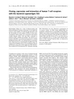
Fig 1. Structure of cholesterol. The structure of cholesterol consists of four fused
rings with the carbons numbered in sequence, and an eight-membered, branched
hydrocarbon chain attached to the D ring. Cholesterol can be esterified by acyl-CoA:
cholesterol acyltransferase (ACAT) to form cholesterol esters. Cholesterol ester has a
fatty acid attached at carbon 3, which makes the structure even more hydrophobic.
However, much less is understood about cellular sterol transport and how a
nonhomogenous distribution of sterols between different internal membranes is
maintained. Sterol homeostasis requires that there must be mechanisms to sense
cellular sterol levels, and although there has been much recent progress in identifying
some of the key regulators of cholesterol metabolism (Brown and Goldstein, 1999),
little is known about how sterol sensing occurs. The intracellular traffic of cholesterol
appears to be important in this feedback (Lange and Steck, 1996). The majority of
cholesterol is found in the plasma membrane, but it is synthesized in the endoplasmic
15
reticulum (ER), where the cholesterol level is low and where changes in cellular
cholesterol levels are sensed. The ER-embedded sterol regulatory element-binding
protein (SREBP) system controls the transcription of genes encoding cholesterol
biosynthetic enzymes (Brown and Goldstein, 1999; Lange et al., 1999). Although it
might be expected that the systems controlling cholesterol metabolism would
recognize cholesterol itself, there has been a long-standing interest in the possibility
that oxysterols, a group of oxidized derivatives of sterols, are important second
messengers in sterol homeostasis (Brown and Goldstein, 1974; Kandutsch and Chen
1974; Accad and Farese, 1998). Indeed, oxysterols such as 25-hydroxycholesterol are
up to a 1000 times more potent than cholesterol itself as down-regulators of cholesterol
synthesis (Kandutch et al., 1978; Goldstein and Brown, 1990).
Baker's yeast, Saccharomyces cerevisiae, makes its own cholesterol-like lipid called
ergosterol, which is a major constituent of yeast membrane, where it is present in 3.3fold molar excess over all phospholipids (Zinser et al., 1991). Ergosterol is the bulk
isoprenoid product of the mevalonate biosynthetic pathway, whose structure is showed
in Fig 2. The products of the mevalonate pathway exert feedback regulation on their
own synthesis at both transcriptional and post-transcriptional levels (Goldstein and
Brown, 1990; Brown and Goldstein, 1997, 1999). Although some specific steps are
unique in yeast, most biosynthetic routes of lipids in yeast are similar to those in
mammalian cells (Basson et al., 1986, 1988; Jennings et al., 1991; Reynolds et al.,
1984; Robinson et al., 1993). Thus, the regulation of sterol homeostasis appears to
require many of the similar genes and proteins in yeast and human and much of the
work in defining the role of sterol in eukaryotic membranes has been done using the
yeast model system. In this project, we use yeast as an experimental organism for
16
studying the role of oxysterol-binding protein homologue in intracellular sterol
metabolism and vesicular trafficking.
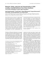
Fig 2. Structure of ergosterol. Ergosterol differs from cholesterol by the presence of
unsaturations at C-7,8 in the ring structure and at C-22 in the side chain and by the
presence of a methyl group at C-24 on the side chain (Zinser et al.,1991).
1.2 OXYSTEROLS
1.2.1 Structure of oxysterols
Oxygenated derivatives of cholesterol (oxysterols) are biosynthetic metabolites of
sterols, steroids and bile acids and they are 27-carbon products of cholesterol oxidation.
Except for 24,25-epoxysterols, most oxysterols arise from cholesterol by autoxidation
or by specific microsomal or mitochondrial oxidations, usually involving cytochrome
P-450 species (Smith, 1987 and Schroepfer, 2000). They can be broadly defined as
compounds which possess (a) a cyclopentanoperhydrophenanthrene nucleus, (b) a
hydrocarbon side chain attached to C17, (c) a hydroxyl group at C3, and (d) one or
more additional oxygens attached to the nucleus or side chain. The structures of
17
cholesterol and some oxysterols are depicted in Fig 3. As cholesterol is insoluble in an
aqueous environment and resides mainly in membranes, it has been difficult to
imagine how it could function as a molecular regulator. Oxysterols, on the other hand,
have emerged as potential sterol homeostatic regulators because of their greater
polarity and aqueous solubility, and because of observations dating from the 1970s that
these compounds are more potent than cholesterol in down-regulating cholesterol
biosynthesis in cultured cells (Brown and Goldstein, 1974; Kandutsch et al., 1974).
18
Fig 3. Structure of the cyclopentanopehydrophenanthrene nucleus, cholesterol
and some oxysterols. (Figure taken from Hwang, 1991)
1.2.2
Roles of oxysterols
Oxygenated derivatives of cholesterol (oxysterols), analogues of cholesterol with
additional polar groups, are widely distributed in nature, being found in the blood and
19
tissues of animals and human as well as in food. As a group, oxysterols have attracted
much attention in recent years on account of their biological activities which are of
potential physiological, pathological or pharmacological importance. Oxysterols
remained to be a biochemical curiosity until it was shown in 1974 that they were
potent inhibitors of sterol biosynthesis by indirect inhibiting the activity of HMG-CoA
reductase, which is the rate-limiting enzyme in cholesterol biosynthesis (Brown and
Goldstein, 1974; reviewed by Hwang, 1991). In the following years, it was found that
oxysterols regulate sterol metabolism by means of nuclear and cytoplasmic actions in
animal cells (Taylor and Kandutch, 1985; Goldstein and Brown, 1990). In the nucleus
oxysterols repress transcription of genes encoding enzymes of sterol biosynthesis,
including 3-hydroxy-3-methyglutaryl (HMG) CoA reductase and HMG-CoA synthase,
and they also repress transcription of the gene encoding low density lipoprotein (LDL)
receptor (Goldstein and Brown, 1990). In the cytoplasm, oxysterols inhibit translation
of the mRNA for HMG-CoA reductase and accelerate the proteolytic degradation of
this ER enzyme (Dawson et al., 1991; Peffley et al., 1988). Furthermore, oxysterols
activated another ER enzyme, acyl-CoA: cholesteryl acyltransferase (ACAT), which
can facilitate the storage of excess sterols as sterol esters (Brown, 1975; Chang and
Doolittle, 1983). These actions limit the biosynthesis and uptake of cholesterol, and
they are part of a coordinated mechanism that prevents the accumulation of
unesterified cholesterol within cells. Research during the ensuing years revealed that
oxysterols participate in several different aspects of lipid metabolism (Fig 4.). In
addition to serving as regulators of gene expression, oxysterols are also substrates for
bile acid synthesis and mediators of sterol transport. As regulatory molecules, they
inhibit the production of transcription factors required for the expression of genes in
the cholesterol supply pathways (Brown and Goldstein, 1997), and they are ligands
20
that activate members of the nuclear hormone receptor gene family (Janowski et al.,
1996). Oxysterols are inactivated by conversion into bile acids, and in some instances,
the essential need for bile acids can be met solely by the metabolism of oxysterols
(Schwarz et al., 1996). They also may be substrates for steroid hormone biosynthesis
(Nes et al., 2000). Tissues such as the lung and brain secrete measurable amounts of
oxysterols into the circulation, which are then transported to the liver and converted
into bile acids (Babiker et al., 1999). This secretion represents a form of reverse
cholesterol transport (Bruce et al., 1998), a mechanism that peripheral tissues use to
return cholesterol to the liver and thus to maintain homeostasis.
The important roles of oxysterols are also in part due to the observation that although
most mammalian cells export cholesterol to high-density lipoprotein particles in the
plasma, at least two cell types, macrophages and neurons, export the bulk of sterol as
27- and 24-hydroxycholesterol, respectively (Bjorkhem et al., 1999). Furthermore,
oxysterols have been shown to play roles in apoptosis, cellular aging, platelets
aggregation and sphingolipids metabolism (reviewed by Schroepfer, 2000).
Fig 4. Physiological roles of oxysterols. Cholesterol is converted into oxysterols that
participate in several aspects of lipid metabolism, including regulation of gene
expression, bile acid synthesis in the liver, and transport of sterol from one tissue to
another (Figure taken from Russell, 2000).
21
If intracellular oxysterols serve as second messengers to regulate lipid metabolism,
there must be particular proteins that recognize them. To date, two protein families
appear to mediate many of the activities attributed to oxysterols. These proteins
include some of the steroid hormone nuclear receptors such as liver X receptor α
(LXRα) and steroidogenic factor 1 (SF-1) (Russell, 1999) and another family known as
the oxysterol-binding proteins (OSBPs). When activated by oxysterols, the liver X
receptors (LXR) regulate the expression of several genes in key positions in the
maintenance of the whole body cholesterol balance and function as a ligand-activated
transcription factor to up-regulate cholesterol catabolism to bile acids (Peet et al.,
1998). These include genes involved in cholesterol absorption in the gut (Repa, et al.,
2000), cholesterol efflux from peripheral cells (Repa et al., 2000; Venkatewaran et al.,
2000), synthesis of fatty acids (Repa et al., 2000; Schultz et al., 2000), remodeling of
lipoproteins in the circulation (Luo and Tall, 2000), and the bile acid synthetic pathway
(Lehmann et al., 1997; Peet et al., 1998). However, in this project, we are more
interested in OSBP, which appears to be the only protein known to bind specifically to
the group of oxysterols that are active in the down-regulation of cholesterol synthesis
(Dawson, 1989). OSBP was identified as being the most abundant cytosolic protein
that bound to such regulatory oxysterols (Taylor et al., 1984, 1985).
1.2.3 Oxysterol-binding protein
The presence of OSBP was first reported in 1977 (Kandutsch et al., 1977). Based on
its high affinity to 25-hydroxycholesterol, cytosolic OSBP was first purified. It binds a
wide variety of oxysterols with affinities that are generally proportional to their
potencies in regulating sterol metabolism (Taylor et al., 1984). The OSBP gene was
cloned from the rabbit and the human (Dawson et al., 1989; Levanon et al., 1990), and
22
the encoded OSBPs are 98% identical. Multiple OSBP homologues have been found in
the genomes of all eukaryotes so far examined, including humans (Levanon et al.,
1990), flies (Alphey et al., 1998), worms (C. elegans Sequencing Consortium, 1998),
and fungi (Jiang et al., 1994; Schmalix and Bandlow, 1994; Fang et al., 1996; Daum et
al., 1999; Hull and Johnson, 1999). These proteins all share a conserved 400 amino
acid domain found at the C-terminus of OSBP, which has been shown to bind
oxysterols (Ridgway et al., 1992). For convenience we will refer to this shared,
characteristic domain as the “oxysterol binding domain”, although its binding
specificity in other species has not been investigated. OSBP homologues can be
divided into two general classes: short ones that comprise an oxysterol-binding domain
alone, and longer ones such as OSBP itself have a pleckstrin homology (PH) domain at
the N-terminus.
The localization of OSBP within cells is governed by lipids. In transfected Chinese
hamster ovarian cells (CHO) overproducing OSBP, the OSBP is found to be
distributed diffusely in the cytoplasm and associated with small perinuclear vesicles. In
the presence of 25-hydroxycholesterol, OSBP translocates to Golgi apparatus through
PH domain where it appears to stimulate conversion of ceramide to sphingomyelin
(Ridgway et al., 1992 and Lagace et al., 1999). Most PH domains in other proteins
direct
localization
to
the
plasma
membrane,
often
by
interaction
with
phosphatidylinositol phosphates (PIPs). We have found that, in contrast, the PH
domain of OSBP specifies targeting to the trans-Golgi network (TGN) of mammalian
cells, and this interaction requires the presence of Golgi PIPs (Levine and Munro,
1998). OSBP localization is also sensitive to concentrations of the lipid sphingomyelin
(Storey et al., 1998; Ridgway et al., 1998). Based on the linkage between OSBP
23
localization and cellular lipid distribution, the function of OSBP likely involves in
maintaining lipid homeostasis in membranes.
Although the precise function of OSBP family has remained elusive, it at least seems
certain that their function is required in all eukaryotes. Because of its binding activity
and the potency of oxysterols as feedback regulators, OSBP was proposed to mediate
feedback control of the mevalonate pathway. Overexpression of OSBP in Chinese
Hamster Ovary (CHO) cells causes pleiotropic effects on both cholesterol synthesis
and expression of genes encoding some mevalonate pathway enzymes (Lagace et al.,
1997). Several other studies have implicated OSBP in the regulation of cellular
cholesterol and sphingomyelin homeostasis (Ridgway et al., 1998; Storey et al., 1998;
Lagace et al., 1999). These data suggested the involvement of OSBP in mediating the
effects of oxysterols on cholesterol metabolism even though OSBP was not found to
be a major controller of transcription of the genes responsible for cellular cholesterol
homeostasis. However, the in vivo role of the OSBP family is still unclear.
Recently, 11 OSBP-related proteins (ORPs) have been cloned based on the highly
conserved OSBP domain. Two of these ORPs, ORP1 and ORP2, with the highest
degree of similarity to yeast Osh4p, were shown to bind to phospholipids instead of
oxysterols (Xu et al., 2001). Osh4p has been implicated in the PI-dependent formation
of Golgi-derived transport vesicles, which will be discussed in more detail in the later
part of this thesis. In Chinese hamster ovary cells, ORP1 localized to a cytosolic
location while ORP2 was associated with the Golgi apparatus, consistent with the
hypothesis that ORP1 and ORP2 function at different steps in the regulation of vesicle
transport. Overexpression of ORP2 protein can cause increase in [14C] cholesterol
24
efflux and decrease in ACAT activity. These results implicated ORP2 as a novel
regulator of cellular sterol homeostasis and intracellular membrane trafficking. The
yeast Saccharomyces cerevisiae has seven OSBP homologues whose functions are
currently not well defined (Jiang et al., 1994).
1.3 YEAST OSBP HOMOLOGUES
1.3.1 Structure of Osh proteins
There are seven OSBP homologues in yeast Saccharomyces cerevisiae, named OSH1
through OSH7, respectively (Beh et al., 2001). The homology of all the seven proteins
was the highest in a small domain of 150 to 200 amino acids. This small domain is
known as the OSBP domain. Beside the OSBP domain, these proteins also contain a
putative coiled-coil motif, which might be important for protein-protein interaction
(Fig 5.). Three of these proteins (Osh1p, Osh2p and Osh3p) have a large N-terminal
region that includes a PH domain, which might regulate protein targeting to
membranes and thereby serve as membrane adaptors by interacting with
phospholipids. Osh1p and Osh2p also have three ankyrin repeats, which are not found
in the mammalian protein. Ankyrin repeats mediate protein-protein interactions and are
generally found in cytoskeleton proteins and transcription factors. Thus the structure of
Osh1p and Osh2p is suggestive of being able to bind both a phosphoinositide lipid
through their PH domain and a protein partner through their anykyrin repeats. A
putative membrane-spanning domain would constitute a contiguous stretch of 19-20
residues predicted to form an α-helix, with a hydrophilicity score of <-1.6 over the
entire length (Kyte and Doolittle, 1982). By these criteria, none of the OSBPs was
likely to be an integral membrane protein (Beh et al., 2001). Although secondary
structure predictions indicated that all the yeast Osh proteins are likely to be soluble
25