Effects and mechanisms of a concentrated goyasaponin fraction in 3t3 l1 cell line
Bạn đang xem bản rút gọn của tài liệu. Xem và tải ngay bản đầy đủ của tài liệu tại đây (1.26 MB, 89 trang )
CHAPTER 1
LITERATURE REVIEW AND OVERALL HYPOTHESES
1. Type 2 Diabetes and Obesity
1.1 Type 2 Diabetes
Diabetes mellitus is characterized by abnormally high blood glucose levels
(hyperglycemia) and caused by an altered secretory amount of insulin from pancreas as well
as decreased effectiveness in insulin action [1, 2]. According to recent classification, Type 1
and Type 2 are two main types of this chronic disease [2, 3]. Type 1 diabetes, also known as
juvenile-onset or insulin-dependent diabetes, is an autoimmune disease in which pancreatic
islet cells are destroyed by antibodies produced by the body [2, 4]. Therefore, absolute
deficiency in insulin production by the pancreas is always associated with Type 1 diabetes
[3]. Type 2 diabetes, also known as adult-onset or non-insulin dependent diabetes, is the
consequence of altered insulin secretion as well as resistance to insulin action [2, 4].
The prevalence of type 2 diabetes, which accounts for approximately 90 - 95% of all
diagnosed cases of diabetes, is increasing worldwide and the global diabetic population is
estimated to double from 151 million in 2000 to 300 million in 2025 [5, 6]. Meanwhile,
annual medical expenditure associated with type 2 diabetes imposed a significant financial
burden on society. In United States, type 2 diabetes accounts for more than $100 billion in
healthcare costs annually [2] while in western European countries, 2-7% of total national
health budgets has been spent on diabetes care [7]. Even more disturbing is the growing
number of children and adolescents diagnosed with type 2 diabetes which was previously
present in adults only [7, 8]. Additionally, as diabetes mellitus can lead to serious long-term
1
complications including heart disease, retinal damage, renal failure and stroke [9], it was
ranked sixth as the leading cause of death in United States in 2004 [10].
In normal subjects, the plasma glucose levels are maintained with the aid of insulin
[11]. Insulin is a multipotent hormone secreted from β-cell in pancreas and its most
important effect is on glucose homeostasis [12]. First of all, insulin can stimulate the
peripheral glucose uptake in muscle (around 60% of insulin-stimulated whole body glucose
uptake), liver (around 30%) and adipose tissue (about 10%) [12]. At the same time, insulin
also stores excess glucose as glycogen in liver and muscle [13, 14]. Insulin resistance,
characterized as defects in insulin signaling in insulin-responsive tissues, occurs many years
prior to diabetes onset and serves as a strong predictor for the type 2 diabetes development
[15, 16]. Therefore, both normal insulin secretion and sensitivity to insulin action are of
importance for maintenance of plasma glucose level.
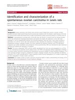
The developmental process of type 2 diabetes is a complex and multifactorial
consequence as shown in Figure 1 proposed by Olefsky et al. in 1995 [16]. Several risk
factors, including both genetic and acquired factors, make significant contributions to type 2
diabetes. In addition, further new insights into mechanism in the future would provide a
better understanding of type 2 diabetes pathogenesis.
2
Figure 1. Pathology of type 2 diabetes concluded by Olefsky et al. in 1995
3
1.2 Obesity
Obesity is another chronic disease associated with the excessive presence of body fat
and it has become a leading public health problem in the world [17]. Similar to type 2
diabetes, increased prevalence of obesity among both adults and children has been observed
in many countries throughout the world [18, 19]. According to National Centre for Health
Statistics, it is reported that 61% of adults are overweight and 26% are obese in United States
in 1999, as defined by body mass index (BMI) [20] . Globally, the prevalence of obesity has
increased more than 75% since 1980 [17].
Obesity is a consequence of an imbalance in energy metabolism resulting from
excessive food intake concurrent with decreased energy expenditure [17]. There are several
explanations for this global increase in obesity rate. First of all, genetic factor makes
dominant contribution to body weight as inheritability for obesity is estimated to be 50-90%
[17]. Moreover, some acquired factors, including obesity-promoting changes in diet and
sedentary lifestyle, also exacerbate this increased prevalence [17].
1.3 Close Connection between Type 2 Diabetes and Obesity
Obesity is closely associated with insulin resistance and is considered to be a leading
risk factor for both type 2 diabetes and cardiovascular disease [21, 22]. Recent
epidemiological studies have shown that BMI over 28 kg/m2 exponentially increase the risk
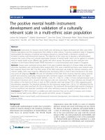
of type 2 diabetes [23] . Various hypotheses have been proposed to explain the causal
relationship between obesity and type 2 diabetes [24], one of the which accepted is the
‘lipotoxicity hypothesis’ as shown in Figure 2: in obese individuals, beta-cell dysfunction,
4
insulin resistance and impaired glucose tolerance develop in response to physiological
dysfunction in muscle, liver and pancreas where excess lipids are deposited [24-26].
1. Increased number of adipocytes (hyperplasia)
Obesity
2. Enlarged size of adipocytes (hypertrophy)
1. Excess lipids cannot be stored in adipose tissue in the
form of triglyceride
Lipotoxicity
2. Excess lipids deposited insulin-sensitive tissues, such as
muscle, liver and pancreas
3. Physiological dysfunction in muscle, liver and pancreas
Insulin Resistance
Beta-cell dysfunction
Impaired glucose tolerance
Type 2 Diabetes
Figure 2. Proposed hypothesis of lipotoxicity
5
2. Adipose Tissue and 3T3-L1 Cell Line
2.1 Adipose Tissue
Mature adipocytes are the main cellular component of adipose tissue. In addition,
adipose tissue also contains other components, such as undifferentiated preadipocytes,
immune cells (leukocytes, macrophages), nerve fibers, vascular stroma, lymph nodes and a
matrix of collagen and reticular fibers [27].
There are essentially two types of adipose tissue in humans referred to as white
adipose tissue (WAT) and brown adipose tissue (BAT) [27, 28]. BAT is found in fetuses and
newborn infants and is practically absent in adults as its principal function is to burn fat to
generate heat for newborns during the initial hours after birth [27, 28]. Uncoupling Protein-1
(UCP-1), exclusively expressed in brown adipocytes, plays a vital role in heat production as
it uncouples electron transport from adenosine-5'-triphosphate (ATP) production allowing
energy to dissipate as heat [27].
However, the predominant type of adipose tissue in humans is WAT [29]. Two types
of WAT exist and are classified as visceral and subcutaneous [30]. Functional differences
exist between these two types and it appears that individuals with visceral fat accumulation
are more likely to develop metabolic and cardiovascular diseases [30]. In response to energy
demands, WAT serves as a site for storing excess energy as triglyceride via lipogenesis and
releasing energy in the form of free fatty acid/glycerol via lipolysis when there is a calories
deficit [27, 29, 31]. In addition to its biological repertoire necessary for storing and releasing
energy [32], WAT has been proved recently to be an essential endocrine organ which
secretes a variety of bioactive proteins termed as adipocytokines, including adiponectin,
leptin, resistin and visfatin, which have been proved to be actively involved in energy
6
regulation, lipid metabolism, insulin resistance, immunological response and vascular disease
[32-34]. Moreover, WAT also expresses and secretes some other cytokines and chemokines,
such as tumor necrosis factor-alpha (TNF-α), interleukin-6 (IL-6) and monocyte
chemoattractant protein-1 [35].
Besides these two roles of adipose tissue as mentioned above, Cao et al. in 2008 found
that palmitoleate, a free fatty acid metabolite generated in and released from adipocytes, is a
circulating factor that promotes the insulin sensitivity in liver and muscle [36]. This latest
finding is a further step to demonstrate that lipid metabolism and glucose homeostasis are
highly interconnected processes [37].
Therefore, due to its multifunctional characters, WAT has been focused by
researchers as a possible central mediator of whole body insulin resistance in recent years
[38].
2.2 3T3-L1 is an established in vitro model of adipocyte biology
The molecular and cellular events during the adipogenesis process have been studied
on various cell culture models, including both preadipocyte cell lines and primary culture of
adipose-derived stromal vascular precursor cells [32]. 3T3-L1 is a substrain of Swiss 3T3
murine cell line derived from disaggregated 17- to 19- mouse embryos [39]. It has been
proved to be an established murine preadipocyte fibroblast which can be induced to
adipocyte differentiation (adipogenesis) and completely convert into oil droplet-containing
mature adipocytes [22]. Therefore, both differentiation process and adipocytokines secretion
from mature adipocyte can be investigated under this cell line condition [22, 40].
7
Progress has been made in understanding adipocyte differentiation process. First of all,
these new findings provided the molecular and cellular basis of the adipose tissue growth of
physiological and pathophysiological state. Additionally, they also provided means of
developing strategies for both prevention and treatment of obesity [39]. Characterized by
increased adipose tissue mass, obesity is determined by both enlarged size and increased
number of adipocytes [41]. Therefore, several applicable anti-obesity mechanisms, including
decreased preadipocyte proliferation, inhibition of adipocyte differentiation, reduced
lipogenesis, increased lipolysis and enhanced free fatty acid oxidation were proposed by
Wang et al. [42].
2.3 Peroxisome Proliferator-Activated Receptor γ (PPARγ)
Peroxisome proliferator-activated receptors (PPARs) constitute a subfamily of nuclear
hormone receptors which regulate storage and catabolism of dietary fats [43-45]. There are
three subunits of PPARs: α, δ and γ [46]. Among those PPAR subunits, the expression of
PPARγ exhibits the predominant specificity in adipose tissue while smaller amounts are also
present in skeletal muscle, liver, pancreatic β-cells, vascular endothelial cells and
macrophages [46, 47].
The roles of PPARγ in both adipogenesis and differentiated adipocytes have been
investigated extensively in vitro and in vivo. First of all, PPARγ plays a role as one key
transcriptional factor in adipocyte differentiation [43, 46]. The expression of PPARγ is
induced early in adipogenesis, subsequently induces the other transcriptional factors and
promotes development of differentiation phenotype [48]. The observations that PPARγdeficient cells failed to differentiate to mature adipocytes demonstrated that PPARγ plays an
8
essential and irreplaceable role in the adipocyte differentiation [49, 50]. Secondly, PPARγ
also play an important role in mature differentiated adipocytes. Tamori et al. found PPARγ
functions at least in part by regulating relevant gene expressions to maintain the
characteristics of mature adipocytes including free fatty acid (FFA) uptake and triglyceride
accumulation [51]. Similarly, Way et al. also found that PPARγ activation with the potent
PPARγ ligand GW1929 stimulated the expression of genes involved in lipogenesis and fatty
acid metabolism in adipose tissue in Zucker diabetic fatty rat [52].
The therapeutic usage of PPARγ agonists is mainly focused on type 2 diabetes
treatment. PPARγ has been identified as the receptor for thiazolidinediones (TZDs) and the
antidiabetic effects of TZDs are mediated through PPARγ [4, 53, 54]. Therefore, adipose
tissue, where PPARγ predominantly expressed, has become a main target tissue of TZDs.
However, PPARγ agonists also bring in a paradox. On one hand, PPARγ agonists effectively
reduce plasma glucose and ameliorate insulin resistance [55]. On the other hand, PPARγ
agonists also promote adipocyte differentiation which potentially leads to obesity, a major
risk factor for the development of type 2 diabetes. One of postulated explanations to this
paradox is that PPARγ activation in rodents induced increased number of small adipocytes
which typically demonstrate greater insulin sensitivity, more glucose uptake and lower rates
of lipolysis when compared to large adipocytes [55-57]. However, further investigations are
still in need to clarify this issue.
2.4 Adiponectin
Adiponectin is predominantly secreted from mature adipocytes to influence insulin
sensitivity by improving glucose and lipid metabolism [58, 59]. Adiponectin has been
9
regarded as a clinical marker of type 2 diabetes as high levels of adiponectin are associated
with reduced risk of diabetes while reduced levels of adiponectin have been observed in type
2 diabetics and obese patients [60, 61]. Moreover, adiponectin also has become one of
therapeutic targets for type 2 diabetes. First of all, adiponectin has been shown to suppress
hepatic glucose production [62]. Moreover, via activating AMP-activated protein kinase
(AMPK) pathway, adiponectin increases glucose uptake and stimulates fatty acid oxidation
[63, 64].
3. Antidiabetic Drugs: Thiazolidinediones (TZDs) and Metformin
Besides the use of insulin in the treatment of diabetes, oral hypoglycemic agents such
as sulphonylureas, thiazolidinediones (TZDs), α-glucosidase inhibitor, and metformin are
also used to regulate plasma glucose level [65]. The principal modes of action of these
antidiabetic agents are listed in Table 1 [4, 54, 66].
10
Table 1. Current therapeutic agents for type 2 diabetes
Class
Molecular Target(s)
Sites of Action
Main mode of action
Adverse Effects
Liver, muscle
Decrease hepatic
glucose output;
Increase peripheral
glucose uptake
Gastrointestinal
Disturbances;
lactic acidosis
PPARγ
Fat, liver, muscle
Increase insulin
sensitivity
Weight gain;
Oedema;
Anaemia
SU receptor/ K+ ATP
channel
β-cell
Increase insulin
secretion
Hypoglycaemia;
Weight gain
Intestine
Decrease rate of
intestinal carbohydrate
digestion
Gastrointestinal
disturbances
Fat, muscle, liver
Decrease hepatic
glucose output;
Increase peripheral
glucose uptake;
Decrease lipolysis
Hypoglycaemia;
Weight Gain
Biguanide (Metformin) Unknown
TZDs
(Pioglitazone,
rosiglitazone)
Sulfonylurea
α-glucosidase inhibitor
(acarbose)
Insulin
α-glucosidase
Insulin receptor
11
TZDs and metformin are two important classes of drugs for the treatment of Type 2
diabetes but they counter insulin resistance via different cellular mechanisms [54]. Currently,
two TZDs, pioglitazone and rosiglitazone remain on the US market while troglitazone was
taken off because of liver toxicity [55]. TZDs function as PPARγ agonists. The main actions
of TZDs include increasing systematic insulin sensitivity, increasing peripheral glucose
uptake, reducing plasma fatty acid concentration and enhancing adiponectin secretion [15,
54]. Adiponectin influences insulin sensitivity by improving glucose and lipid metabolism
and its decreased expression has been reported in models of obesity and diabetes [58, 59].
Moreover, TZDs also inhibited secretion of TNF-α, IL-6 and resistin which promoted muscle
insulin resistance [67]. In addition, TZDs were proved to activate AMPK in rat liver and
adipose tissue but it remains unclear whether this is a direct effect and/or mediated by
PPARγ via increasing plasma level of adiponectin [68]. However, the safety of earlier
generation TZD has been questioned as well as concerns over common side effects of newer
generation of TZD such as weight gain, edema and heart failure [69].
Metformin, on the other hand, achieve hypoglycemic effects principally via
suppressing hepatic glucose output [54]. It was found that metformin might mediate its
insulin-sensitizing effect by directly activating AMPK pathway in rat liver and muscle [66].
In addition, a clinical study on type 2 diabetes patients demonstrated that metformin caused a
significant increase in AMPK α2 activity in muscle after 10-week treatment [70]. These
findings provide strong evidences that AMPK is the mediator, in part at least, of metabolic
effects of metformin. However, unlike TZDs, metformin could remain weight stable [71],
enhance lipolysis [53] and reduce triglyceride accumulation in adipocytes [72]. Moreover, it
12
was observed that metformin could not enhance secretion of adiponectin in both in vivo [73]
and in vitro experiments [74].
4. Momordica Charantia and Goyasaponins
Although current therapeutic agents for type 2 diabetes are effective in controlling
hyperglycemia, they also cause significant side effects as shown in Table 1. Some of the
drugs such as sulphonylureas and TZDs frequently lead to weight gain which may further
exacerbate the hyperglycemic conditions [23]. In view of these undesirable side effects as
well as the increasing prevalence of type 2 diabetes, there is demands to search more
efficacious agents with fewer side effects [38]. This fueled the search for alternative
therapeutic substances [75] and led to a rising interest in dietary adjuncts and herbal products
that exhibit hypoglycemic properties [76].
4.1 Momordica Charantia
The use of traditional functional foods, such as plants and herbal remedies to treat
disease and symptoms has a long history of use in Asia and other developing countries [77,
78]. Momordica charantia is commonly known as bitter gourd or bitter melon [77]. It is a
tropical climber belonging to the Cucurbitaceae family and has a unique bitter taste and is
cultivated worldwide for its edible fruit [38]. Besides being consumed as a vegetable, M.
charantia has been used to maintain health, prevent illnesses as well as manage chronic
diseases, most notably diabetes, in the traditional medicinal systems of many cultures
worldwide, including those of the Asian Indians, Chinese and South Americans [79]. Besides
anti-diabetic properties, M. charantia has also been credited with antiviral, antitumor,
13
antileukemic, antibacterial, anthelmintic, antimutagenic, antimycobacterial, antioxidant,
antiulcer, anti-inflammatory, hypocholesterolemic, hypotriglyceridemic, hypotensive,
immunostimulant, and insecticidal properties [77]. However, it has been used extensively as
a folk remedy for diabetes in Asia and is most widely studied with regards to its antidiabetic
effects [78, 80, 81].
Previous studies on hypoglycemic activity of M. charantia have mainly focused on
chemically-induced diabetic rodents, such as streptozotocin (STZ)-induced rats [82-84].
Momordica charantia extracts (methanol, ethanol and aqueous extracts, or fresh juice) were
found to depress the level of plasma glucose [84, 85], stimulate glucose uptake into skeletal
muscle cells after incubation with glucose [86], improve insulin sensitivity by ameliorating
insulin signaling cascade in both skeletal muscle [87] and liver [88] of high-rat-red rats,
enhance glucose transporter (GLUT4) protein content of plasma membrane in muscle tissue
of rats [89], improve blood glucose tolerance [90], increase the number of pancreatic betacells [82], regenerate beta cells in islets of Langerhans in pancreas [91], reduce triglyceride
content, decrease LDL-cholesterol and increase HDL-cholesterol [92, 93]. Clinical dietary
trials using fruit juice of M. charantia have also shown similar serum hypoglycemic effects,
such as decreased serum glucose concentration [94], improved glucose tolerance [94, 95],
and a reduction of both fasting and postprandial serum glucose levels [92].
Therefore, the biochemical, pharmacological and histopathological profiles of M.
charantia extracts in both in vivo and in vitro studies clearly indicate its potential antidiabetic
activity and other beneficial effects against associated complications [96]. However, the
active antidiabetic component of M. charantia has not been adequately identified, although a
wide range of compounds have been isolated, notably steroidal-like saponins such as
14
cucurbitane-triterpene glycosides, oleanane-triterpene saponins [97] and proteins including
insulin-like polypeptide-p and napin-like protein [98, 99].
4.2 Saponins
Saponins are widely distributed in the plant kingdom and include a structurally
diverse group of compounds [100]. They are amphiphilic in nature, consisting of triterpenoid,
steroidal or steroid alkaloidal aglycones that are substituted with a varying number of sugar
side chains [101-103]. M. charantia provides a rich source of triterpenoid saponins [104].
The triterpene aglycones share a similar basic structure where the 30 carbon atoms of the 6
linked isoprene units are arranged into 4- or 5- ring structures. The sites of glycone
attachment may be one (monodesmosides), two (bisdesmosides) or three (tridesmosides)
[103]. Triterpene saponins are typically bidesmosidic saponins, often with one sugar chain
attached through an ether linkage at C-3 and one attached through an ester linkage at C28[103]. The unsubstituted, non-polar aglycones are classified as sapogenins.
Triterpenes can be subdivided into around 20 groups depending on their molecular
structures, including oleananes and cucurbitanes [104]. Saponins were initially a rather
neglected area of research primarily because of great difficulties in their isolation and
characterization [103]. With the advent of more sophisticated methods of isolation and
structure elucidation through the last two decades, there has been great interest in these
compounds [103]. Saponins occur as major constituents in active fractions isolated from
many plants used in traditional medicine, and have been shown to possess a large variety of
physiobiological activity, including anti-inflammatory, hemolytic, cholesterol lowering, and
15
anticancer properties [101-103, 105]. However, due to the structural complexity of saponins,
only a few of these properties are common to all members of this diverse group [100].
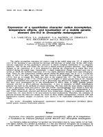
4.3 Goyasaponins
Goyasaponin (in Japanese) means saponins extracted from bitter melon.
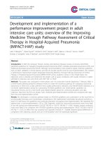
Goyasaponin fraction, as elucidated by Murakami et al. [97], includes abundant types of
cucurbitane-triterpene glycosides and oleanane-triterpene saponins, such as goyaglycoside
group (a, b, c, d, e, f, g, h), goyasaponin group (I, II, III) and momordicoside group (A, C, F1,
F2, I, K, L). The molecular formulae and structures of these compounds are shown in
Figures 3 – 5. Over 40 different cucurbitane-type and oleanane-type triterpene saponins
have been isolated from various parts of the of M. charantia plant [106] including the
fruits[97, 106-108], seeds [109] and vines [110].
Recently, significant research interest has focused on isolation, identification and
purification of cucurbitane-type triterpenoids from M. charantia. Utilizing mass spectrogram
(MS) and nuclear magnetic resonance spectroscopy (NMR), there have been a number of
reported new discoveries of cucurbitane-type triterpenoids isolated from M. charantia [106108, 110-112]. Moreover, it is noteworthy that one recent report documented that n-butanol
soluble M. charantia goyasaponin fraction inhibited sucrose-loading serum glucose elevation
in rats [81] which was an in vivo evidence demonstrating the hypoglycemic property of these
compounds.
However, mainly due to significantly low concentration of each identified single
compounds as shown in Table 2 [97] and no commercial HPLC standards available on the
market, research progress in this field is limited and much less is known about the bioactivity
16
of goyasaponins. In addition, there are very few reports identifying the possible mechanisms
related to the anti-diabetic action at the cellular level of adipocyte, especially on regulation of
adipocyte differentiation and adiponectin secretion.
17
Table 2. Yield percentage of each single goyasaponin compounds from fresh fruit [97]
Compound
Yield % from
Fresh Fruit
Compound
Yield % from
Fresh Fruit
Compound
Yield % from
Fresh Fruit
Goyaglycoside a
0.00008%
Goyaglycoside h
0.00008%
Goyasaponin I
0.00010%
Goyaglycoside b
0.00005%
Momordicoside A
0.0013%
Goyasaponin II
0.00027%
Goyaglycoside c
0.00007%
Momordicoside C
0.00016%
Goyasaponin III
0.00009%
Goyaglycoside d
0.00008%
Momordicoside F1
0.00011%
Goyaglycoside e
0.00010%
Momordicoside I
0.00006%
Goyaglycoside f
0.00009%
Momordicoside K
0.00003%
Goyaglycoside g
0.00006%
18
OR1
R2
H
GlcO
Goyaglycoside a, b, c, d, e, g
Goyaglycoside
R1
R2
Molecular
Formula
a
H
OCH3
C37H60O9
b
H
OCH3; 3’-epimer
C37H60O9
c
CH3
OCH3
C38H62O9
d
CH3
OCH3; 3’-epimer
C38H62O9
H
C42H68O13
OCH3; 3’-epimer
C43H70O14
β-Dglucopyranosyl
β-Dglucopyranosyl
e
g
OH
O
OH
OGlc
OH
H
H
O
GlcO
Glc - Glc - O
Goyaglycoside f (C42H68O13)
Goyaglycoside h (C42H70O15)
Figure 3. Molecular formulae and structures of goyaglycosides a – h
19
C
O
O
CH 3
OH
COOH
O O
OH
O
OH
CHO
H
OH
O
OH
H
O
H
OH
O
O
CH3
OH
O
H
O
CH 3
O
OH
OH
O
H
OH
OH
H
OH
OH
OH
OH
Goyasaponin I (C65H102O31)
C
O
O
CH 3
COOH
O O
OH
H
OH
O
OH
O
OH
OH
H
OH
OH
O
O
OH
CHO
CH 3
O
OH
H
OH
OH
O
H
OH
O
O
CH3
OH
O
H
O
H
O
H
OH
OH
OH
OH
Goyasaponin II (C70H110O35)
COOH
O
COOH
O O
C
H3 C
O
H
O
OH
O
O
OH
H
OH
O
OH
OH
OH
H
OH
Goyasaponin III (C49H76O19)
Figure 4. Molecular formulae and structures of goyasaponins I, II, III
20
OH
OH
R1
OH
OH
H
H
R 2O
Glc - Glc - O
Momordicoside A
(C42H72O15)
Momordicoside
R1
C
CH3
D
H
R2
βgentiobiosyl
βgentiobiosyl
Molecular
Formula
C42H72O14
C42H70O13
OR 1
O
2
R O
Momordicoside
R1
R2
F1
CH3
β-D-glucopyranosyl
Molecular
Formula
C37H60O8
F2
H
β-D-allopyranosyl
C36H58O8
G
CH3
β-D-allopyranosyl
C37H60O8
I
H
β-D-glucopyranosyl
C36H58O8
Figure 5. Molecular formulae and structures of selected momordicosides
21
OR
OHC
HO
OGlc
Momordicoside
R
K
CH3
Molecular
Formula
C37H60O9
L
H
C36H58O9
Figure 5 (Continued). Molecular formulae and structures of selected momordicosides
22
5. Overall Hypotheses, Objectives and Implications of This Study
5.1 Overall Hypotheses
A concentrated goyasaponin fraction (CGF) from M. Charantia fruit was obtained
using an extraction and concentration method. It is hypothesized that this fruit CGF
shows a potential as a PPARγ agonist and plays a similar role as TZDs which can
influence preadipocyte proliferation, process of adipocyte differentiation and adiponectin
secretion of differentiated adipocytes by modulating cell signaling. In addition, neither
the saponin contents in M. Charantia seed nor bioactivity has been investigated
thoroughly. It is proposed that seed CGF shows similar effects as fruit CGF on this in
vitro model. These findings may be important to elucidate the effects and relevant
mechanisms of M. Charantia on anti-obesity and type 2 diabetes prevention and/or
management.
5.2 Overall Objectives
M. charantia is widely used as a traditional functional food for the treatment of
type 2 diabetes while adipose tissue is one of the targets for anti-diabetic drugs. Based on
proven hypoglycemic effect of goyasaponin fraction on rats [81], the overall objective of
this study is to investigate whether a concentrated saponin fraction (CGF) extracted from
both M. charantia fruit and seed show a potential as PPARγ agonist by evaluating their
effects and relevant mechanisms of in the 3T3-L1 murine cell line.
(1) To optimize methods of extracting and concentrating saponin fractions from both M.
Charantia fruit and seed and to obtain lyophilized CGF in powder for further in vitro
23
experiments.
(2) To investigate the effect of CGF extracted from Momordica charantia fruit and seed
in 3T3-L1 cell model related to diabetes and obesity.
(3) To elucidate related mechanisms of these observed activities.
5.3 Implications of This Study
There are two implications of this study. First of all, because adipose tissue is one
of the target tissues for antidiabetic agents, assessments of both fruit CGF and seed CGF
effects on 3T3-L1 preadipocyte proliferation, adipocyte differentiation and adiponectin
secretion can shed more light on the underlying mechanisms of the bioactive compounds
responsible for the extensively investigated antidiabetic properties of M. charantia.
Secondly, the discovery of any enhanced bioactivity of M. charantia goyasaponins from
fruit or seed will constitute a basis for determination of any potential for its development
into a nutraceutical product.
24
CHAPTER 2
CONCENTRATION OF GOYASAPONIN FRACTIONS
1. Objectives
(1) Sample preparation of CGF from both M. Charantia fruit and seed for further in vitro
experiments.
(2) Component analysis of CGF by HPLC-MS based on molecular weight confirmation.
2. Materials and Methods
2.1 Preparation of Concentrated Goyasaponin Fraction from M. charantia Fruit
M. charantia fruit was purchased from a local supermarket, washed, separated
from the seeds and aerial fibers, cut into small pieces and lyophilized. Dried fruit pieces
were powdered and stored at -15℃ until extraction. Lyophilized samples were refluxed
in methanol for 4 h, filtered (No.1 Whatman paper, Maidstone, England) and vacuum
evaporated. The residue was dissolved in deionized water and centrifuged for 5 minutes
at 500 x g. The supernatant was then applied equally to five Amberlite XAD4 columns
(Sigma, St. Louis, MO) with a bed volume of 140 cm3 each at a flow rate of 3 bed
volume / hour (BV/H) followed by 1 L water wash at a rate of 5 BV/H. The sample was
eluted by ethanol (500 mL) in each column at a rate of 3 BV/H, and concentrated by
vacuum evaporation. The residue was dissolved in water and lyophilized and is herein
referred to as the fruit concentrated goyasaponin fraction (Fruit CGF) as shown in Figure
6.
25