THE STUDY OF THE EFFECTS OF a CHANGE IN THE EXPRESSION OF MIXED LINEAGE LEUKEMIA 5 ON TRANSCRIPTION REGULATION
Bạn đang xem bản rút gọn của tài liệu. Xem và tải ngay bản đầy đủ của tài liệu tại đây (2.34 MB, 122 trang )
THE STUDY OF THE EFFECTS OF A CHANGE IN THE
EXPRESSION OF MIXED LINEAGE LEUKEMIA 5 ON
TRANSCRIPTION REGULATION
LEE PEI
BSc (Hons), National University of Singapore
A THESIS SUBMITTED FOR THE
DEGREE OF MASTER OF SCIENCE
DEPARTMENT OF BIOCHEMISTRY
NATIONAL UNIVERSITY OF SINGAPORE
2012
1
Acknowledgements
I would like to express my utmost gratitude to my supervisor Dr Deng Lih-Wen for
her guidance despite her other academic and professional commitments and her
generous funding for the project. I would also like to thank my lab members, Yew
Chow Wenn, Cheng Fei, Liu Jie for guiding me on the technical and analytical skills
as wells as their encouragement and companionship all this while. I would like to
offer special thanks to everyone who has helped me in one way or another in the
course of my research project.
I would also want to express my sincere thanks to the Department of Biochemistry for
providing me the opportunity to do my research work.
Lastly, I am grateful to my family for their constant encouragement and support
throughout my graduate studies.
2
TABLE OF CONTENTS
LIST OF FIGURES ················································································5
LIST OF TABLES ·················································································7
LIST OF ABBREVIATIONS ····································································8
LIST OF PUBLICATIONS ·····································································10
SUMMARY·························································································11
CHAPTER 1: INTROUDCTION
1.1 Nuclear speckles ················································································13
1.1.1 Discovery of nuclear speckles ···························································13
1.1.2 Characterization and dynamics of nuclear speckles ···································14
1.2 Splicing ··························································································15
1.2.1 An overview ···············································································15
1.3 Transcription ····················································································19
1.3.1 An overview ···············································································19
1.3.2 Coordination between transcription and splicing ······································20
1.3.3 Chromatin organization and transcription ··············································23
1.4 Mixed Lineage Leukemia (MLL) Protein Family ·········································24
1.4.1 A summary of MLL protein family ·····················································24
1.4.2 MLL protein family as human H3K4 specific methyltransferases ··················26
1.4.3 MLL protein family and transcription ··················································27
1.4.4 MLL protein family and pre-mRNA processing ······································29
1.5 Mixed Lineage Leukemia 5 (MLL5) ·······················································30
1.5.1 A summary of MLL5 ·····································································30
1.5.2 Current findings on MLL5 ·······························································31
1.5.2.1 MLL5 and cell cycle regulation ··················································31
1.5.2.2 MLL5 and DNA damage response ···············································31
1.5.2.3 MLL5 and animal studies ·························································32
1.5.2.4 MLL5 and epigenetic regulation ·················································33
1.6 Aims and objectives of the study ····························································34
CHAPTER 2: MATERIALS AND METHODS
2.1 Cell lines and culture conditions ·····························································37
2.2 RNA interference and delivery ······························································38
2.3 Cloning ··························································································40
2.4 Calcium-phosphate mediated DNA plasmid transfection·································42
2.5 Cell lysate preparation, Immunoprecipitation and Western blot ·························43
2.6 Immunofluorescence microscopy ···························································49
2.7 Nuclease digestion ·············································································49
3
2.8 RNA extraction, cDNA synthesis and semi -quantitative real-time PCR ··············50
2.9 Splicing assay ··················································································52
2.10 Bromo-uridine triphosphate incorporation in permeabilized cells ·····················55
2.11 Micrococcal nuclease (MNase) accessibility assay ······································55
CHAPTER 3: RESULTS
3.1 Co-localization of MLL5 with the spliceosome components ····························59
3.2 Localization of MLL5 and spliceosome components in response to nuclease and
heat-shock treatment ············································································64
3.3 Association of MLL5 and SC35······························································67
3.4 Alteration in MLL5 protein level induced the redistribution of SC35 to enlarged
speckle domains ················································································70
3.5 Multiple transcription inhibitors induce MLL5 to redistribute to enlarged speckles ··76
3.6 Intra-nuclear reorganization of MLL5 speckles is reversible and temperature
dependent ························································································78
3.7 Alteration in MLL5 expression triggered transcription block ····························79
3.8 Association of MLL5 and RNAPII ··························································85
3.9 MLL5 overexpression resulted in a slower migration of Cyclin T1 ·····················87
3.10 MLL5 knockdown does not affect the phosphorylation state of RNAPII ·············89
3.11 MLL5 knockdown affects chromatin structure ···········································91
3.12 MLL5 and chromatin remodelling complex ··············································93
3.13 MLL5 and splicing activity ··································································95
CHATPER 4: DISCUSSION
4.1 An overview ····················································································98
4.2 Importance of maintaining MLL5 at a homeostatic level ································98
4.3 Plausible roles of MLL5 in transcription regulation ·····································105
4.3.1 MLL5 and its involvement in histone modifications································105
4.3.2 MLL5 and its involvement in chromatin organization······························107
CHAPTER 5: FUTURE DIRECTIONS AND CONCLUSION
5.1 Chromatin remodelling, histone modifications and DNA methylation –
How does it all fit together? ·······························································109
5.2 Histone modifying properties of MLL5 – When does it occur? ·······················111
5.3 Cell cycle arrest or transcription inhibition – Which comes first? ····················112
5.4 Conclusion ····················································································113
REFERENCES ··················································································115
4
LIST OF FIGURES
Figure 1: A simplified representation of the spliceosome assembly pathway and premRNA splicing …………………………………………………………...18
Figure 2: Integration of transcription and pre-mRNA processing…………………..21
Figure 3: Bi-directional coupling: a splicing factor regulates transcription, which in
turn regulates alternative splicing ………………………………………..22
Figure 4: A schematic presentation of MLL family proteins……………………….26
Figure 5: Co-localization of MLL5 with the spliceosome components …………...60
Figure 6: Different anti-MLL5 antibodies and their co-localization with SC35 …..62
Figure 7: Co-localization of MLL5 with the spliceosome components in different cell
lines ……………………………………………………………………....64
Figure 8: Association of MLL5 with splicing factor SC35 under RNase A digestion
and heatshock ……………………………………………………………..67
Figure 9: Association of MLL5 with splicing factor SC35 …………………………69
Figure 10: SC35 protein expression remains unaltered in MLL5 depleted cells …..71
Figure 11: Alteration in MLL5 protein levels by RNA interference induced the redistribution of SC35 to enlarged speckle domains ……………………...73
Figure 12: Exogenous introduction of MLL5 induced the re-distribution of SC35 to
enlarged speckle domains …………………………………………….....75
Figure 13: Multiple transcription inhibitors induce MLL5 to redistribute to enlarge
Speckles…………………………………………………………………77
Figure 14: Re-distribution of MLL5 speckles is temperature dependent ……….....79
Figure 15: Gene expression of S14 ribosomal subunit after MLL5 knockdown ….80
Figure 16: Alteration in MLL5 expression by RNA interference triggers transcription
block …………………………………………………………………....82
Figure 17: Exogenous introduction of MLL5 triggered transcription block ……...84
Figure 18: Distribution pattern of MLL5 and RNAPII …………………………….85
Figure 19: Association of MLL5 and RNAPII ……………………………………..87
5
Figure 20: MLL5 overexpression resulted in a slower migration of Cyclin T1…...89
Figure 21: MLL5 knockdown does not affect the phosphorylation state of
RNAPII…………………………………………………………………..90
Figure 22: Analysis of chromatin modifications in MLL5 knockdown cells ……..92
Figure 23: Analysis of chromatin organization in MLL5 knockdown cells ……....93
Figure 24: Effect of MLL5 knockdown on SWI/SNF protein complex …………..94
Figure 25: A test system for determining the splicing efficiency in mammalian
cells …………..………………………………………………………....96
Figure 26: Analysis of splicing efficiency in MLL5 knockdown cells ……………97
Figure 27: A model illustrating the participation of MLL5 in transcription and
splicing processes ………………………………………………………104
Figure 28: Possible epigenetic modifications on the chromatin …………………..111
6
LIST OF TABLES
Table 1: Nucleotide sequences of the siRNA used for MLL5 or SC35 gene
Silencing ……………………………………………………………………39
Table 2: Optimised volumes as well as concentrations of Lipofectamine™
RNAiMAX (Invitrogen) and siRNAs used in preparation of the transfection
mixes for MLL5 gene silencing ……………………………………………40
Table 3: PCR reaction composition and conditions of pXJ-HA-SC35 ………….....41
Table 4: Digestion reaction composition of pXJ-HA-SC35 ……………………......42
Table 5: Reaction composition for ligation of SC35 into pXJ-HA vector ………....42
Table 6: Transfection mixture using calcium-phosphate method for a typical 60mm
dish …………………………………………………………………….......43
Table 7: Buffers used in Western Blot ……………………………………………...45
Table 8: Conditions for Western Blot ………………………………………………45
Table 9: Self-generated or commercial MLL5 antibodies used in Western blot,
immunofluorescence and immunoprecipitation …...…………………......46
Table 10: Commercial antibodies and beads used in Western blot,
immunofluorescence and immunoprecipitation ………………………....47
Table 11: cDNA synthesis conditions ……………………………………………..51
Table 12: Primers used in qPCR ……………………………………………….......51
Table 13: qPCR reaction mixture and conditions ...........................................…....52
Table 14: Preparation of media and reagents required for β-galactosidase activity
activity ………………………………………………………………......54
Table 15: RT-PCR conditions ……………………………………………………...55
Table 16: Components of buffers used in MNase assay …………………………..58
7
LIST OF ABBREVATIONS
Abbreviations
ASCOM
ASH2L
ATCC
BSA
CBP
CD
CGBP
ChIP
CIP
CT
DAPI
DMEM
DRB
FBS
Br-UTP
Gal
H3
H4
HBS
HCF
HMT
HOX
HP1
HSC
KD
GO
IGCs
LAR II
LT-HSC
Luc
MBD
miRNA
MLL5
Mnase
NC-siRNA
ONPG
PcG
PHD
PF
PML
PS
PS1
PS2
P-TEFb
PTIP
Full Names
ASC-2-containing co-activator complexes
Absent, Small or Homeotic-like
American Type Culture Collection
Bovine serum albumin
CREB binding protein
Central domain
CpG-binding protein
Chromatin immunoprecipitation
Calf intestinal alkaline phosphatase
C terminus
4’ 6-diamidino-2-phenylindole, dihydrochloride
Dulbecco’s Modified Eagles Medium
5,6-dichlorobenzimidazole riboside
Fetal bovine serum
Bromo-uridine Triphosphate
Galactosidase
Histone 3
Histone 4
Hanks Buffered Salt
Host cell factor
Histone methyltransferase
Homeobox
Heterchromatin protein 1
Hematopoietic stem cells
Knockdown
Gene Ontology
Interchromatin granule clusters
Luciferase Assay Reagent II
Long-term hematopoietic stem cells
Luciferase
Methyl-CpG-binding domain
microRNA
Mixed Lineage Leukemia 5
Micrococcal nuclease
Negative control-siRNA
o-Nitrophenyl-β-D-galactopyranoside
Polycomb
Plant homeodomain
Perichromatin fibrils
Promyelocytic leukaemia
PHD SET
Permeabilization solution 1
Permeabilization solution 2
Positive transcription elongation factor b
Pax transactivation domain-interacting protein
8
qPCR
RbBP5
RNA
RNAPI
RNAPII
RNAPIIa
RNAPIIo
RNAPII CTD
RT
RT-PCR
SC
SET
Sm
snRNA
snRNP
SR
SS
SWI/SNF
TrxG
WB
WDR5
Semi-quantitative polymerase chain reaction
Retinoblastoma Binding protein 5
Ribonucleic acid
RNA polymerase I
RNA polymerase II
Hypo-phosphorylated RNAPII
Hyper-phosphorylated RNAPII
RNA polymerase II C-terminal domain
Room temperature
Reverse transcription polymerase chain reaction
Scrambled
Su(var)3-9, enhancer-of-zeste and trithorax
smith antigens
small nuclear RNA
Small nuclear ribonucleoproteins
Serine / arginine
Splice sites
SWItch/Sucrose Non Fermentable
Trithorax group
Western blot
WD Repeat Domain 5
9
LIST OF PUBLICATIONS
Journal Articles
1. Yew CW, Lee P, Chan WK, Lim VK, Tay SK, Tan TM, Deng LW (2011). A
Novel MLL5 Isoform That Is Essential to Activate E6 and E7 Transcription in
HPV16/18-Associated Cervical Cancers. Cancer Res 2011 Nov 1;71(21):6696-707.
2. Lee P, Yew CW, Wu Q, Deng LW (2012) Impact of altering the basal level of
Mixed Lineage Leukemia 5 on global chromatin organization and transcription
regulation. (Manuscript to be submitted)
10
SUMMARY
Mixed Lineage Leukaemia 5 (MLL5) is a mammalian Trithorax group (TrxG) gene
located at chromosome band 7q22, a frequently deleted region in myeloid
malignancies. MLL5 was discovered and subsequently cloned in year 2002. Currently,
there are a total of fifteen publications dedicated to MLL5.
MLL5 is identified as a nuclear protein and either over-expression or depletion of
MLL5 resulted in dual-phase cell cycle arrest. In interphase cells, MLL5 exhibits
distinct irregular, punctate intra-nuclear speckles but with uncharacterized biological
functions. Intrigued by the complexities of nuclear speckles, which are dynamic
structures enriched with a reservoir of factors that participate in transcription and premRNA processing, we attempted to unravel the biological functions of MLL5 within
the nuclear speckles. To begin with, we examined the co-staining pattern of MLL5
with several well-characterized proteins that were known to display nuclear speckle
pattern by immunofluorescence staining. Interestingly, we found that MLL5 nuclear
speckles exhibited extensive co-localization with the spliceosome protein SC35 which
has recently been reported to be involved in the bi-directional coupling of
transcription and splicing. Given the fact that alterations in MLL5 level through
ectopic over-expression or siRNA-mediated knockdown resulted in the enlargement
and aggregation of nuclear speckles, a phenotype that indicated a defect in cotranscriptional splicing process, we therefore speculate a novel biological role of
MLL5 involving in the transcription and splicing processes. We tested this hypothesis
by examining if MLL5 is sensitive to transcription inhibitors and whether MLL5 is
associated with RNA Polymerase II (RNAPII) transcription machinery. Results
11
showed that MLL5 not only physically interacted with RNAPII but also affected the
progression of RNAPII along the DNA template as MLL5 depletion resulted in
chromatin compaction and affected the subunits of chromatin remodelling proteins. In
addition, histone signatures signifying transcription activation, namely H3K4 trimethylation and H4 acetylation, were largely reduced in MLL5-kockdown cells.
Splicing activity was also reduced as a result of a disruption in the transcription
process. Taken together, our findings suggest that MLL5 participates in transcription
regulation, which consequently affects gene regulation and cell-cycle progression.
12
CHAPTER 1 – INTRODUCTION
1.1 Nuclear speckles
1.1.1 Discovery of nuclear speckles
The pioneer work for nuclear speckles was reported by Santiago Ramo´n y Cajal in
1910 [reviewed in (Lafarga et al., 2009)]. In this study, Ramo´n used acid aniline
stains to identify structures he described as “grumos hialinas”, which literally meant
“translucent clumps”. In 1959, through the use of electron microscopy, Hewson Swift
(Swift, 1959) observed particles in the cells to be localized in “clouds” instead of
being randomly distributed. Further investigations by Swfit through cyto-chemical
analysis suggested that these particles harboured ribonucleic acid (RNA). Swift
termed these particles as interchromatin particles. It was only in 1961 when researcher
J. Swason Beck (Beck, 1961), upon examining rat liver sections that were immunelabelled with serum from auto-immune disorder patients, coined the term “speckles”
for the interchromatin particles that were discovered two years ago. However, it was
only after several years later that the first connection between pre-mRNA splicing and
nuclear speckles or interchromatin granules emerged. This was found through an
examination of the distribution of small nuclear ribonucleoproteins (snRNP antigens)
using anti-splicing factor-specific antibodies that illustrated a speckled distribution of
snRNPs in the cell nuclei (Perraud et al., 1979; Lerner et al., 1981; Spector et al.,
1983). These distinct classes of sub-nuclear bodies have always been an area of
intense research even till present.
13
1.1.2 Characterization and dynamics of nuclear speckles
The mammalian cell nucleus is a multi-functional and complex organelle where a
plethora of cellular mechanisms occur in sub-nuclear compartments collectively
termed as foci. These foci, approximately 20-50 of them diffusely distributed in the
nucleoplasm, appeared as irregular, punctate structures with interconnections existing
in variable shapes and sizes (Lamond and Spector, 2003). These distinct foci,
identified as nuclear speckles and Cajal (coiled) bodies, are dynamic structures
involved in transcription and pre-mRNA splicing (Spector, 1993; Matera, 1999).
Further characterizations by electron microscopy revealed these nuclear speckles to
co-localize in nuclear regions designated as interchromatin granules clusters (IGCs)
and perichromatin fibres (PFs) (Fakan et al., 1984; Raska et al., 1990; Spector et al.,
1993). Active pre-mRNA transcription pre-dominates at the PFs that are enriched
with nascent DNA, RNA, RNA polymerase II (RNAPII) and histone modifiers for
transcriptionally active chromatin. Splicing speckles observed in IGCs signifed the
sites for splicing factor assembly and storage as well as the sites for splicing processes
such as RNA editing and transport (Carter et al., 1991; Wang et al., 1991; Spector and
Lamond, 2011).
Nuclear speckles are dynamic structures and there is a continuous shuttling of splicing
factors in and out of the speckles. In the event of transcription inhibition, either
through the use of inhibitors or as a consequence of heat-shock, nuclear speckles
became enlarged and rounded as splicing factors aggregate in them (Spector et al.,
1991; Melcak et al., 2000). However, when the expression of intron-containing genes
is high (Huang and Spector, 1996; Misteli et al., 1997) or during a viral infection
14
when transcription activity increases (Jimenez-Garcia and Spector, 1993; Bridge et al.,
1995), the accumulation of splicing factors within the speckles decrease as they get
distributed to the transcription sites in the nucleoplasm. Undeniably, much progress
has been made in recent years towards a better understanding of the structure and
function of the nuclear speckles. However, given the dynamic nature of the speckle
morphology, answers to a number of questions remain. In particular, the detailed
molecular mechanism on how the components of the nuclear speckles efficiently
coordinate the complex events in the cell, how splicing factors systematically execute
the splicing process, consequently giving rise to the different splice forms of the gene
transcript.
1.2 Splicing
1.2.1 An overview
Nuclear pre-mRNA splicing is an essential and important process that governs
eukaryotic gene expression. It is a process where introns are excised and this occurs in
the spliceosome complexes that constitute two different classes of snRNP antigens U1, U2, U4/U6, U5 (Bindereif and Green, 1990) and non-snRNP antigens like SC35
(Reed, 1990). Both groups belong to the serine/arginine (SR) family and share
structural features including an RNA binding domain and a SR-rich domain that is
responsible for their targeting to nuclear speckles (Zahler et al., 1992; Birney et al.,
1993). These proteins function cooperatively to catalyse the excision of the
intervening sequences in the pre-messenger RNA (pre-mRNA).
15
Among the SR protein family, SC35, discovered through a monoclonal antibody
against partially purified spliceosomes, is commonly used to define splicing nuclear
speckles (Fu and Maniatis, 1990). The group discovered that SC35 co-localized well
with snRNPs within the speckled nuclear domains, thereby providing the first
evidence that these speckled regions constituted both types of snRNPs. It has been
reported that nuclear extracts depleted of SC35 was incapable of splicing exogenous
pre-mRNA. However, this was a reversible process as splicing activity could be
restored by complementing the extracts with SC35 antigen or other members of the
SR family (Zahler et al., 1992).
The process of pre-mRNA splicing constituted two trans-esterification reactions,
namely lariat intron formation and exon ligation. Briefly, this occurred in an orderly
step-wise manner, involving the interaction between the spliceosomal snRNPs and
non-snRNPs such as splicing factors SC35. Briefly, U1-snRNP first associated with
the 5' splice site, thereafter, the attachment of the U2-snRNP near the branch-point
enable the entry of the U4/U5/U6 tri-snRNP complex to complete the spliceosome
assembly. Structural rearrangements then occurred and this resulted in U1 and U4
expulsion, catalytic activation, lariat formation, exon ligation, spliced product release
and the eventual association of the remaining components that constitute the
spliceosome assembly. A simplified representation of the spliceosome assembly
pathway and pre-mRNA splicing is illustrated in Figure 1. Over the years, extensive
research has revealed that the splicing of pre-mRNA in eukaryotes is also tightly
coupled to the transcription process and this occurs as nascent transcripts are
synthesized from RNA polymerase II. In fact, unravelling the splicing process not
only aid in having a better understanding of gene expression at the molecular level;
16
even at the medical level, it allows for better treatment and prognosis as aberrant premRNA splicing has been associated with the onset of human diseases.
17
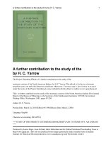
Figure 1: A simplified representation of the spliceosome assembly pathway and
pre-mRNA splicing. The pre-mRNA is depicted with rectangular boxes (blue) as
exons, linked by a single intron (black line) from the 5’to the 3’ splice sites (SS). For
simplicity, only the ordered interactions of the snRNPs (indicated by circles), but not
those of non-snRNP proteins are illustrated. During the assembly phase, the
spliceosomal snRNP U1 first assembles onto the pre-mRNA before the systematic
recruitment of U2, followed by the other snRNPs. During activation, the Prp28associated complex joins the spliceosome while the U1 and U4 snRNPs depart.
Catalysis proceeds in two steps: lariat formation and exon ligation. Eventually, the
mRNA is released and the spliceosome is disassembled. Backward arrows indicate the
reversibility of process as the cycle begins. [Adapted from (Will and Luhrmann,
2011)]
18
1.3 Transcription
1.3.1 An overview
RNA polymerase II (RNAPII) is a key player in the transcription process. Prior to
splicing, nascent RNA transcripts are generated by RNAPII. The RNAPII harbours 52
tandem consensus heptapeptide (YSPTSPS) repeats at its C-terminal domain
(RNAPII CTD) (Corden, 1990) and phosphorylation on the multi-sites controls the
state of transcription. RNAPII with un-phosphorylated CTD is recruited to the preinitiation site at the promoters while the transition between transcription initiation and
elongation is mediated by multi-phosphorylation events that are catalysed by proteinkinase complexes. Cdk7-cyclinH phosphorylates RNAPII CTD at Serine-5,
generating a hypo-phosphorylated RNAPII (RNAPIIa) that participates in
transcriptional initiation. Phosphorylation at Serine-2 is catalysed by Cdk9-cyclinT,
forming
hyper-phosphorylated
RNAPII
(RNAPIIo)
that
associates
with
transcriptional elongation (Zawel et al., 1995). RNAPIIo has also been reported to
exist in splicing factor-rich nuclear speckles (Bregman et al., 1995; Mortillaro et al.,
1996) and significant enrichment and co-localization has been observed for Cyclin T1
with the nuclear speckles than Cdk9 (Herrmann and Mancini, 2001). A growing body
of evidence has also suggested that Cdk9 not only regulates RNAPII activity, but also
participates in co-transcriptional histone modifications and pre-mRNA processing like
splicing and 3’ end processing (Pirngruber et al., 2009a; Pirngruber et al., 2009b).
19
1.3.2 Coordination between transcription and splicing
Emerging evidence has proved that functional integration of transcription by RNAPII
and RNA processing machineries are mutually beneficial for efficient and regulated
gene expression. The transcription process progresses from the initiation phase to the
elongation phase and finally, the termination phase and these coordinated events
within the cell nucleus are briefly summarized in Figure 2. Research over the years
has also suggested that RNAPII CTD is critical in coupling the transcription and
splicing processes as several observations have associated the elongating RNAPII to
pre-mRNA splicing (Corden and Patturajan, 1997; Bentley, 1999; Hirose and Manley,
2000). Phosphorylated CTD serves as a recruitment and docking site for mRNA
processing factors (Greenleaf, 1993) and stimulates the early steps of spliceosome
assembly (Hirose et al., 1999). Besides, the phosphorylated CTD also recruits
chromatin modifiers such as histone methyltransferases Set 1/2 (Phatnani and
Greenleaf, 2006; Yoh et al., 2008) and histone acetyltransferases p300 and PCAF
(p300/CBP-associated factor) (Cho et al., 1998). Hence, the cycle of phosphorylation
and de-phosphorylation at the CTD during each round of transcription may coordinate
the recruitment of these processing factors at different states of mRNA formation.
20
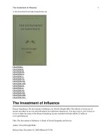
Figure 2: Integration of transcription and pre-mRNA processing. RNAPII is
modified on its CTD with Serine-5 phosphorylation predominately at the start of the
gene (blue line) and Serine-2 phosphorylation in the middle and end of the gene
(yellow line). 5’-Capping enzymes are recruited through direct interactions with
Serine-5 phosphorylated CTD to catalyse the co-transcriptional capping reaction.
Various splicing factors are recruited during the elongation phase of transcription to
facilitate co-transcriptional splicing. These splicing factors are dependent on Serine-2
phosphorylation on the CTD. The 3’-end formation is functionally coupled to
transcription termination. Importantly, increasing evidence now suggests that the
transcription and RNA processing machineries are functionally integrated in a
reciprocal fashion such that individual co-transcriptional processing events can
influence transcription at different phases. [Adapted from (Pandit et al., 2008)].
Recently, Lin and colleagues (Caslini et al., 2009) has uncovered a new and important
role in transcription for a splicing regulator protein, SC35, that has previously been
thought to be involved primarily in the splicing process. In the study, SC35 is needed
to promote RNAPII elongation in a subset of genes where depletion in SC35
dramatically caused a decrease in nascent RNA synthesized by RNAPII but has no
effect on the transcription by RNA polymerase I. Through the use of chromatin
21
immunoprecipitation combined with microarrays (ChIP-chip), the group observed that
RNAPII was accumulated within the gene body upon SC35 depletion, indicating
RNAPII stalling before it reached the end of the gene. This stalling led to a decrease
in RNAPII elongation, which was confirmed by measuring the nascent transcripts
using a run-on assay that utilized non-radioactive nucleotides. In short, these findings
confirm the involvement of SC35 in the bi-directional coupling between transcription
and splicing. A schematic diagram of this bi-directional coupling is illustrated in
Figure 3.
Figure 3: Bi-directional coupling: a splicing factor regulates transcription, which
in turn regulates alternative splicing. The splicing factor SC35 interacts with RNA
polymerase II (Pol II) and the elongation factor P-TEFb and, via phosphorylation of
the C-terminal domain (CTD) of Pol II at Serine2 (Ser2), stimulates transcriptional
elongation. In parallel, high elongation rates allow the simultaneous presentation to
the splicing machinery of strong and suboptimal 3’ splice sites, which favours the use
of the stronger one, leading to skipping of an alternative exon. [Adapted from (Fededa
and Kornblihtt, 2008)]
22
In summary, the continuous shuttling of splicing factors to active transcription sites
brings the elongating and splicing complexes into close proximity to facilitate cotranscriptional splicing. Given the tight coupling of transcription with the downstream
RNA processing steps, transcription inhibition may halt a chain of gene expression
events and arrest complexes at various RNA metabolism stages. Such disruption in
transcription activity causes nuclear speckles to accumulate in the cell nucleus in an
aggregate manner.
1.3.3 Chromatin organization and transcription
Extensive chromatin research over the years indicates that chromatin structure is a
primary regulator of gene transcription. The dynamics of chromatin structure is tightly
regulated through multiple mechanisms which include histone modifications,
chromatin remodelling, histone variant incorporation and histone eviction. In this
study, we will examine how histone modifications and chromatin remodelling affect
transcription.
Histone tails are susceptible to numerous post-translational modifications (Li et al.,
2007). These modifications include methylation of arginine (R) residues; methylation,
acetylation, ubiquitination, ADP-ribosylation, and sumoylation of lysines (K); and
phosphorylation of serines and threonines. Among them, modifications pertaining to
active transcription include acetylation of histone 3 and histone 4 (H3 and H4) or dior tri-methylation of H3K4; and these are classified as euchromatin modifications.
Heterochromatin modifications are associated with inactive transcription, and
methylation occurs on H3K9 or H3K27. These histone modifications consequently
23
cause a change in the net charge of the nucleosomes, which in turn could strengthen
or weaken inter-or intranucleosomal DNA-histone interactions. These effects
eventually affect RNAPII progression along the chromatin, thereby affecting
transcription.
Chromatin remodelling is an energy-dependent process which involves a transient
unwrapping of DNA from histone octamers. This facilitates transcription factors to
become accessible to nucleosomal DNA. An example of chromatin modellers are the
SWItch/Sucrose Non-Fermentable (SWI/SNF) proteins, which are a group of highly
conserved DNA-stimulated ATPase complex (Muchardt and Yaniv, 1999). Taken
together, chromatin architecture and its dynamic nature has a crucial role in dictating
the
fate
of
DNA-related
metabolic
processes
which
include
DNA
repair/recombination/replication, in particular, transcription by RNAPII that will be
highlighted in this thesis.
1.4 Mixed Lineage Leukemia (MLL) Protein Family
1.4.1 A summary of MLL protein family
The mammalian mixed lineage leukemia (MLL) family comprises five members
(MLL1, MLL2, MLL3, MLL4/ALR and MLL5) and these proteins are human
homologues of the Drosophila Trithorax group (TrxG) gene. Vertebrate and
Drosophila TrxG genes encode transcriptional regulators that are postulated to be
involved in the maintenance of gene expression. Proteins that are encoded by TrxG
repress Homeobox (HOX) gene expression while their other antagonistic parties,
24
polycomb group (PcG) proteins, maintain the HOX gene expression (Ziemin-van der
Poel et al., 1991). The mechanisms by which these two evolutionally conserved genes
maintain the HOX gene expressions occur at the epigenetic level by chromatin
remodeling and histone modifications, upon the formation of multi-protein complexes
(Muller et al., 2002; Schuettengruber et al., 2007). Since HOX gene expressions are
essential in determining the fates of embryonic development and haematopoiesis,
aberrant HOX gene expression may represent a major molecular consequence of
leukaemia-associated genetic lesions (Orlando and Paro, 1995; Look, 1997; Dorrance
et al., 2006)
MLL protein family possesses variable number of cysteine-rich plant homeodomain
(PHD), zinc fingers and a highly-conserved Su(var)3-9, enhancer-of-zeste and
trithorax (SET) domain. A schematic representation of MLL protein family is
illustrated in Figure 4. Structural and biochemical analysis show that PHD finger and
SET domain are involved in protein-protein interactions (Gould, 1997; van Lohuizen,
1999). PHD finger is usually present in chromatin-associated proteins and has been
reported to be associated with nucleosomes or specific nuclear protein partners
(Aasland et al., 1995) or serve as binding or recognition modules for histone
modifications (Mellor, 2006) while the SET domain possesses methyltransferase
activity (Nakamura, et al. 2002). Among the MLL family, MLL1 is the most
extensively studied. For instance, the existence of PHD fingers within MLL1 regulate
homodimerization and are indispensable for the interaction with cyclophilin Cyp33
(Fair et al., 2001).
25