Molecular biology of glutamate dehydrogenase and glutamine synthetase in two air breathing teleosts
Bạn đang xem bản rút gọn của tài liệu. Xem và tải ngay bản đầy đủ của tài liệu tại đây (2.24 MB, 220 trang )
MOLECULAR BIOLOGY OF GLUTAMATE
DEHYDROGENASE AND GLUTAMINE SYNTHETASE
IN TWO AIR BREATHING TELEOSTS
TOK CHIA YEE
(B. Sc. (Hons), NUS)
A THESIS SUBMITTED
FOR THE DEGREE OF MASTER OF SCIENCE
DEPARTMENT OF BIOLOGICAL SCIENCES
NATIONAL UNIVERSITY OF SINGAPORE
2011
ACKNOWLEDGEMENTS
I wish to express my heartfelt thanks and gratitude to my mentor, Professor
Ip Yuen Kwong, for his guidance, advices and teachings. It is through his wisdom
that I have learnt a lot during my time as a student, and I want to try my best to put
into practice what he has taught me. Many thanks to Madam Wong Wai Peng for
her help whenever I needed it, and for all the advices she has given me as a colleague
and a senior. Thanks to my senior Dr. Loong Ai May for all the advices that she has
given me. A big thank you, to my fellow lab mate, friend and colleague Ching
Biyun, for being there to lend a helping hand and to encourage me during the course
of my study. Finally, thanks to all the undergraduate lab mates; it has been a joy
working and learning with all of you.
i
TABLE OF CONTENTS
ACKNOWLEDGEMENTS………………………………………………….
TABLE OF CONTENTS…………………………………………………….
SUMMARY………………………………………………………………….
LIST OF TABLES…………………………………………………………...
LIST OF FIGURES………………………………………………………….
LIST OF ABBREVIATIONS………………………………………………..
Literature Review…………………………………………………………….
Ammonia production, ammonia toxicity and excretory nitrogen
metablolism……………………………………………………………
Ammonia production …………………………………………...
Ammonia toxicity……………………………………………….
Excretory nitrogen metabolism………………………………….
Functional roles of glutamate dehydrogenase and glutamate in
nitrogen metabolism…………………………………………………...
Functional roles of glutamine synthetase and glutamine in nitrogen
metabolism…………………………………………………………….
Air-breathing fishes and defense against ammonia toxicity during
emersion………………………………………………………………..
Reduction in ammonia production by suppressing amino acid
catabolism………………………………………………………..
Partial amino acid catabolism leading to the formation of
alanine……………………………………………………………
Glutamine synthesis……………………………………………..
Detoxification of ammonia to urea………………………………
Ammonia volatilization………………………………………….
Active transport of NH4+………………………………………...
Monopterus albus and Misgurnus anguillicaudatus…………………...
Introduction………………………………………………………….
Materials and methods……………………………………………………….
Fish…………………………………………………………………….
Exposure of M. anguillicaudatus to experimental conditions and
collection of samples…………………………………………………..
Exposure of M. albus to experimental conditions and collection of
samples……………………………………………………...………….
Extraction of total RNA………………………………………………..
Obtaining gdh and gs partial fragments from PCR…………………….
Cloning of gs partial fragments………………………………………...
Sequencing of PCR products and plasmid DNA inserts……………….
RACE PCR to obtain sequences upstream and downstream of gdh and
gs partial fragments…………………………………………………….
Cloning and sequencing of RACE PCR products……………………...
Phylogenetic analysis…………………………………………………..
Designing primers for quantitative real-time PCR on M.
anguillicaudatus gdh and gs and M. albus gdh………………………..
Designing primers for semi-quantitative PCR and quantitative realtime PCR on M. albus gs isoforms…………………………………….
cDNA synthesis for semi-quantitative PCR and quantitative real-time
PCR…………………………………………………………………….
i
ii
v
viii
x
xviii
1
1
1
2
7
9
10
11
11
12
12
13
14
14
15
23
31
31
31
32
33
34
37
37
37
42
42
43
44
47
ii
Tissue expression study of gs1 in M. albus…………………………….
Relative quantification of gs1 by semi-quantitative PCR……………...
Relative quantification by quantitative real-time PCR………………...
Statistical analyses……………………………………………………..
1. Molecular biology of glutamate dehydrogenase in Misgurnus
anguillicaudatus…………………………………………………………..
1.1 Results……………………………………………………………..
1.1.1 RACE PCR and cloning of gdh……………………………
1.1.2 Analyses of gdh and the deduced Gdh sequences…………
1.1.3 The phylogenetic analysis of Gdh…………………………
1.1.4 mRNA expression of gdh in the liver and intestine of M.
anguillicaudatus…………………………………………...
1.2 Discussion………………………………………………………….
1.2.1 A single gdh was elucidated from the liver of M.
anguillicaudatus…………………………………………...
1.2.2 Phylogeny and conservation of M. anguillicaudatus Gdh...
1.2.3 mRNA expression of gdh in the liver and intestine of M.
anguillicaudatus exposed to terrestrial conditions were
differentially regulated…………………………………….
1.2.4 mRNA expressions of gdh in the liver and intestine of M.
anguillicaudatus exposed to elevated environmental
ammonia remained unchanged…………………………….
Conclusion……………………………………………………….
2. Molecular biology of glutamine synthetase in Misgurnus
anguillicaudatus…………………………………………………………..
2.1 Results……………………………………………………………...
2.1.1 RT-PCR, cloning of partial gs fragment and RACE PCR…
2.1.2 Analyses of gs and the deduced Gs sequences…………….
2.1.3 The phylogenetic analysis of Gs…………………………...
2.1.4 mRNA expression of gs in the liver and intestine of M.
anguillicaudatus…………………………………………...
2.2 Discussion………………………………………………………….
2.2.1 Multiple forms of gs were absent in the liver of M.
anguillicaudatus…………………………………………...
2.2.2 The liver of M. anguillicaudatus expresses Gs in the
cytosol……………………………………………………..
2.2.3 Phylogeny and conservation of M. anguillicaudatus Gs
sequence…………………………………………………...
2.2.4 Expressions of gs mRNA in the liver and intestine of M.
anguillicaudatus were down-regulated after 2 days of
exposure to terrestrial conditions………………………….
2.2.5 Exposure to elevated envieonmental ammonia led to
changes in the expressions of gs mRNA in the liver and
intestine of M. anguillicaudatus…………………………..
Conclusion……………………………………………………….
3. Molecular biology of glutamate dehydrogenase in Monopterus albus…...
3.1 Results……………………………………………………………...
3.1.1 RT-PCR for gdh partial fragment………………………….
3.1.2 RACE PCR………………………………………………...
3.1.3 Analyses of gdh and the deduced Gdh sequences…………
47
47
48
49
51
51
51
51
52
62
65
65
65
68
70
71
73
73
73
73
74
83
86
86
86
88
89
91
92
93
93
93
93
94
iii
3.1.4 The phylogenetic analysis of Gdh…………………………
3.1.5 mRNA expression of gdh in the liver, intestine and brain
of M. albus………………………………………………...
3.2 Discussion………………………………………………………….
3.2.1 A single gdh was elucidated from the liver, intestine and
brain of M. albus…………………………………………..
3.2.2 Phylogeny and conservation of Gdh……………………….
3.2.3 mRNA expressions of gdh in the liver, intestine and brain
of M. albus exposed to terrestrial conditions and elevated
ammonia were differentially regulated……………………
3.2.4 mRNA expression of gdh in the intestine of M. albus
exposed to elevated ambient salinity was up-regulated…...
Conclusion……………………………………………………….
4. Molecular biology of glutamine synthetase in Monopterus albus………..
4.1 Results……………………………………………………………...
4.1.1 RT-PCR and cloning for gs partial fragments……………..
4.1.2 RACE PCR and cloning of RACE products………………
4.1.3 Analyses of gs and the deduced Gs isoforms……………...
4.1.4 The phylogenetic analysis of Gs isoforms…………………
4.1.5 mRNA expression of gs1 in the liver, intestine and brain of
M. albus……………………………………………….…...
4.1.6 Semi-quantitative analysis of gs1 mRNA expression in the
intestine and brain of M. albus…………………………….
4.1.7 mRNA expression of gs2 and gs3 in the liver, intestine and
brain of M. albus by quantitative real-time PCR………….
4.2 Discussion………………………………………………………….
4.2.1 Multiple gs were present in the organs of M. albus……….
4.2.2 Expression of gs1, gs2 and gs3 in M. albus……………….
4.2.3 Phylogeny and conservation of Gs isoforms in M. albus….
4.2.4 The Gs isoforms, Gs1, Gs2 and Gs3 are cytosolic enzymes
4.2.5 Differential expressions of gs isoforms in the liver of M.
albus exposed to terrestrial conditions or elevated
environmental ammonia suggest differing kinetic
properties between Gs1, Gs2 and Gs3…………………….
4.2.6 Expression of gs isoforms in the brain and intestine of M.
albus exposed to terrestrial conditions or elevated
environmental ammonia were differentially regulated……
4.2.7 Increased protein abundance of Gs in M. albus exposed to
salinity stress was not correlated to the mRNA expressions
of gs isoforms……………………..……………………….
Conclusion……………………………………………………….
5. Integration, Synthesis and Conclusions…………………………………...
5.1 gdh in M. anguillicaudatus and M. albus: a comparison…………..
5.2 Comparing gdh expression in the liver and intestine of M.
anguillicaudatus and M. albus…………………………………….
5.3 gs in M. anguillicaudatus and M. albus: a comparison……………
5.4 gs expression in the liver and intestine of M. anguillicaudatus and
M. albus…………………………………………………………...
References…………………………………………………………………….
Appendix……………………………………………………………………..
102
102
111
111
111
113
115
116
117
117
117
117
125
126
133
133
137
151
151
153
153
154
155
157
158
159
161
161
162
163
164
167
201
iv
SUMMARY
Air-breathing fishes such as the weatherloach Misgurnus anguillicaudatus
and the swamp eel Monopterus albus often encounter the problem of endogenous
ammonia buildup leading to ammonia toxicity during emersion or exposure to
increased environmental ammonia. Occasionally, M. albus also faces hyperosmotic
stress when it inhabits swamps. Both M. anuguillicaudatus and M. albus are capable
of coping with the various adverse conditions by synthesizing glutamine, which is a
product of ammonia detoxification. Moreover, glutamine may also act as an organic
osmolyte in M. albus. As glutamine synthesis involves glutamate dehydrogenase
(Gdh) and glutamine synthetase (Gs), this study was undertaken to examine the
molecular biology of Gdh and Gs in M. anguillicaudatus and M. albus, so as to
better understand the mechanisms affecting and regulating their function in these two
air-breathing fishes.
Results obtained from this study reveal that M. anguillicaudatus and M.
albus each express one form of gdh in the liver, which may be influenced by
different transcriptional and translational controls.
Early phases of terrestrial
exposure induced increased hepatic gdh mRNA expression in both M.
anguillicaudatus and M. albus.
On the other hand, increased environmental
ammonia led to an initial increase in hepatic gdh mRNA expression in M. albus but
not in M. anguillicaudatus. Additionally, intestinal gdh mRNA expression was
down-regulated in M. anguillicaudatus exposed to terrestrial conditions, but upregulated in M. albus exposed to increased ambient salinity. As such, it appears that
unlike M. albus, the intestine of M. anguillicaudatus was unlikely to be involved in
increased glutamate synthesis to facilitate increased glutamine synthesis
v
This study also reveals for the first time that a single form of gs is expressed
in the liver of M. anguillicaudatus, but three isoforms of gs are expressed in the liver,
intestine and brain of M. albus. Terrestrial exposure resulted in a significant downregulation of gs mRNA expression in the liver and intestine of M. anguillicaudatus.
Furthermore, even though ammonia loading conditions led to an initial up-regulation
of hepatic and intestinal gs mRNA expression in M. anguillicaudatus, gs mRNA
expressions in both organs were subsequently down-regulated. In contrast, M. albus
exposed to terrestrial conditions up-regulated hepatic gs1 mRNA expression and
intestinal and hepatic gs2 mRNA expression. Additionally, exposure to elevated
environmental ammonia also induced a significant up-regulation of hepatic gs1
mRNA expression. This differential regulation of gs between M. anguillicaudatus
and M. albus is indicative of the latter utilizing mainly the strategy of glutamine
synthesis while the former relying on more than one strategy to deal with increased
endogenous ammonia during terrestrial exposure and ammonia loading.
vi
NOTE:
The following gene sequences have been submitted to GenBank, and the respective
accession numbers are given in the table below.
Genbank Accession
Fish
Gene
Misgurnus
gdh
JF694443
anguillicaudatus
gs
JF694444
gdh
JF694445
gs1
JF694448
gs2
JF694447
gs3
JF694446
number
Monopterus albus
vii
LIST OF TABLES
Table 1
Degenerate PCR primer pairs designed to amplify glutamate
dehydrogenase (gdh) from the liver of Misgurnus
anguillicaudatus and the liver, intestine and brain of Monopterus
albus.…………………………………………………………..…. 36
Table 2
Gene specific primers designed to amplify and sequence
glutamate dehydrogenase (gdh) and glutamine synthetase (gs)
from the liver of Misgurnus anguillicaudatus in the direction of
the 5’ UTR or 3’ UTR……………………..………………….…. 40
Table 3
Gene specific primers designed to amplify and sequence
glutamate dehydrogenase (gdh) and glutamine synthetase (gs)
from the liver, intestine and brain of Monopterus albus in the
direction of the 5’ UTR or 3’ UTR………………………………. 41
Table 4
Gene specific primer pairs designed for quantitative real-time
PCR on actin, glutamate dehydrogenase (gdh) and glutamine
synthetase (gs) from the liver and intestine of Misgurnus
anguillicaudatus…………………………………………………. 45
Table 5
Gene specific primer pairs designed for quantitative real-time
PCR on actin, glutamate dehydrogenase (gdh), glutamine
synthetase isoform 1 (gs1) and 2 (gs2) and for semi-quantitative
PCR on glutamine synthetase isoform 3 (gs3) from the liver,
intestine and brain of Monopterus albus.……………………….. 46
Table 6
Sequence identity matrix of GDH from various organisms and
Misgurnus anguillicaudatus obtained using Cluster W multiple
alignment. The sequences used their respective accession
number in either GenBank or Ensembl databases were as
follows: Oncorhynchus mykiss Gdh1 (AAM73775.1) and Gdh3
(AAM73777.1),
Tetraodon
nigroviridis
Gdh1
(ENSTNIP00000008014) and Gdh2 (ENSTNIP00000016349),
Danio rerio Gdh1a (NP_997741.1) and Gdh1b (NP_955839.2),
Salmo salar Gdh1 (CAD89353.1), Gdh2 (CAD58714.1) and
Gdh3
(CAD58715.1),
Tribolodon
hakonensis
Gdh
(BAD83654.1), Chaenocephalus aceratus Gdh (P82264.1),
Litopenaeus vannamei Gdh (ACC95446.1), Xenopus laevis GDH
(NP_001087023.1),
Xenopus
tropicalis
GDH
(NP_001011138.1), Mus musculus GDH (NP_032159.1), Homo
sapiens GLUD1 (NP_005262.1) and Rattus norvegicus GDH
(NP_036702.1). Protein sequences for Bostrychus sinensis Gdh1
and Gdh2 were obtained from Peh (2008)…………..…….
58
Table 7
Sequence identity matrix of GS from various organisms and
Misgurnus anguillicaudatus obtained using Cluster W multiple
alignment. The sequences used and their respective accession
number in GenBank database were as follows: Oncorhynchus
viii
mykiss Gs1 (AAM73659.1), Gs2 (AAM73660.1) and Gs4
(AAM73662.2), Opsanus beta liver Gs (AAD34720.1) and gill
Gs (AAN77155.1), Bostrichthys sinensis liver Gs (AAL62447.1)
and stomach Gs (AAL62448.1), Salmo salar Gs
(NP_001134684.1), Heterodontus francisci Gs (AAD34721.1),
Squalus acanthias Gs (AAA61871.1), Paracentrotus lividus Gs
(AAC41562.1), Xenopus laevis GS (NP_001080899.1), Xenopus
tropicalis GS (AAH64190.1), Mus musculus GS (NP_032157.2),
Homo sapiens GS (NP_002056.2) and Rattus norvegicus GS
(AAC42038.1). Protein sequence for Oxyeleotris marmoratus
Gs
was
obtained
from
Tng
(2008)……………………………………………………………. 79
Table 8
Sequence identity matrix of GDH from various organisms and
Monopterus albus obtained using Cluster W multiple alignment.
The sequences used their respective accession number in either
GenBank or Ensembl databases were as follows: Oncorhynchus
mykiss Gdh1 (AAM73775.1) and Gdh3 (AAM73777.1),
Tetraodon nigroviridis Gdh1 (ENSTNIP00000008014) and
Gdh2
(ENSTNIP00000016349),
Danio
rerio
Gdh1a
(NP_997741.1) and Gdh1b (NP_955839.2), Salmo salar Gdh1
(CAD89353.1), Gdh2 (CAD58714.1) and Gdh3 (CAD58715.1),
Tribolodon hakonensis Gdh (BAD83654.1), Chaenocephalus
aceratus Gdh (P82264.1), Litopenaeus vannamei Gdh
(ACC95446.1), Xenopus laevis GDH (NP_001087023.1),
Xenopus tropicalis GDH (NP_001011138.1), Mus musculus
GDH (NP_032159.1), Homo sapiens GLUD1 (NP_005262.1)
and Rattus norvegicus GDH (NP_036702.1) . Protein sequences
for Bostrychus sinensis Gdh1 and Gdh2 were obtained from Peh
(2008)…………………………………………………………….. 100
Table 9
Sequence identity matrix of GS from various organisms and
Monopterus albus obtained using Cluster W multiple alignment.
The sequences used and their respective accession number in
GenBank database were as follows: Oncorhynchus mykiss Gs1
(AAM73659.1), Gs2 (AAM73660.1) and Gs4 (AAM73662.2),
Opsanus beta liver Gs (AAD34720.1) and gill Gs
(AAN77155.1), Bostrichthys sinensis liver Gs (AAL62447.1)
and stomach Gs (AAL62448.1), Salmo salar Gs
(NP_001134684.1), Heterodontus francisci Gs (AAD34721.1),
Squalus acanthias Gs (AAA61871.1), Paracentrotus lividus Gs
(AAC41562.1), Xenopus laevis GS (NP_001080899.1), Xenopus
tropicalis GS (AAH64190.1), Mus musculus GS (NP_032157.2),
Homo sapiens GS (NP_002056.2) and Rattus norvegicus GS
(AAC42038.1). Protein sequence for Oxyeleotris marmoratus
Gs was obtained from Tng (2008)…………….…………………. 129
ix
LIST OF FIGURES
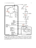
Fig. 1
The complete nucleic acid sequence and the corresponding
deduced amino acid sequence of the complete CDS of glutamate
dehydrogenase (gdh) from the liver of Misgurnus
anguillicaudatus. “*” indicates the stop codon. The start and
the end of the CDS are indicated in boldface type, and the
priming positions of the RACE primers used are underlined and
indicated in boldface type. Pentameric motifs corresponding to
AU-rich elements (AREs) are highlighted in grey……………… 53
Fig. 2
The alignment of the deduced amino acid sequence of glutamate
dehydrogenase (Gdh) from the liver of Misgurnus
anguillicaudatus and the amino acid sequences of Tribolodon
hakonensis Gdh (BAD83654.1), Oncorhynchus mykiss Gdh1
(AAM73775.1), Chaenocephalus aceratus Gdh (P82264.1),
Xenopus laevis GDH (NP_001087023.1) and Homo sapiens
GLUD1 (NP_005262.1). Identical residues in the alignment are
indicated by “*”; similar amino acids in the alignment are
indicated by “:”; dissimilar amino acids in the alignment are
indicated by “.”. Residues involved in adenine binding domain
are boxed; residues contributing to the antenna domain are
shaded grey……………………………………………………… 56
Fig. 3
The phylogenetic tree of several vertebrate glutamate
dehydrogenase (Gdh) protein sequences and Misgurnus
anguillicaudatus Gdh sequence. Litopenaeus vannamei Gdh
sequence was used as the outgroup. Bootstrap values are
indicated at the nodes of tree branches. The sequences used in
the tree and their respective accession number in either
GenBank or Ensembl databases were as follows: Oncorhynchus
mykiss Gdh1 (AAM73775.1) and Gdh3 (AAM73777.1), Danio
rerio Gdh1a (NP_997741.1) and Gdh1b (NP_955839.2), Salmo
salar Gdh1 (CAD89353.1), Gdh2 (CAD58714.1) and Gdh3
(CAD58715.1), Tribolodon hakonensis Gdh (BAD83654.1),
Chaenocephalus aceratus Gdh (P82264.1), Xenopus laevis
GDH (NP_001087023.1), X. (Silurana) tropicalis GDH
(NP_001011138.1), Gallus gallus GDH (P00368.1), Rattus
norvegicus GDH (NP_036702.1), Mus musculus GDH
(NP_032159.1), Bos taurus GDH (AAI03337.1), Homo sapiens
GLUD1 (NP_005262.1) and GLUD2 (NP_036216.2),
Litopenaeus vannamei Gdh (ACC95446.1), Tetraodon
nigroviridis Gdh1 (ENSTNIP00000008014) and Gdh2
(ENSTNIP00000016349),
Takifugu
rubripes
Gdh1
(ENSTRUP00000009100) and Gdh2 (ENSTRUP00000000720)
and Taeniopygia guttata GDH (ENSTGUP00000005951).
Protein sequences for Bostrychus sinensis Gdh1 and Gdh2 were
obtained from Peh (2008). Protein names in parenthesis are
non-indicative of the orthologous and paralogous relationships
between the Gdh isoforms………………………………………. 60
x
Fig. 4
Changes (log2 of fold change) in mRNA expression of
glutamate dehydrogenase (gdh) in the liver of Misgurnus
anguillicaudatus. (A) Fish kept in freshwater for 12 h (12 h
control), or after 12 h of terrestrial exposure, or after exposure to
50 mmol l-1 NH4Cl for 12 h. (B) Fish kept in freshwater for 2
days (2 days control), or after 2 days of terrestrial exposure, or
after exposure to 50 mmol l-1 NH4Cl for 2 days. *Significantly
different from the corresponding control value, P<0.05. Means
of changes in expression not sharing the same letter are
significantly different, P<0.05. Results represent mean +
S.E.M. (N=4)……………………………………………………. 63
Fig. 5
Changes (log2 of fold change) in mRNA expression of
glutamate dehydrogenase (gdh) in the intestine of Misgurnus
anguillicaudatus. (A) Fish kept in freshwater for 12 h (12 h
control), or after 12 h of terrestrial exposure, or after exposure to
50 mmol l-1 NH4Cl for 12 h. (B) Fish kept in freshwater for 2
days (2 days control), or after 2 days of terrestrial exposure, or
after exposure to 50 mmol l-1 NH4Cl for 2 days. Means of
changes in expression not sharing the same letter are
significantly different, P<0.05. Results represent mean +
S.E.M. (N=4)……………………………………………………. 64
Fig. 6
The complete nucleic acid sequence and the corresponding
deduced amino acid sequence of the complete CDS of glutamine
synthetase (gs) from the liver of Misgurnus anguillicaudatus.
“*” indicates the stop codon. The start and the end of the CDS
are indicated in boldface type, and the priming positions of the
RACE primers used are underlined and indicated in boldface
type……………………………………………………………… 75
Fig. 7
The alignment of the deduced amino acid sequences of
glutamine synthetase (Gs) from the liver of Misgurnus
anguillicaudatus and the amino acid sequences of Gs in
Oreochromis niloticus (AAM28589.1), Bostrychus sinensis
(AAL62447.1), Squalus acanthias (AAA61871.1), Xenopus
laevis (NP_001085867.1) and Homo sapiens (AAS57904.1).
Identical residues in the alignment are indicated by “*”; similar
amino acids in the alignment are indicated by “:”; dissimilar
amino acids in the alignment are indicated by “.”. Residues
contributing to the active site of GS are shaded grey…………… 77
Fig. 8
The phylogenetic tree of several vertebrate glutamine synthetase
(Gs) protein sequences and Misgurnus anguillicaudatus Gs
sequence. Paracentrotus lividus Gs sequence was used as the
outgroup. Bootstrap values are indicated at the nodes of tree
branches. The sequences used in the tree and their respective
accession number in either GenBank or Ensembl databases were
as follows: Oncorhynchus mykiss Gs1 (AAM73659.1), Gs2
xi
(AAM73660.1) and Gs4 (AAM73662.2), Salmo salar Gs
(NP_001134684.1),
Bostrichthys
sinensis
liver
Gs
(AAL62447.1) and stomach Gs (AAL62448.1), Opsanus beta
liver Gs (AAD34720.1) and gill Gs (AAN77155.1), Squalus
acanthias Gs (AAA61871.1), Heterodontus francisci Gs
(AAD34721.1), Danio rerio Gs (NP_001068582.1), Xenopus
laevis GS (NP_001085867.1), X. (Silurana) tropicalis GS
(NP_989297.1), Gallus gallus GS (NP_990824.1), Rattus
norvegicus GS (AAA65095.1), Mus musculus GS
(NP_032157.2), Bos taurus GS (NP_001035564.1), Canis lupus
familiaris GS (NP_001002965.1), Homo sapiens GS
(AAS57904.1), Paracentrotus lividus Gs (AAC41562.1),
Takifugu rubripes Gs1 (ENSTRUP00000002875) and Gs2
(ENSTRUP00000005906),
Anolis
carolinensis
GS
(ENSACAP00000008277),
Taeniopygia
guttata
GS
(ENSTGUP00000017624) and Meleagris gallopavo GS
(ENSMGAP00000002947). Protein sequence for Oxyeleotris
marmoratus Gs was obtained from Tng (2008). Protein names
in parenthesis are non-indicative of the orthologous and
paralogous relationships between the Gdh isoforms.…………… 81
Fig. 9
Changes (log2 of fold change) in mRNA expression of
glutamine synthetase (gs) in the liver of Misgurnus
anguillicaudatus. (A) Fish kept in freshwater for 12 h (12 h
control), or after 12 h of terrestrial exposure, or after exposure to
50 mmol l-1 NH4Cl for 12 h. (B) Fish kept in freshwater for 2
days (2 days control), or after 2 days of terrestrial exposure, or
after exposure to 50 mmol l-1 NH4Cl for 2 days. *Significantly
different from the corresponding control value, P<0.05. Means
of changes in expression not sharing the same letter are
significantly different, P<0.05. Results represent mean +
S.E.M. (N=4)……………………………………………………. 84
Fig. 10
Changes (log2 of fold change) in mRNA expression of
glutamine synthetase (gs) in the intestine of Misgurnus
anguillicaudatus. (A) Fish kept in freshwater for 12 h (12 h
control), or after 12 h of terrestrial exposure, or after exposure to
50 mmol l-1 NH4Cl for 12 h. (B) Fish kept in freshwater for 2
days (2 days control), or after 2 days of terrestrial exposure, or
after exposure to 50 mmol l-1 NH4Cl for 2 days. *Significantly
different from the corresponding control value, P<0.05. Means
of changes in expression not sharing the same letter are
significantly different, P<0.05. Results represent mean +
S.E.M. (N=4)……………………………………………………. 85
Fig. 11
The complete nucleic acid sequence and the corresponding
deduced amino acid sequence of the complete CDS of glutamate
dehydrogenase (gdh) from the liver of Monopterus albus. “*”
indicates the stop codon. The start and the end of the CDS are
indicated in boldface type, and the priming positions of the
xii
RACE primers used are underlined and indicated in boldface
type. Pentameric motifs corresponding to AU-rich elements
(AREs) are highlighted in grey. ………………………………… 95
Fig. 12
The alignment of the deduced amino acid sequence of glutamate
dehydrogenase (Gdh) from the liver of Monopterus albus and
the amino acid sequences of Chaenocephalus aceratus Gdh
(P82264.1), Oncorhynchus mykiss Gdh1 (AAM73775.1),
Tribolodon hakonensis Gdh (BAD83654.1), Xenopus laevis
GDH (NP_001087023.1) and Homo sapiens GLUD1
(NP_005262.1).
Identical residues in the alignment are
indicated by “*”; similar amino acids in the alignment are
indicated by “:”; dissimilar amino acids in the alignment are
indicated by “.”. Residues involved in adenine binding domain
are boxed; residues contributing to the antenna domain are
shaded grey……………………………………………………… 98
Fig. 13
The phylogenetic tree of several vertebrate glutamate
dehydrogenase (Gdh) protein sequences and Monopterus albus
Gdh sequence. Litopenaeus vannamei Gdh sequence was used
as the outgroup. Bootstrap values are indicated at the nodes of
tree branches. The sequences used in the tree and their
respective accession number in either GenBank or Ensembl
databases were as follows: Oncorhynchus mykiss Gdh1
(AAM73775.1) and Gdh3 (AAM73777.1), Danio rerio Gdh1a
(NP_997741.1) and Gdh1b (NP_955839.2), Salmo salar Gdh1
(CAD89353.1), Gdh2 (CAD58714.1) and Gdh3 (CAD58715.1),
Tribolodon hakonensis Gdh (BAD83654.1), Chaenocephalus
aceratus
Gdh
(P82264.1),
Xenopus
laevis
GDH
(NP_001087023.1),
X.
(Silurana)
tropicalis
GDH
(NP_001011138.1), Gallus gallus GDH (P00368.1), Rattus
norvegicus GDH (NP_036702.1), Mus musculus GDH
(NP_032159.1), Bos taurus GDH (AAI03337.1), Homo sapiens
GLUD1 (NP_005262.1) and GLUD2 (NP_036216.2),
Litopenaeus vannamei Gdh (ACC95446.1), Tetraodon
nigroviridis Gdh1 (ENSTNIP00000008014) and Gdh2
(ENSTNIP00000016349),
Takifugu
rubripes
Gdh1
(ENSTRUP00000009100) and Gdh2 (ENSTRUP00000000720)
and Taeniopygia guttata GDH (ENSTGUP00000005951).
Protein sequences for Bostrychus sinensis Gdh1 and Gdh2 were
obtained from Peh (2008). Protein names in parenthesis are
non-indicative of the orthologous and paralogous relationships
between the Gdh isoforms...…..………………………………… 103
Fig. 14
Changes (log2 of fold change) in mRNA expression of
glutamate dehydrogenase (gdh) in the liver of Monopterus
albus. (A) Fish kept in freshwater for 1 day (1 day control), or
after 1 day of terrestrial exposure, or after 1 day of exposure to
75 mmol l-1 NH4Cl. (B) Fish kept in freshwater for 6 days (6
day control), or after 6 days of terrestrial exposure, or after 6
xiii
days of exposure to 75 mmol l-1 NH4Cl. (C) Fish kept in
freshwater for 4 days (4 day control) or after exposure to
progressive increase in salinity from freshwater (1‰) to 20‰
water for 1 day. Means of changes in expression not sharing the
same letter are significantly different, P<0.05. Results represent
mean + S.E.M. (N=4)…………………………………………… 105
Fig. 15
Changes (log2 of fold change) in mRNA expression of
glutamate dehydrogenase (gdh) in the intestine of Monopterus
albus. (A) Fish kept in freshwater for 1 day (1 day control), or
after 1 day of terrestrial exposure, or after 1 day of exposure to
75 mmol l-1 NH4Cl. (B) Fish kept in freshwater for 6 days (6
day control), or after 6 days of terrestrial exposure, or after 6
days of exposure to 75 mmol l-1 NH4Cl. (C) Fish kept in
freshwater for 4 days (4 day control) or after exposure to
progressive increase in salinity from freshwater (1‰) to 20‰
water for 1 day. *Significantly different from corresponding
control, P˂0.05. Results represent mean + S.E.M. (N=4)……… 107
Fig. 16
Changes (log2 of fold change) in mRNA expression of
glutamate dehydrogenase (gdh) in the brain of Monopterus
albus. (A) Fish kept in freshwater for 1 day (1 day control), or
after 1 day of terrestrial exposure, or after 1 day of exposure to
75 mmol l-1 NH4Cl. (B) Fish kept in freshwater for 6 days (6
day control), or after 6 days of terrestrial exposure, or after 6
days of exposure to 75 mmol l-1 NH4Cl. (C) Fish kept in
freshwater for 4 days (4 day control) or after exposure to
progressive increase in salinity from freshwater (1‰) to 20‰
water for 1 day. Results represent mean + S.E.M. (N=4)……… 109
Fig. 17
The complete nucleic acid sequence and the corresponding
deduced amino acid sequence of the complete CDS of glutamine
synthetase (A) isoform 1 (gs1) from the intestine, (B) isoform 2
(gs2) and (C) isoform 3 (gs3) from the liver of Monopterus
albus. “*” indicates the stop codon. The start and the end of the
CDS are indicated in boldface type, and the priming positions of
the RACE primers used are underlined and indicated in boldface
type. Pentameric motifs corresponding to AU-rich elements
(AREs) are highlighted in grey…………………………………. 119
Fig. 18
The alignment of the deduced amino acid sequences of
glutamine synthetase (Gs) isoforms Gs1, Gs2 and Gs3 from the
liver of Monopterus albus and the amino acid sequences of Gs
in Opsanus beta (AAN77155.1), Bostrychus sinensis
(AAL62447.1), Squalus acanthias (AAA61871.1), Xenopus
laevis (NP_001085867.1) and Homo sapiens (AAS57904.1).
Identical residues in the alignment are indicated by “*”; similar
amino acids in the alignment are indicated by “:”; dissimilar
amino acids in the alignment are indicated by “.”. Residues
contributing to the active site of GS are shaded grey…………… 127
xiv
Fig. 19
The phylogenetic tree of several vertebrate glutamine synthetase
(Gs) protein sequences and Monopterus albus Gs sequences.
Paracentrotus lividus Gs sequence was used as the outgroup.
Bootstrap values are indicated at the nodes of tree branches.
The sequences used in the tree and their respective accession
number in either GenBank or Ensembl databases were as
follows: Oncorhynchus mykiss Gs1 (AAM73659.1), Gs2
(AAM73660.1) and Gs4 (AAM73662.2), Salmo salar Gs
(NP_001134684.1),
Bostrichthys
sinensis
liver
Gs
(AAL62447.1) and stomach Gs (AAL62448.1), Opsanus beta
liver Gs (AAD34720.1) and gill Gs (AAN77155.1), Squalus
acanthias Gs (AAA61871.1), Heterodontus francisci Gs
(AAD34721.1), Danio rerio Gs (NP_001068582.1), Xenopus
laevis GS (NP_001085867.1), X. (Silurana) tropicalis GS
(NP_989297.1), Gallus gallus GS (NP_990824.1), Rattus
norvegicus GS (AAA65095.1), Mus musculus GS
(NP_032157.2), Bos taurus GS (NP_001035564.1), Canis lupus
familiaris GS (NP_001002965.1), Homo sapiens GS
(AAS57904.1), Paracentrotus lividus Gs (AAC41562.1),
Takifugu rubripes Gs1 (ENSTRUP00000002875) and Gs2
(ENSTRUP00000005906),
Anolis
carolinensis
GS
(ENSACAP00000008277),
Taeniopygia
guttata
GS
(ENSTGUP00000017624) and Meleagris gallopavo GS
(ENSMGAP00000002947). Protein sequence for Oxyeleotris
marmoratus Gs was obtained from Tng (2008). Protein names
in parenthesis are non-indicative of the orthologous and
paralogous relationships between the Gs isoforms.…………..… 131
Fig. 20
Expressions of glutamine synthetase isoform 1 (gs1) in the
liver, intestine and brain of Monopterus albus exposed to
terrestrial conditions for 1 day or 6 days, or exposed to 75 mmol
l-1 NH4Cl for 1 day or 6 days, or exposed to increasing salinity
from freshwater (1‰) to 20‰ water for 1 day. Controls were
maintained in freshwater (1‰) for 1 day, 4 days or 6 days…….. 135
Fig. 21
Semi-quantitation of mRNA expression of glutamine synthetase
isoform 1 (gs1) in the (A) intestine and (B) brain of Monopterus
albus exposed to terrestrial conditions for 1 day or 6 days, or
exposed to 75 mmol l-1 NH4Cl for 1 day or 6 days, or exposed to
increasing salinity from freshwater (1‰) to 20‰ water for 1
day. Controls were maintained in freshwater (1‰) for 1 day, 4
days or 6 days…………………………………………………… 136
Fig. 22
Changes (log2 of fold change) in mRNA expression of
glutamine synthetase isoform 2 (gs2) in the liver of Monopterus
albus. (A) Fish kept in freshwater for 1 day (1 day control), or
after 1 day of terrestrial exposure, or after 1 day of exposure to
75 mmol l-1 NH4Cl. (B) Fish kept in freshwater for 6 days (6
day control), or after 6 days of terrestrial exposure, or after 6
xv
days of exposure to 75 mmol l-1 NH4Cl. (C) Fish kept in
freshwater for 4 days (4 day control) or after exposure to
progressive increase in salinity from freshwater (1‰) to 20‰
water for 1 day. Means of changes in expression not sharing the
same letter are significantly different, P<0.05. Results represent
mean + S.E.M. (N=4)…………………………………………… 139
Fig. 23
Changes (log2 of fold change) in mRNA expression of
glutamine synthetase isoform 3 (gs3) in the liver of Monopterus
albus. (A) Fish kept in freshwater for 1 day (1 day control), or
after 1 day of terrestrial exposure, or after 1 day of exposure to
75 mmol l-1 NH4Cl. (B) Fish kept in freshwater for 6 days (6
day control), or after 6 days of terrestrial exposure, or after 6
days of exposure to 75 mmol l-1 NH4Cl. (C) Fish kept in
freshwater for 4 days (4 day control) or after exposure to
progressive increase in salinity from freshwater (1‰) to 20‰
water for 1 day. Means of changes in expression not sharing the
same letter are significantly different, P<0.05. Results represent
mean + S.E.M. (N=4)…………………………………………… 141
Fig. 24
Changes (log2 of fold change) in mRNA expression of
glutamine synthetase isoform 2 (gs2) in the intestine of
Monopterus albus. (A) Fish kept in freshwater for 1 day (1 day
control), or after 1 day of terrestrial exposure, or after 1 day of
exposure to 75 mmol l-1 NH4Cl. (B) Fish kept in freshwater for
6 days (6 day control), or after 6 days of terrestrial exposure, or
after 6 days of exposure to 75 mmol l-1 NH4Cl. (C) Fish kept in
freshwater for 4 days (4 day control) or after exposure to
progressive increase in salinity from freshwater (1‰) to 20‰
water for 1 day. Means of changes in expression not sharing the
same letter are significantly different, P<0.05. Results represent
mean + S.E.M. (N=4)…………………………………………… 143
Fig. 25
Changes (log2 of fold change) in mRNA expression of
glutamine synthetase isoform 3 (gs3) in the intestine of
Monopterus albus. (A) Fish kept in freshwater for 1 day (1 day
control), or after 1 day of terrestrial exposure, or after 1 day of
exposure to 75 mmol l-1 NH4Cl. (B) Fish kept in freshwater for
6 days (6 day control), or after 6 days of terrestrial exposure, or
after 6 days of exposure to 75 mmol l-1 NH4Cl. (C) Fish kept in
freshwater for 4 days (4 day control) or after exposure to
progressive increase in salinity from freshwater (1‰) to 20‰
water for 1 day. Results represent mean + S.E.M. (N=4)……… 145
Fig. 26
Changes (log2 of fold change) in mRNA expression of
glutamine synthetase isoform 2 (gs2) in the brain of Monopterus
albus. (A) Fish kept in freshwater for 1 day (1 day control), or
after 1 day of terrestrial exposure, or after 1 day of exposure to
75 mmol l-1 NH4Cl. (B) Fish kept in freshwater for 6 days (6
day control), or after 6 days of terrestrial exposure, or after 6
xvi
days of exposure to 75 mmol l-1 NH4Cl. (C) Fish kept in
freshwater for 4 days (4 day control) or after exposure to
progressive increase in salinity from freshwater (1‰) to 20‰
water for 1 day. *Significantly different from corresponding
control, P<0.05. Results represent mean + S.E.M. (N=4)……… 147
Fig. 27
Changes (log2 of fold change) in mRNA expression of
glutamine synthetase isoform 3 (gs3) in the brain of Monopterus
albus. (A) Fish kept in freshwater for 1 day (1 day control), or
after 1 day of terrestrial exposure, or after 1 day of exposure to
75 mmol l-1 NH4Cl. (B) Fish kept in freshwater for 6 days (6
day control), or after 6 days of terrestrial exposure, or after 6
days of exposure to 75 mmol l-1 NH4Cl. (C) Fish kept in
freshwater for 4 days (4 day control) or after exposure to
progressive increase in salinity from freshwater (1‰) to 20‰
water for 1 day. Means of changes in expression not sharing the
same letter are significantly different, P<0.05. Results represent
mean + S.E.M. (N=4)…………………………………………… 149
Fig. 28
Comparing the putative mitochondrial targeting sequence of
Opsanus beta gs (AF118103) with the partial 5’UTR sequences
of glutamine synthetase (gs) gene gs3 from Monopterus albus,
and the partial 5’UTR sequences of gs in Oreochromis niloticus
(AF503208) and Oxyeleotris marmoratus. The start codon for
M. albus gs3, O. niloticus gs, O. marmoratus gs and the second
start codon of O. beta mitochondrial gs are indicated in bold.
Conserved sequences are indicated with “*”.………………..….. 156
xvii
LIST OF ABBREVIATIONS
UTR: untranslated region
CDS: coding sequence
RACE: rapid amplification of cDNA ends
Gs: glutamine synthetase protein
Gdh: glutamate dehydrogenase protein
gs: glutamine synthetase gene
gdh: glutamate dehydrogenase gene
xviii
LITERATURE REVIEW
Ammonia production, ammonia toxicity and excretory nitrogen metabolism
Ammonia production
In animals, the major source of amino acids comes from dietary proteins.
While carbohydrates and lipids can be stored as glycogen and triglycerides,
respectively, animals are unable to store excess amino acids (Ip and Chew, 2010).
Therefore, excess dietary amino acids that are not utilized for growth and
maintenance of protein turnover are preferentially degraded over carbohydrates and
lipids in the liver (Campbell, 1991). Dietary carbon may be extracted from the
carbon chain of amino acids following removal of the α-amino group in fishes
dependent on high protein diets (Ip and Chew, 2010). Apart from dietary proteins,
muscle proteins can also serve as a source of amino acids in fasting fishes, which are
then catabolized to produce ATP or carbohydrates (Houlihan et al., 1995).
Catabolism and transamination of amino acids results in the production of ammonia.
In mammals, the small intestine is a major organ implicated in ammonia production,
with approximately 40% being produced through the activities of bacterial urease
and amino acid oxidases while the rest is produced from amino acid transamination
and glutamine metabolism (Shawcross et al., 2005; Lemberg and Fernandez, 2009).
Ammonia is also produced from enzymatic pathways catalyzed by glutamate
dehydrogenase (GDH) and AMP-deaminase (Szerb and Butterworth, 1992).
For fish, ammonia is mainly produced from the α-amino group of amino
acids that are catabolized (Ip and Chew, 2010). Liver is a main site of ammonia
production in fish. For goldfish, the liver accounts for 50-70% (Van den Thillart and
van Raaji, 1995), or even up to 99% (van Warde, 1981) of ammonia produced. The
mechanism of ammonia production can occur in the cytosol of hepatocytes through
1
the activities of specific deaminases (histidase, asparaginase, serine dehydratase and
threonine dehydratase; Youngson et al., 1982) or via transdeamination, involving the
combined actions of cytosolic aminotransferases and mitochondrial GDH (Walton
and Cowey, 1977, 1982; French et al., 1981; Campbell et al., 1983). Nonetheless,
transdeamination is the primary mechanism through which amino acids are
catabolized in fish liver (Ballantyne, 2001). The rate of glutamate deamination by
intact catfish liver mitochondria can account for 160% of the rate of ammonia
excretion (Campbell et al., 1983). On the other hand, the rates of alanine and
glutamine deamination by catfish hepatocytes account for only 50% and 85%,
respectively, of the total ammonia excreted by live fish (Campbell et al., 1983). As
GDH is localized exclusively in the matrix of fish liver mitochondria,
transdeamination releases ammonia into this compartment. Some fish species also
possess glutaminase, which release NH3 from the amide-function of glutamine, in
the mitochondrial matrix. Thus, at the cellular level, the excretion of ammonia
involves its permeation of the hepatic mitochondrial membranes (Ip and Chew, 2010)
into the cell cytoplasm.
Ammonia toxicity
Ammonia is toxic as it can disrupt the normal functioning and homeostasis of
several cellular processes (Campbell, 1991; Lemberg and Fernandez, 2009). At the
molecular level, NH4+ can substitute for K+ in neurons and permeate through K+
background channels, affecting the membrane potential (Binstock and Lecar, 1969).
Additionally, NH4+ can also substitute for K+ in Na+, K+-ATPase and in Na+/K+/2Cl
-
co-transporter (see Wilkie, 1997, 2002 for reviews; Person-Le Ruyet et al., 1998),
and for H+ in Na+/ H+ exchanger (Randall et al., 1999) in gills, upsetting the ionic
balance in fish in the process. At the cellular level, ammonia inhibits key glycolytic
2
enzymes, such as isocitrate dehydrogenase, α-ketoglutarate dehydrogenase and
pyruvate dehydrogenase (see review by Cooper and Plum, 1987). This leads to the
impairment of the tricarboxylic acid cycle (Arillo et al., 1981), and can result in
brain energy failure (Lemberg and Fernandez, 2009). In the mammalian brain,
ammonia also lowers the response of the central nervous system by inhibiting
excitatory post-synaptic potentials (Szerb and Butterworth, 1992).
At the organismal level, ammonia affects the central nervous system of
vertebrates, including fish, causing hyperventilation (Hillaby and Randall, 1979;
McKenzie et al., 1993), hyperexcitability, coma and convulsions, which eventually
leads to death (Ip et al., 2004a). Ammonia is also implicated in the pathology of
acute hepatic encephalopathy in mammals (Brusilow, 2002; Felipo and Butterworth,
2002; Rose, 2002; Shawcross et al., 2005; Chastre et al., 2010).
Cranial
hyperammonemia (3 mmol L-1; Kosenko et al., 1994) resulting from acute liver
failure leads to astrocyte swelling and brain edema (Norenberg et al., 2005; Vaquero
and Butterworth, 2008), intracranial hypertension (Master et al., 1999) as well as
brainstem herniation (Clemmesen et al., 1999) and glutamatergic dysfunction
(Michalak et al., 1996; Hilgier et al., 1999 ). It is thought that hyperammonemiainduced glutamine synthesis is the causative process that brings about astrocyte
swelling and dysfunction and cerebral edema (Takahashi et al., 1991; Zwingmann et
al., 2000; Brusilow, 2002; Tanigami et al., 2005; Albrecht and Norenberg, 2006;
Tofteng et al., 2006). Excess glutamine can cause mitochondrial dysfunction (Bai et
al., 2001; Rao and Norenberg, 2001) and induces mitochondrial permeability
transition in cultured astrocytes (Bai et al., 2001; Rama Rao et al., 2003; Jayakumar
et al., 2004).
3
The theories explaining the mechanisms for acute ammonia toxicity in
mammalian brains have yet to be established in fish (Ip and Chew, 2010). However,
it is known that the mechanisms of ammonia toxicity in the brains of some fishes
with high ammonia tolerance apparently differ from those in mammalian brains
(Opsanus
beta,
Veauvy et
al.,
2005;
Periophthalmodon
schlosseri
and
Boleophthalmus boddarti, Ip et al., 2005a; Clarias gariepinus, Wee et al., 2007;
Monopterus albus, Tng et al., 2009). Monopterus albus that succumbed to a lethal
dose (16 μmol g−1 fish) of ammonium acetate (CH3COONH4) has an extraordinary
high content of ammonia in the brain (Tng et al., 2009).
L-methionine S-
sulfoximine (MSO) is an irreversible inhibitor of glutamine synthetase (GS)
(Folbergrova, 1964). For two species of mudskippers, P. schlosseri and B. boddarti,
MSO at a dosage (100 μg g−1 fish) protective for rats does not reduce the mortality of
fish injected with a lethal dose of CH3COONH4 (Ip et al., 2005a). Taken together,
results from M. albus (Tng et al., 2009), P. schlosseri and B. boddarti (Ip et al.,
2005a) indicates that unlike mammals, increased glutamine synthesis in the brain is
not the major cause of death for these fishes. MSO exhibits a partial protective
effect against acute ammonia toxicity in C. gariepinus (Wee et al., 2007) and M.
albus (Tng et al., 2009). The mortality of C. gariepinus injected with a lethal dose
of CH3COONH4 reduces from 100 to 80% with prior administration of MSO (100
μg g−1 fish) and the time of death is prolonged from 27 to 48 min (Wee et al., 2007).
Similarly, prior administration of MSO (100 μg g−1 fish) in M. albus reduces
mortality from 100 to 80% and extends the time of death from 85.3 min to 133 min
(Tng et al., 2009). The protective effect of MSO in both C. gariepinus and M. albus
is probably not related to the inhibition of GS and prevention of glutamine
accumulation. Instead, it reduces the rate of ammonia accumulation in the brain
4
through its effects on GDH, increasing the amination of α-ketoglutarate and/or
decreasing deamination of glutamate (Wee et al., 2007; Tng et al., 2009).
In mammals, acute ammonia intoxication is often associated with brain
edema and the generation of oxidative and/or nitrosative stress (Master et al., 1999;
Schliess et al., 2002, 2006; Haussinger and Gorg, 2010). Glutamate exocytosis in rat
astrocytes in response to ammonia toxicity (Gorg et al., 2010) facilitates increases in
extracellular glutamate (Michalak et al., 1996). This leads to the overactivation of
NMDA receptors, leading to cerebral production of reactive oxygen and nitrogen
species (ROS/RNOS) (Marcaida et al., 1992; Miñana et al., 1996; Kosenko et al.,
1999), protein tyrosine nitration (Kosenko et al., 2004; Schliess et al., 2002, 2006),
oxidation of RNA (Gorg et al., 2008; Schliess et al., 2009), and death (Miñana et al.,
1996; Hermenegildo et al., 1996). In addition, oxidative and nitrosative stress brings
about the activation of nuclear factor kappaB, resulting in the up-regulation of
inducible nitric oxide synthase (iNOS) expression (Sinke et al., 2008). Subsequently,
production of nitric oxide – one of the causal agents of astrocyte swelling – increased
(Sinke et al., 2008), contributing to nitric oxide-induced blood brain barrier damage
(Tan et al., 2004).
The brain of B. boddarti also experiences ammonia-induced oxidative stress
(Ching et al., 2009). Fish exposed to 8 mmol l−1 NH4Cl for 12 or 24 h increases
cranial superoxide dismutase activity, decreases glutathione reductase and catalase
activity, and there are increases in oxidized glutathione content and oxidized:reduced
glutathione ratio (Ching et al., 2009). However, cranial ammonia-induced oxidative
stress does not bring about excessive activation of NMDA receptors (Ip et al., 2005a).
Ching et al. (2009) also noted that ammonia can induce oxidative stress in the gills,
an organ that lacks NMDA receptors, of B. boddarti, leading to the conclusion that
5
there could be multiple routes through which ammonia induces oxidative stress in
brain or non-brain tissues. As such, it is proposed that ammonia may increase
intracellular NO and/or Ca2+ concentrations, causing increased production of free
radicals (Hernández-Fonseca et al., 2008), in the gills and brain of B. boddarti.
Gills are the main site of respiration in fish (Evans et al., 2005) which would
be directly in contact with exogenous ammonia during environmental ammonia
exposure (Ip and Chew, 2010). As such, ammonia must permeate through the
branchial and cutaneous epithelia before being transported through the blood to the
brain and other organs (Ip and Chew, 2010).
Environmental ammonia has
deleterious effects on branchial ion transport not associated with endogenous
ammonia accumulation, which is absent in fish simply exposed to terrestrial
conditions or to fish injected/infused with exogenous ammonia (Ip et al., 2004b).
Acute exposure to environmental ammonia results in inhibition of Na+ influx in the
goldfish Carassius auratus (Maetz and Garcia Romeu, 1964; Maetz, 1973) and the
temperate rainbow trout Oncorhynchus mykiss (Avella and Bornancin, 1989). In C.
auratus, the deleterious effect is specific to Na+ uptake and not general to the
epithelium or all ion uptake mechanisms (Maetz and Garcia Romeu, 1964).
However, no deleterious effect of ammonia exposure (up to 28.2 μmol l -1 NH3-N or
5.2 mmol l-1 total ammonia) is seen on Na+ uptake in juvenile rainbow trout, but Na+
efflux is stimulated by ammonia levels greater than 6.4 μmol l-1 NH3-N (1.2 mmol l-1
total ammonia) (Twitchen and Eddy, 1994).
It is likely that an increased Na+
permeability of the gills brings about the increases in Na+ efflux (Gonzalez and
McDonald, 1994), which is mediated through a modulation of the paracellular
pathway (Madara, 1998).
Additionally, exposure to environmental ammonia
predisposes the gills to histopathological changes that may disrupt ion transport
6