Nanoparticles of biodegradable polymers for delivery of diagnostic therapeutic agents their potential application in brain cancer therapy
Bạn đang xem bản rút gọn của tài liệu. Xem và tải ngay bản đầy đủ của tài liệu tại đây (1.84 MB, 117 trang )
NANOPARTICLES OF BIODEGRADABLE
POLYMERS FOR DELIVERY OF
DIAGNOSTIC/THERAPEUTIC AGENTS: THEIR
POTENTIAL APPLICATION IN BRAIN CANCER
THERAPY
YU QIANRU
NATIONAL UNIVERSITY OF SINGAPORE
2005
NANOPARTICLES OF BIODEGRADABLE
POLYMERS FOR DELIVERY OF
DIAGNOSTIC/THERAPEUTIC AGENTS: THEIR
POTENTIAL APPLICATION IN BRAIN CANCER
THERAPY
YU QIANRU
(B.Eng, Southeast University)
A THESIS SUBMITTED
FOR THE DEGREE OF MASTER OF SCIENCE
GRADUATE PROGRAM IN BIOENGINEERING
NATIONAL UNIVERSITY OF SINGAPORE
2005
Acknowledgement
At the point of finishing my master candidature in Singapore and completing my
thesis, I would like to thank the following organizations and people.
Firstly, I would like to thank National University of Singapore and GPBE to give me
such a great chance to pursue my research in Singapore. Being exposed to the frontier
of bioengineering field, I have thus enriched my knowledge and enhanced my ability
for future work.
Secondly, I would like to thank my supervisors, A/P Feng Si-Shen, A/P Wang ShihChang, A/P Sheu Fwu-Shan for their useful advice and continuous guidance
throughout my graduate study at the department of bioengineering, NUS.
Thirdly, I will thank all my colleagues in Chemotherapeutic Engineering Lab and
National University Hospital. I am especially grateful to Ms. Chen Lirong who is my
mentor in two lab rotations and Mr Shuter, Borys who kindly helped me with the MRI
imaging.
Last but not least, I owe my thanks to my parents and all my friends. Thanks for your
help and kind encouragement. You are the most precious treasure all my life.
i
Table of Contents
Acknowledgement
i
Table of Contents
ii
Summary
vi
Nomenclature
viii
List of Figures
x
List of Tables
xii
Chapter 1: Introduction
1
1.1 Background
1
1.2 Objectives
3
1.3 Thesis Organization
5
Chapter 2: Literature Review
6
2.1 Paclitaxel and Its Limitations in Modern Chemotherapy
6
2.2 Brain Cancers and Blood Brain Barrier (BBB)
8
2.2.1 Brain cancers and cancer treatment
8
2.2.2 Introduction to the blood brain barrier
9
2.2.2.1 History of blood brain barrier
9
2.2.2.2 Structure and function of the blood brain barrier
10
2.2.2.3 In vitro and in vivo models of blood brain barrier
12
2.2.2.4 Strategies to conquer the blood brain barrier
14
2.3 Nanoparticles of Biodegradable Polymers for Drug Delivery
15
2.3.1 Basic information of biodegradable polymers
16
2.3.2 Manufacture techniques of nanoparticles
18
2.3.3 Current research on biodegradable nanoparticles across BBB
21
2.4 Magnetic Resonance Image(MRI) and MRI Contrast Agent
23
2.4.1 Basic principles of MRI
23
2.4.2 Important parameters of MRI
24
ii
2.4.3 Introduction to MRI contrast agent
26
Chapter 3: Materials and Methods
28
3.1 Materials
28
3.2 Methods
29
3.2.1 Preparation of nanoparticles
29
3.2.1.1 Preparation of paclitaxel/fluorescence loaded nanoparticles-single
29
emulsion
3.2.1.2 Preparation of Gd-DTPA loaded nanoparticles-nanoprecipitation
30
3.2.1.3 Preparation of Gd-DTPA loaded nanoparticles-double emulsion
31
3.2.2 Characterization of nanoparticles
31
3.2.2.1 Size and size distribution
31
3.2.2.2 Particle morphology
31
3.2.2.3 Surface charge
32
3.2.3 Encapsulation efficiency and drug entrapment
33
3.2.3.1 Encapsulation efficiency and drug entrapment of paclitaxel loaded
33
nanoparticles
3.2.3.2 Encapsulation efficiency and drug entrapment of Gd-DTPA loaded
34
nanoparticles
3.2.4 In vitro release
34
3.2.4.1 In vitro release of paclitaxel loaded nanoparticles
34
3.2.4.2 In vitro release of Gd-DTPA loaded nanoparticles
35
3.2.5 Cell line experiments
35
3.2.5.1 Cell culture
35
3.2.5.2 Trypsinization procedures of the cells
36
3.2.5.3 Cell viability study/cototoxity study
36
3.2.5.4 Cell uptake study
37
3.2.5.5 Fluorescence microscopy and confocal study
38
3.2.6 Animal Study
39
iii
3.2.7 MRI Characterization
39
Chapter 4: In Vitro Study of Paclitaxel Loaded PLGA Nanoparticles to Treat Brain
41
Cancer Cells
4.1 Novel Formulation of PLGA Nanoparticles with Natural Emulsifiers
41
4.2 Size, Size Distribution and Surface Charge
43
4.2.1 Particle size and size distribution
44
4.2.2 Surface charge study
46
4.3 Surface and Bulk Morphology
47
4.4 Encapsulation Efficiency of Paclitaxel Loaded Nanoparticles
50
4.5 In Vitro Release Profile of Paclitaxel from Nanoparticles
52
4.6 Cell Culture of Rat Brain Tumor Cell Line C6
54
4.7 Cell Viability Study
55
Chapter 5: In Vitro and In Vivo Uptake Study of Fluorescence Loaded Polymeric
59
Nanoparticles to Cross the Blood Brain Barrier
5.1 MDCK Cell Line as In Vitro BBB Model
59
5.2 Cell Uptake Study
60
5.2.1 Surfactant effect
60
5.2.2 Particle size effect
62
5.3 Confocal Study
65
5.4 In Vivo Study with Rat Models
66
Chapter 6: Formulation and Characterization of Gadolinium-DTPA Encapsulated
69
Nanoparticles for Potential In Vivo Imaging
6.1 Significance to Develop MRI Contrast Agent Gd-DTPA Encapsulated Biodegradable
69
Nanoparticles
6.2 Size, Size Distribution, Zeta Potential Study
70
iv
6.3 Morphology of Gd-DTPA Encapsulated Nanoparticles
72
6.4 Drug Entrapment and In Vitro Release Profile of Gd-DTPA Encapsulated
73
Nanoparticles
6.4.1 Drug entrapment study
73
6.4.2 In vitro release kinetics
75
6.5 MRI Characterization
76
6.5.1 Calibration curve of pure Gd-DTPA in vitro
76
6.5.2 Relaxation rate characteristics of Gd-DTPA encapsulated nanoparticles
79
Chapter 7: Conclusions and Recommendations
83
7.1 Conclusions
83
7.2 Recommendations
85
Reference
86
v
Summary
Made up of brain micro-vessel endothelial cells, blood brain barrier (BBB) is a
physiologic barrier between the blood and the central nervous system (CNS). It
provides neurons with nutrition and isolates the CNS from toxic chemicals in the
blood. However, it also severely restricts the delivery of therapeutic agents into the
brain. Paclitaxel, one of the most widely used anti-cancer drugs, has limited
application in treating brain tumor because of the existence of BBB. Of various
strategies developed to enhance drug delivery to the brain, nanoparticles of
biodegradable polymers show great potential because they can conquer BBB
non-invasively and achieve prolonged pharmacological action of drug molecules.
In this research, paclitaxel loaded poly (D,L-lactide-co- glicolide) (PLGA)
nanoparticles were fabricated using single emulsion technique. The emphasis was put
on the effect of surfactants of nanoparticles. Chemical surfactant polyvinyl alcohol
(PVA) and natural surfactants DPPC, vitamin E TPGS were used. Nanoparticles of
sizes around 250nm with narrow size distribution and negative surface charge were
achieved. Scanning electron microscopy (SEM) and atomic force microscopy (AFM)
images showed the morphologies of these nanoparticles. It was found that vitamin E
TPGS emulsified nanoparticles had much higher encapsulation efficiency than the
other two batches. All batches of nanoparticles had sustained in vitro release in about
a month. Cell viability study was carried out using rat glioma cell line C6 to test
paclitaxel loaded nanoparticles’ potential to treat brain tumor. It was found that time
vi
and concentration had effect on the viability.
Cell uptake and confocal laser scanning microscopic studies revealed that fluorescent
marker coumarin-6 loaded PLGA nanoparticles were ready to cross the in vitro BBB
model- Madin-Darby Canine Kidney (MDCK) cell line, but the uptake percentage
was affected by surfactants. Particle size effect on cellular uptake was also studied
using fluorescent polystyrene nanoparticles with uniform particle sizes. In vivo
experiment was carried out subsequently. PLGA nanoparticles were overcoated with
tween 80 before injecting to the tail vein of the rats. Fluorescence was detected both
in rat brain vessels and tissues under fluorescence microscope.
MRI contrast agent Gadolinium-DTPA loaded biodegradable nanoparticles were also
developed for future non-invasive in vivo imaging. Besides size, morphology, drug
entrapment and in vitro release study, MRI characteristics of Gd-DTPA encapsulated
nanoparticles were also investigated.
Overall, this research conducted systematic investigation on feasibility of
nanoparticles of biodegradable polymers for drug delivery across the blood brain
barrier. It was found that emulsifiers and particle size played an important part on
nanoparticles’ ability to cross BBB. Preliminary research on MRI contrast agent
Gd-DTPA encapsulated nanoparticles for future non-invasive in vivo imaging was
also investigated. These results will provide comprehensive information on
nanoparticles of biodegradable polymers as potential drug carriers to treat brain
cancer and brain related diseases such as AIDS.
vii
Nomenclature
AFM
Atomic Force Microscopy
BBB
Blood brain barrier
CNS
Central nervous system
DCM
Dichloromethane
DMEM
Dulbecco’s Modification of Eagle’s Medium
DPPC
1,2-dipalmitoyl-sn-glycerol-3-phospatidylchlorine
EE
Encapsulation efficiency
FBS
Fetal Bovine Serum
Gd-DTPA
Gadolinium Diethylenetriaminepenta-acetic Acid
HBSS
Hank’s balanced salt solution
HPLC
High performance liquid chromatography
ICP-OES
Inductively Coupled Plasma - Optical Emission Spectrometer
LLS
Laser Light Scattering
MDCK
Madin-Darby Canine Kidney
MDR
Multidrug Resistance
MPEG-PLA
Methoxy poly(ethylene glycol)-poly(lactide)
MRI
Magnetic Resonance Imaging
MRP
Multidrug resistance protein
MTT
3-(4,5-dimethylthiazol-2-yl)-2,5-diphenyltetrazolium bromide
PBS
Phosphate Buffer Saline
P-gp
P-glycoprotein
viii
PLA-PEG
Poly (Lactic acid) - poly(ethylene glycol)
PLGA
Poly (D, L-lactide-co-glicolide)
PS
Polystyrene
PVA
Polyvinyl alcohol
SEM
Scanning Electron Microscopy
Vitamin E TPGS
Vitamin E succinate with polyethylene glycol 1000
ix
List of Figures
Fig 2.1 Chemical structure of paclitaxel
Fig 2.2 Chemical structures of PEG (a), MPEG-PLA(b), PLA(c) and PLGA(d)
Fig 2.3 Chemical structure of Gd-DTPA and Fe3O4-dextran
Fig 4.1 chemical structure of PVA, vitamin E TPGS and DPPC
Fig 4.2 SEM and AFM images of PVA emulsified PLGA nanoparticles (5% drug
loading)
Fig 4.3 SEM and AFM images of PVA & DPPC co-emulsified PLGA nanoparticles
(5% drug loading)
Fig 4.4 SEM and AFM images of vitamin E TPGS emulsified PLGA nanoparticles
(5% drug loading)
Fig 4.5 Encapsulation efficiency of PVA, DPPC and vitamin E TPGS emulsified
PLGA nanoparticles(5% paclitaxel loading, n=3)
Fig 4.6 In vitro release profile of PVA, DPPC, vitamin E TPGS emulsified
nanoparticles (5% paclitaxel loading)
Fig 4.7 Morphology of rat brain glioma cell line C6 reaching ~50% confluence after ~
3 days’ culture
Fig 4.8 C6 cell viability study of pure taxol, 5% paclitaxel loaded and no-drug loaded
(placebo) PLGA nanoparticles with different emulsifiers in different concentrations,
incubation time=24h. (n=6)
Fig 4.9 C6 cell viability study of pure taxol, 5% paclitaxel loaded and no-drug loaded
(placebo) PLGA nanoparticles with different emulsifiers in different time intervals.
Concentration=0.25 µg/mL (n=6)
Fig 5.1 Morphology of MDCK cell line reaching ~80% confluence after ~5 days’
culture
Fig 5.2 MDCK cellular uptake of PLGA nanoparticles with different emulsifiers,
incubation time = 4 hours, concentration = 250 µg/mL. (n=6)
Fig 5.3 MDCK Cellular uptake profile of fluorescent polystyrene nanoparticles with
uniform particle sizes (n=6), the concentration unit is µg/mL, the size unit is nm.
x
Fig 5.4 Confocal images of fluorescence loaded PLGA nanoparticles with different
emulsifiers. (a) PVA emulsified nanoparticles (b) DPPC& PVA emulsified
nanoparticles (c) vitamin E TPGS emulsified nanoparticles.
Fig 5.5 Fluorescent microscope image of rat brain tissue after injecting with tween-80
coated PLGA nanoparticles. (bar =10µm)
Fig 6.1 FESEM images of Gd-DTPA encapsulated nanoparticles.(a) PLGA
nanoparticles using double emulsion, bar=1µm (b) PLGA nanoparticles using
nanoprecipitation, bar=1µm (c) mPEG-PLA nanoparticles using double emulsion,
bar=100nm (d)mPEG-PLA nanoparticles using nanoprecipitation, bar=1µm
Fig 6.2 In vitro release profile of Gd-DTPA encapsulated MPEG-PLA nanoparticles.
DE=double emulsion, NP=nanoprecipitation
Fig 6.3 Calibration curve of Gd concentration to R1 relaxation rate in water using
pure Gd-DTPA
Fig 6.4 Calibration curve of Gd concentration to R1 relaxation rate in gelatin using
pure Gd-DTPA
Fig 6.5 R1 relaxation rate of water, blank MPEG-PLA nanoparticles without
Gd-DTPA, Gd-DTPA encapsulated nanoparticles using nanoprecipitation and
Gd-DTPA encapsulated nanoparticles using double emulsion suspended in water
Fig 6.6 R1 relaxation rate of gelatin, blank MPEG-PLA nanoparticles without
Gd-DTPA, Gd-DTPA encapsulated nanoparticles using nanoprecipitation and
Gd-DTPA encapsulated nanoparticles using double emulsion suspended in gelatin
Fig 6.7 MRI images of Gd-DTPA encapsulated MPEG-PLA nanoparticles, from left
to right: 1. blank nanoparticle in water, 2. blank nanoparticles in gelatin, 3. Gd-DTPA
loaded nanoparticles using nanoprecipitation in water, 4.
Gd-DTPA loaded
nanoparticles using nanoprecipitation in gelatin, 5. Gd-DTPA loaded nanoparticles
using double emulsion in water, 6. Gd-DTPA loaded nanoparticles using double
emulsion in gelatin. TR= 800ms; TE =12ms, 256x256, 0.7mm in-plane and 5mm slice
thickness
xi
List of Tables
Table 2.1 Summary of BBB History
Table 2.2 Summary of in vivo techniques to study BBB
Table 2.3 Summary of biodegradable polymers for drug delivery
Table 2.4 Summary of drug loaded biodegradable nanoparticles across BBB
Table 4.1 Size, polydispersity and zeta potential of 5% paclitaxel loaded PLGA
nanoparticles with different emulsifiers
Table 6.1 Size, size distribution and surface charge of Gd-DTPA encapsulated
nanoparticles with various formulations
Table 6.2 Drug entrapment of Gd-DTPA encapsulated nanoparticles with various
formulations
xii
Chapter 1
Introduction
1.1 Background
Brain cancer is caused by uncontrolled cell growth in the brain. It can be divided into two
categories: the primary brain cancer which is originated within the brain and the
secondary brain cancer which is originated from cells in other parts of the body and
migrate to the brain (oncology channel). Although a lot of efforts have been exerted,
brain cancer still remains one of the most difficult diseases to treat mainly because the
existence of the blood brain barrier. The blood brain barrier (BBB) is a physiological
mechanism that alters the permeability of the brain capillaries so that some substances
such as the toxins and drugs are prevented from entering the brain while necessary
nutrition is allowed to enter freely. Although BBB plays an important role in maintaining
a homeostatic environment for the brain, it also represents a main obstacle for
chemotherapy of brain diseases. Paclitaxel, a widely used anticancer drug, has limited
application in treating brain tumors because of its poor solubility and BBB permeability.
Due to its low solubility, paclitaxel is often administered together with Cremophor EL as
a co-solvent which can cause a lot of side effects (Weiss, 1990; Kongshaug, 1991; Dorr,
1994; Fjallskog, 1993). P-glycoprotein, which is abundantly distributed in the BBB,
serves as a biochemical barrier and is responsible for paclitaxel’s poor brain permeability.
Various carriers have been developed to formulate paclitaxel without the toxic co-solvent
(Singla et al., 2002), among which nanoparticles of biodegradable polymers seem to be
1
an ideal option. However, little information can be found from literature about efficient
brain delivery of paclitaxel loaded nanoparticles. On the contrary, nanoparticle
formulation for enhanced brain drug delivery often uses water soluble drugs such as
darlagin, doxorubicin as model drug due to their poor bioavailability (Schroeder et al.,
1998; Gulyaev et al., 1999).
In previous studies, poly(butylcyanoacrylate) (PBCA) was often used as working
polymer for enhanced drug delivery to cross the blood brain barrier. However, PBCA is
not authorized and may have toxicity effects on CNS (Oliver, et al, 1999; Davis, 2000).
Therefore, it is of significance to choose a polymer with more favorable properties to
develop nanoparticles. Poly (D,L-lactide-co-glycolide) (PLGA), a widely used
biodegradable polymer which has been approved by Food and Drug Administration
(FDA), is a good candidate. Due to its unique advantages over other polymers such as
biodegradability, biocompatibility and ability for sustained release, PLGA has been
broadly applied in drug delivery.
Apart from the nature of polymers, proper surfactant and particle size are two important
factors that can affect nanoparticles’ fate both in vitro and in vivo. It was found that
nanoparticles overcoated with some chemical surfactants such as poloxamer 407,
poloxamer 188 and polysorbate 80 could yield much higher uptake by bovine brain
microvessel endothelial cells (Borchard et al, 1994). Researchers also found that particle
size could significantly affect cellular and tissue uptake. The uptake efficiency of
nanoparticles was much higher than that of microparticles (Panyam and Labhasetwar,
2
2003). However, very limited studies have been carried out for the application of natural
surfactant and particle size effect on brain delivery.
When in vivo experiments are carried out to evaluate drug delivery to the brain, indirect
or invasive methods such as the hot-plate test, tail-flick test and fluorescent brain slice are
often adopted (Kreuter et al., 1995; Ramge et al., 1999; Sun et al., 2003). These methods
help us to know the efficiency of drug carriers in vivo qualitatively. However, the
specific delivery site can not be assessed readily. As a high contrast imaging instrument,
magnetic resonance imaging (MRI) is very useful in medical field. Contrast agent such as
iron oxide and gadolinium-DTPA can be used to enhance the imaging significantly. By
encapsulating MRI contrast agent into the nanoparticles, it is possible to visualize the
exact site of nanoparticles in vivo with a noninvasive way. Up to now, only two very
recent papers presented similar ideas of using Gd-DTPA encapsulated microparticles for
bladder imaging (Faranesh et al., 2004; Chen et al., 2005). No literature has been found
about using positive contrast agent Gd-DTPA loaded nanoparticles for brain imaging.
1.2 Objectives
A series of experiments will be carried out to investigate the feasibility of PLGA
nanoparticles to cross the BBB both in vitro and in vivo. The research will be focused on
surfactant coating technique and particle size effect. The potential for treating brain
cancers with therapeutic agent paclitaxel loaded PLGA nanoparticles will also be
investigated by cell line experiment. Besides, MRI contrast agent Gd-DTPA loaded
nanoparticles of biodegradable polymers are also developed for future investigation of
non-invasive imaging of nanoparticles in vivo.
3
In the therapeutic agent/fluorescence loaded nanoparticles study, paclitaxel or fluorescent
marker coumarin-6 loaded PLGA nanoparticles will be fabricated using the
extraction/evaporation method. Two natural surfactants: vitamin E TPGS and DPPC (1,2dipalmitoyl-sn-glycerol-3-phospatidylchlorine) will be tried as novel emulsifiers and
surface coating during the fabrication process compared with traditional emulsifier PVA
(polyvinyl alcohol). Particle size and size distribution will be measured with the laser
light scattering (LLS) system. Surface charge will be determined by the zeta potential
analyzer. Scanning electron microscopy (SEM) and atomic force microscopy (AFM)
allow us to get a close look at the particle morphology. Encapsulation efficiency and in
vitro release of paclitaxel from the nanoapheres are measured by the high performance
liquid chromatography (HPLC). MDCK (Madin-Darby canine kidney) cell line will be
used as a simple in vitro BBB model for uptake study of fluorescence loaded PLGA
nanoparticles. Direct evidence of cellular uptake of nanoparticles will be presented by
confocal study. Particle size effect will also be detected by MDCK cell uptake
experiment using commercially available fluorescent polystyrene nanoparticles with
uniform particle sizes. The potential for drug loaded PLGA nanoparticles to treat brain
tumors will be verified by cell viability study using MTT assay with rat brain tumor cell
line C6 as the model. Finally, preliminary in vivo study will also be carried out by
observing brain tissue slice under fluorescence microscopy after injection of fluorescence
loaded PLGA nanoparticles to the rats.
In the MRI contrast agent loaded nanoparticles study, Gd-DTPA loaded nanoparticles of
biodegradable polymers such as PLGA and MPEG-PLA will be developed with different
fabrication methods. The achieved nanoparticles with favorable properties will be
4
characterized by LLS, zeta potential analyzer and SEM. Inductively coupled plasma optical emission spectrometer (ICP-OES) will be used to measure drug entrapment and in
vitro release profiles. MRI characteristics of the contrast agent loaded nanoparticles will
also be investigated.
1.3 Thesis Organization
The body of this thesis is made up of seven chapters. Chapter 1 gives a brief introduction
to the project. It comprises of the general background as well as the objectives of the
proposed project. Chapter 2 is literature review on brain cancer, blood brain barrier,
paclitaxel and various technologies to fabricate nanoparticles. Known research on
nanoparticles to enhance CNS drug delivery will also be described in this chapter. In
chapter 3, the materials and methods used in all experiments are recorded. The
experimental results and discussions are presented in chapter 4 and chapter 5. In chapter
4 and 5, we present the results of applying paclitaxel and fluorescence marker loaded
nanoparticles of biodegradable polymers respectively for treating brain cancer cells and
enhancing brain drug delivery. In chapter 6, we develop novel MRI contrast agent loaded
nanoparticles for imaging purpose. Conclusion drawn from the project and
recommendations for future work are presented in chapter 7.
5
Chapter 2
Literature Review
2.1 Paclitaxel and Its Limitations in Modern Chemotherapy
Discovered in 1971(Wani et al., 1971) and first approved by US FDA in 1992 for
treatment of ovarian cancer, paclitaxel becomes one of the most promising anti-cancer
drugs that can deal with a wide spectrum of cancers such as ovarian, breast and non-small
cell lung cancers. It also has application in treating brain related diseases like AIDS (Feng
& Shu, 2003; Lopes et al., 1993; Donehower et al., 1987; Panchagnula, 1998). Paclitaxel
exerts its effect by blocking the replication of cancer cells in the late G2-mitotic phase.
The interaction between paclitaxel and cells makes microtubules dysfunctional and leads
to apoptosis of cancer cells (Horwitz, 1992).
Despite the effectiveness of paclitaxel in chemotherapy, it also has quite a few limitations.
These are also limitations in current chemotherapy.
The reasons mainly lie in the
following four aspects:
(1) Availability. Paclitaxel was extracted from the bark of very slow-growing west
yew with low extraction rate (<0.04%) (Cragg, 1991). In order to solve the
problem, alternative sources for preparation of semi-synthetic taxol and taxol
analogues have been found, such as needles and twigs of English yew trees or
Chinese red bean yew trees. Unlike the bark, the needles can regenerate and
provide a continuous source for production (Horwitz, 1992). However, efficient
6
and low-cost ways of large-scale synthesis of paclitaxel still remains a challenge.
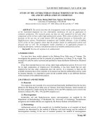
(2) Dosage form and toxicity. In 1971, Wall and his colleagues first reported the
structure of taxol and its cytotoxicity to KB cell line and mouse leukemia cells
(Wani, 1971). It is obvious that paclitaxel has some benzene rings and other
hydrophobic structures (refer to Fig 2.1 below), which lead to its low water
solubility of less than 0.5mg/L. There is no way for direct injection of paclitaxel
by dissolving it in distilled water, the only dosage form available in clinical
administration uses Cremophor EL and dehydrated alcohol as adjuvant, which is
rather toxic and can cause serious side effects such as hypersensitivity reaction,
neurotoxicity, cardiotoxicity and nephrotoxicity (Weiss, 1990; Kongshaug, 1991;
Dorr, 1994; Fjallskog, 1993 ).
Fig 2.1 Chemical structure of paclitaxel
(3) Drug Resistance and bioavailability. It has been found that paclitaxel is able to
induce the multidrug resistance (MDR) phenotype with overexpression of Pglycoprotein (P-gp) (Horwitz, 1992; Roy & Horwitz, 1985; Greenberger et al.,
1987; Drion et al., 1996). P-gp exists in the cell membrane and serves as a kind of
efflux pump that can prevent drugs and other toxic substances from entering cells
7
(Gatmaitan & Arias, 1993). P-gp is widely distributed in many tissues, such as
gastro-intestinal tract, kidney and blood brain barrier. It has already been found
that paclitaxel has a rather high affinity for P-gp transporter. Another problem is
when drugs are administered, especially orally, they have to withstand metabolic
barriers before reaching the blood system. There are a lot of digestive enzymes
throughout the GI tract which can degrade drugs and further result in a low
bioavailability.
(4) Targeted and controlled release. Although paclitaxel has excellent effect on tumor
cells, it can also harm normal cells, especially cells that divide quickly such as the
bone marrow and lining of the GI tract. Many dangerous side effects may be
caused by this kind of non-specific action, such as loss of hair, fussy thinking and
difficult concentrating. Another problem is that in order to achieve therapeutic
effect, drug concentration should be between the therapeutic level, i.e., above the
minimum effective level but below the toxic level. Thus the initial burst should be
lowered to achieve a prolonged and sustained release. Besides, drugs may be cellcycle or cell-growth-phase specific. Thus, cell-cycle specific drugs can be
developed to achieve maximum effect (Ratain et al., 1990). Briefly, the desired
pharmacokinetics is to release a sufficient quantity of drugs at the right time, the
correct location and over a long period of time.
2.2 Brain Cancers and Blood Brain Barrier (BBB)
2.2.1 Brain Cancers and Cancer Treatment
Cancer is a group of diseases characterized by uncontrolled cell division leading to
8
growth of abnormal tissues. Cancer can spread from its original site to other parts of the
body and can be fatal (Web definitions for cancer). Every year, more than 10 million
people are diagnosed with cancer and 6 million people die of cancer, which accounts for
12% of deaths worldwide. Although brain cancers are rare cancers which represent only
1.5% of all cancers, the death rate of brain cancers is very high. Moreover, brain cancer
also ranks second in all childhood cancers, representing 21% of childhood cancer cases
(American Cancer Society). There are basically two kinds of brain tumors. One is
primary brain tumors which start in the brain, the other is metastatic brain tumors which
are cancers from other parts of the body that can spread to the brain and cause secondary
tumor through a process called metastasis. The cells of a metastatic brain tumor resemble
the cells of the organ where the tumor startes, not brain cells.
Like other cancers, effective treatments of brain cancers include surgery, radiotherapy,
chemotherapy, hormone therapy, biotherapy, and immunotherapy (Oncology, 2002). Two
or more methods are often used in combination to achieve better effects. Surgery is the
primary method for treatment of brain tumors that can be removed without damaging
critical neurological functions. Radiation therapy and chemotherapy are often combined
with surgery as secondary and adjuvant treatment. However, severe side effects often
accompany these treatments. One of the most important factors that limit brain cancer
chemotherapy is due to the existence of the blood brain barrier.
2.2.2 Introduction to the Blood Brain Barrier
2.2.2.1 History of Blood Brain Barrier
9
The concept of blood brain barrier was first raised by the German scientist Paul Ehrlich
in 1885(Enrlich, 1885). After that, many studies have been carried out on this important
physiologic barrier. Table 2.1 gives a brief summary of the research history on BBB.
Discoverer
Ehrlich P
Table 2.1 Summary of BBB History
Time
Main Point
1885
i.v. injection of acidic vital dyes stain all rabbits body
except brain and spinal cord (Enrlich, 1885)
Lewandowsky
1900
Goldmann EE
1909
Gautier& Stern
1920s
Broman
1941
Reese& Karnovsky 1967
Reese et al.
1970
Weiler-Guttler
1989
Muldoon LL
1999
coin the term blood-brain barrier while studying potassium
ferrocyannide penetration into the brain (BBB history)
i.v. injection of trypan blue to cerebrospinal fluid stains
entire brain but not the internal organs(Goldmann, 1909)
bile salt, morphine and bromide appear in CSF while bile
pigment, epinephrine and curare not after i.v. injection
tight junction not the astrocytic end forms barrier function
of BBB (BBB history)
visulize BBB using electron microscope & traceable
proteins, revealing the protein diffuse past astrocytic
end feet and stop at tight junction (Reese & karnovsky,
1967; Reese et al., 1970)
characteristics of BBB; studies in molecular biology of
BBB, cloning and sequencing glucose transporter gene
(weiler-Guttler et al., 1989)
BBB is a physiologic barrier (Muldoon et al., 1999)
2.2.2.2 Structure and Function of the Blood Brain Barrier
The blood brain barrier is created by tight apposition of endothelial cells lining blood
vessels in the brain and is surrounded by astrocyte foot process. A thin basement
membrane surrounds the endothelial cells and associated pericytes, providing mechanical
support as well as a barrier function. The part in the circle in Fig 2.2 is the BBB site.
There are quite a few important differences between the ultrastructure of brain blood
vessels and systemic blood vessels. The brain capillaries lack fenestration that exists in
10
other systemic capillaries, instead, the membrane of the endothelial cells in the brain is
fused into tight junctions, forming continuous, uninterrupted structures. These endothelial
tight junctions are the anatomical site of BBB and play an important role in preventing
the free exchange of substances between blood and brain (Brightman & Reese, 1969;
Reese et al., 1970).The tight junctions result in a much higher transendothelial electrical
resistance than other tissues (>50 times), which makes the BBB more hydrophobic and
reduces the aqueous based paracellular transport (Lo et al., 2001). BBB also possesses
specific enzyme systems, glucose transporters and protein receptors, which indicates its
special mechanisms in exchanging substances.
Moreover, blood brain barrier is
incorporated with many efflux proteins such as P-glycoprotein (P-gp), multidrug
resistance protein (MRP). These proteins are responsible for ATP-dependent outward
transport of a wide range of substances, including many therapeutic agents (Crone, 1971).
The major function of the blood brain barrier is to protect the brain from possible toxins
while supply it with necessary nutrients. Thus it acts both as an impermeable wall and a
selective sieve (Betz, 1992). Due to its special structures mentioned above, blood brain
barrier effectively filters most ionized, water-soluble molecules greater than 180 Daltons
and substances that are substrates of its efflux system. Only small, lipophilic molecules
can cross the BBB (Lee,2001;Kroll& Neuwelt, 1998). Several mechanisms are known to
be involved in the transport of substances across BBB. Only a few substances such as
water can enter the brain using the paracellular route because of the existence of the tight
junction. Lipophilic molecules can cross BBB by simple diffusion through transcellular
pathway. Lipophilicity and hydrogen bonding potential determine the ability of molecules
to cross BBB (Egleton& Davis, 1997). However, a large family of lipid soluble
11