Macleods clinical diagnosis 2013
Bạn đang xem bản rút gọn của tài liệu. Xem và tải ngay bản đầy đủ của tài liệu tại đây (8.79 MB, 291 trang )
Macleod’s
Clinical
Diagnosis
For Elsevier
Commissioning Editor: Laurence Hunter
Development Editor: Helen Leng
Project Manager: Caroline Jones
Designer/Design Direction: Miles Hitchen
Illustration Manager: Jennifer Rose
Illustrator: Antbits
Macleod’s
Alan G Japp MBChB(Hons) BSc(Hons) MRCP
Cardiology Registrar
Royal Inirmary of Edinburgh
Edinburgh, UK
Colin Robertson
BA(Hons) MBChB FRCPEd FRCSEd FSAScot
Honorary Professor of Accident and Emergency Medicine
University of Edinburgh
Edinburgh, UK
Associate Editor
Iain Hennessey MBChB(Hons) BSc(Hons) MRCS
Speciality Trainee in Paediatric Surgery
Alder Hey Children’s Hospital
Liverpool, UK
Edinburgh London New York Oxford Philadelphia St Louis Sydney Toronto
Clinical
Diagnosis
Edited by
© 2013 Elsevier Ltd. All rights reserved.
No part of this publication may be reproduced or transmitted in any form or by
any means, electronic or mechanical, including photocopying, recording, or any
information storage and retrieval system, without permission in writing from
the publisher. Details on how to seek permission, further information about the
publisher’s permissions policies and our arrangements with organizations such
as the Copyright Clearance Center and the Copyright Licensing Agency, can be
found at our website: www.elsevier.com/permissions.
This book and the individual contributions contained in it are protected
under copyright by the publisher (other than as may be noted herein).
First published 2013
ISBN 9780702035432
International ISBN 9780702035449
eBook ISBN 9780702051227
British Library Cataloguing in Publication Data
A catalogue record for this book is available from the British Library
Library of Congress Cataloging in Publication Data
A catalog record for this book is available from the Library of Congress
Notices
Knowledge and best practice in this ield are constantly changing. As new
research and experience broaden our understanding, changes in research
methods, professional practices, or medical treatment may become necessary.
Practitioners and researchers must always rely on their own experience and
knowledge in evaluating and using any information, methods, compounds, or
experiments described herein. In using such information or methods they
should be mindful of their own safety and the safety of others, including parties
for whom they have a professional responsibility.
With respect to any drug or pharmaceutical products identiied, readers are
advised to check the most current information provided (i) on procedures
featured or (ii) by the manufacturer of each product to be administered, to
verify the recommended dose or formula, the method and duration of
administration, and contraindications. It is the responsibility of practitioners,
relying on their own experience and knowledge of their patients, to make
diagnoses, to determine dosages and the best treatment for each individual
patient, and to take all appropriate safety precautions.
To the fullest extent of the law, neither the publisher nor the authors,
contributors, or editors, assume any liability for any injury and/or damage to
persons or property as a matter of products liability, negligence or otherwise,
or from any use or operation of any methods, products, instructions, or ideas
contained in the material herein.
Working together to grow
libraries in developing countries
www.elsevier.com | www.bookaid.org | www.sabre.org
Printed in China
The
publisher’s
policy is to use
paper manufactured
from sustainable forests
Preface
‘Ninety per cent of diagnoses are made from the
history.’
‘Clinical examination is the cornerstone of
assessment.’
These, or similar platitudes, will be familiar
to most students in clinical training. Many,
however, will have noticed a glaring ‘disconnect’ between the importance ascribed
to basic clinical skills during teaching and
the apparent reliance on sophisticated
investigations in the parallel world of clinical practice. Modern diagnostics have radically altered the face of medical practice;
clinical training is still catching up. We
recognise that both teachers and textbooks
frequently fall into the trap of eulogising the
clinical assessment rather than explaining
its actual role in contemporary diagnosis.
Yet we come to praise the clinical assessment not to bury it. The history may not, by
itself, deliver the diagnosis in 90% of cases
but it is essential in all cases to generate a
logical differential diagnosis and to guide
rational investigation and treatment. Some
physical signs are now vanishingly rare and
certain aspects of the clinical examination
have been marginalised by the emergence
of novel imaging techniques and disease
biomarkers. However, a focused clinical
examination is critical to recognising the
sick patient, raising red lags, identifying
unsuspected problems and, in some cases,
revealing signs that cannot be identiied
with tests (for example, the mental state
examination).
Our aim is to show you how to use your
core clinical skills to maximum advantage.
We offer a grounded and realistic approach
to clinical diagnosis with no bias towards
any particular element of the assessment.
Where appropriate, we acknowledge the
limitations of the history and examination
and direct you to the necessary investigation. However, we also highlight those
instances where diagnosis is critically
dependent on basic clinical assessment,
thereby demonstrating its vital and enduring importance. We wish you every success
in your training and practice, and hope that
this book provides at least some small
measure of assistance.
AJ
CR
v
Acknowledgements
On behalf of the editors and authors, I
would like to thank Laurence Hunter for his
trust and support; Helen Leng for her
careful scrutiny and remarkable tolerance;
Caroline Jones and Wendy Lee for their
hard work and support; and everyone else
who has volunteered ideas, comments,
assistance or a friendly ear.
On a more personal note, I would like
to acknowledge the teaching of Mike Ford
as a major inspiration for this book and
to thank Deepa Japp for her encouragement, optimism and extraordinary patience
throughout the long months of writing and
editing.
AJ
Figure sources
Figs 1.1, 2.1A&B, 4.2, 4.3, 6.1, 12.4, 12.5, 12.6,
17.1, 18.1, 22.2, 27.2, 29.2: Douglas G,
Nicol F, Robertson C. Macleod’s Clinical
Examination, 12th edn. Edinburgh:
Churchill Livingstone, 2009.
Figs 2.2A–C, 4.1, 5.1, 11.1, 12.2, 19.1, 22.1,
23.1, 23.2, 23.3, 23.4: Ford MJ, Hennessey I,
Japp A. Introduction to Clinical Examination,
8th edn. Edinburgh: Churchill Livingstone,
2005.
Fig. 4.5A&B: Begg JD. Abdominal X-rays
Made Easy, 2nd edn. Edinburgh: Churchill
Livingstone, 2006.
vi
Figs 6.10, 6.12, 25.1: Hampton JR. 150 ECG
Problems, 3rd edn. Edinburgh: Churchill
Livingstone, 2008.
Figs 10.1, 12.3, 17.3, 25.6, 29.1, 29.4:
Boon NA, Colledge NR, Walker BR.
Davidson’s Principles & Practice of Medicine,
20th edn. Edinburgh: Churchill Livingstone,
2006.
Figs 12.1, 12.8, 17.2, 17.4: Corne J,
Pointon K. Chest X-ray Made Easy,
3rd edn. Edinburgh: Churchill Livingstone,
2010.
Figs 6.2, 6.7, 6.11, 25.3, 25.4, 25.5, 29.3, 29.5:
Hampton JR. The ECG in Practice, 5th edn.
Edinburgh: Churchill Livingstone, 2008.
Figs 26.1, 26.2, 26.4, 26.6, 26.7, 26.11, 26.12,
26.13, 26.14, 26.15, 26.17: Gawkrodger DJ.
Dermatology ICT, 4th edn. Edinburgh:
Churchill Livingstone, 2008.
Fig. 6.3: Grubb NR, Newby DE. Churchill’s
Pocketbooks
Cardiology,
2nd
edn.
Edinburgh: Churchill Livingstone, 2006.
Fig. 26.3: Kumar P, Clark M. Kumar and
Clark Clinical Medicine, 7th edn. Edinburgh:
Churchill Livingstone, 2009.
Figs 6.6, 6.8, 25.2: Hampton JR. The ECG
Made Easy, 7th edn. Edinburgh: Churchill
Livingstone, 2008.
Figs 26.5, 26.8, 26.9, 26.10: Bolognia J,
Jorizzo J, Rapini R. Dermatology, 1st edn.
London: Mosby, 2003.
Contributors
R Benjamin Aldridge
Colin Mitchell
MBChB MSc MRCP MRCS
MBChB MRCP
Clinical Research Fellow in Dermatology,
University of Edinburgh; Specialty
Registrar in Plastic and Reconstructive
Surgery, Canniesburn Unit, Glasgow
Royal Inirmary, UK
Consultant Geriatrician, St Mary’s
Hospital, London, UK
Roland C Aldridge
Jonathan C L Rodrigues
BSc(Hons) MBChB(Hons) MRCP(UK)
Specialist Registrar in Clinical Radiology,
Severn School of Radiology, Bristol, UK
MSc MRCS MRCP
Clinical Lecturer, University of Edinburgh;
Honorary Specialty Registrar in General
Surgery, South East Scotland, Edinburgh,
UK
J Kenneth Baillie
Lynn M Urquhart
MBChB, MRCP, DTMH
Specialty Registrar in Infectious Diseases
and Clinical Research Fellow Medical
Education, Ninewells Hospital and
Medical School, Dundee, UK
BSc(Hons) MRCP FRCA
Clinical Lecturer, Department of Critical
Care Medicine, University of Edinburgh,
Edinburgh, UK
Xavier L Grifin
Hugh B Waterson
MRCSEd
Specialist Registrar Trainee in
Orthopaedics, Royal Inirmary of
Edinburgh, Edinburgh, UK
MA MSc MRCS
Specialist Registrar in Trauma and
Orthopaedic Surgery, Warwick Medical
School, University of Warwick, Warwick,
UK
vii
This page intentionally left blank
Contents
Abbreviations . . . . . . . . . . . . . . . . . . . . . . . . . . . . . . . . . . . . . . . . . . . . . .xi
Part 1 Principles of clinical assessment
1 What’s in a diagnosis? . . . . . . . . . . . . . . . . . . . . . . . . . . . . . . . 3
2 Assessing patients: a practical guide . . . . . . . . . . . . . . . . . . . . . . . . 7
3 The diagnostic process . . . . . . . . . . . . . . . . . . . . . . . . . . . . . 19
Part 2 Assessment of common presenting problems
4 Abdominal pain . . . . . . . . . . . . . . . . . . . . . . . . . . . . . . . . . 26
5 Breast lump . . . . . . . . . . . . . . . . . . . . . . . . . . . . . . . . . . . 44
6 Chest pain . . . . . . . . . . . . . . . . . . . . . . . . . . . . . . . . . . . . 48
7 Coma and altered consciousness . . . . . . . . . . . . . . . . . . . . . . . . 70
8 Confusion: delirium and dementia . . . . . . . . . . . . . . . . . . . . . . . . 76
9 Diarrhoea . . . . . . . . . . . . . . . . . . . . . . . . . . . . . . . . . . . . 88
10 Dizziness . . . . . . . . . . . . . . . . . . . . . . . . . . . . . . . . . . . . 94
11 Dysphagia . . . . . . . . . . . . . . . . . . . . . . . . . . . . . . . . . . . .106
12 Dyspnoea . . . . . . . . . . . . . . . . . . . . . . . . . . . . . . . . . . . .110
13 Fatigue. . . . . . . . . . . . . . . . . . . . . . . . . . . . . . . . . . . . . .128
14 Fever. . . . . . . . . . . . . . . . . . . . . . . . . . . . . . . . . . . . . . .136
15 Gastrointestinal haemorrhage: haematemesis and rectal bleeding . . . . . . . .148
16 Haematuria . . . . . . . . . . . . . . . . . . . . . . . . . . . . . . . . . . .156
17 Haemoptysis. . . . . . . . . . . . . . . . . . . . . . . . . . . . . . . . . . .160
18 Headache . . . . . . . . . . . . . . . . . . . . . . . . . . . . . . . . . . . .164
19 Jaundice . . . . . . . . . . . . . . . . . . . . . . . . . . . . . . . . . . . . .172
20 Joint swelling . . . . . . . . . . . . . . . . . . . . . . . . . . . . . . . . . .182
21 Leg swelling . . . . . . . . . . . . . . . . . . . . . . . . . . . . . . . . . . .186
22 Limb weakness . . . . . . . . . . . . . . . . . . . . . . . . . . . . . . . . .196
23 Low back pain . . . . . . . . . . . . . . . . . . . . . . . . . . . . . . . . . .210
24 Mobility problems: falls and ‘off legs’ . . . . . . . . . . . . . . . . . . . . . .216
25 Palpitation . . . . . . . . . . . . . . . . . . . . . . . . . . . . . . . . . . . .224
26 Rash: acute generalised skin eruption . . . . . . . . . . . . . . . . . . . . . .232
27 Scrotal swelling . . . . . . . . . . . . . . . . . . . . . . . . . . . . . . . . .242
28 Shock . . . . . . . . . . . . . . . . . . . . . . . . . . . . . . . . . . . . . .246
29 Transient loss of consciousness: syncope and seizures . . . . . . . . . . . . .252
Appendix . . . . . . . . . . . . . . . . . . . . . . . . . . . . . . . . . . . . . . . . . . . . . . 264
Index . . . . . . . . . . . . . . . . . . . . . . . . . . . . . . . . . . . . . . . . . . . . . . . . 267
ix
This page intentionally left blank
Abbreviations
Abbreviations that do not appear in this list are spelled out in the main text.
ABG
ACE
ACPA
AIDS
arterial blood gases
angiotensin-converting enzyme
anti-citrullinated protein antibodies
acquired immunodeiciency
syndrome
ALP
alkaline phosphatase
ALT
alanine aminotransferase
ANA
antinuclear antibody
ANCA
antineutrophil cytoplasmic
antibodies
APTT
activated partial thromboplastin time
ASMA
anti-smooth muscle antibodies
ASO
anti-streptolysin O
AST
aspartate aminotransferase
BM
blood glucose meter reading
BMI
body mass index
BP
blood pressure
bpm
beats per minute
BS
breath sounds
CK
creatine kinase
CNS
central nervous system
CPET
cardiopulmonary exercise test
CRP
C-reactive protein
CRT
capillary reill time
CSF
cerebrospinal luid
CSU
catheter specimen of urine
CT
computed tomogram/tomography
CTPA
computed tomographic pulmonary
angiography
CVP
central venous pressure
CXR
chest X-ray
DC
direct current
DMARDs disease-modifying anti-rheumatic
drugs
ECG
electrocardiogram/
electrocardiography
EEG
electroencephalogram/
electroencephalography
ENA
extractable nuclear antigen
ENT
ear, nose and throat
ERCP
endoscopic retrograde
cholangiopancreatography
ESR
erythrocyte sedimentation rate
FBC
full blood count
GCS
Glasgow Coma Scale (score)
GFR
glomerular iltration rate
GGT
gamma-glutamyl transferase
GI
gastrointestinal
GP
Hb
HIV
HR
ICP
ID
IM
INR
IV
IVU
JVP
LDH
LFTs
LIF
LKM
LLQ
LP
LUQ
MRA
MRCP
MRI
MSU
NSAID
PCR
PEFR
PFTs
PR
PRN
PSA
PT
PV
RF
RIF
RLQ
RR
RUQ
SC
SIRS
SSRI
TFTs
TNF
U+E
UGIE
USS
WBC
general practitioner
haemoglobin
human immunodeiciency virus
heart rate
intracranial pressure
infectious disease(s)
intramuscular(ly)
International Normalised Ratio
intravenous(ly)
intravenous urogram/urography
jugular venous pulse
lactate dehydrogenase
liver function tests
left iliac fossa
liver kidney microsomal (antibodies)
left lower quadrant
lumbar puncture
left upper quadrant
magnetic resonance angiography
magnetic resonance
cholangiopancreatography
magnetic resonance imaging
midstream urine (specimen)
non-steroidal anti-inlammatory drug
polymerase chain reaction
peak expiratory low rate
pulmonary function tests
per rectum
pro re nata; whenever required
prostate-speciic antigen
prothrombin time
per vaginam
rheumatoid factor
right iliac fossa
right lower quadrant
respiratory rate
right upper quadrant
subcutaneous(ly)
systemic inlammatory response
syndrome
selective serotonin re-uptake
inhibitor
thyroid function tests
tumour necrosis factor
urea and electrolytes
upper gastrointestinal endoscopy
ultrasound
white blood count
xi
This page intentionally left blank
PART
1
Principles of clinical
assessment
What’s in a diagnosis?...........................................3
Assessing patients: a practical guide ....................7
The diagnostic process........................................19
This page intentionally left blank
WHAT’S IN A DIAGNOSIS?
From differential diagnosis
to inal diagnosis
A diagnosis is simply shorthand for a
patient’s condition or disease process. The
ability to diagnose accurately is fundamental to clinical practice. Only with a correct
diagnosis, or a short-list of possible diagnoses, can you:
• formulate an appropriate sequence of
investigations
• begin correct treatment and assess its
effectiveness
• give an informed prognosis and make
follow-up arrangements.
In most cases, the construction of a differential diagnosis is a stepping-stone to the
inal diagnosis. This is a list of diagnoses,
usually placed in order of likelihood, which
may be causing the presentation. This list
may be lengthy at the outset of assessment
but will become progressively shorter as
you accumulate information about the
patient’s condition through your historytaking, examination and investigations.
When one diagnosis begins to stand out
from the rest as the most likely cause of the
patient’s presentation, it is often referred to
as the working diagnosis. Investigations are
then directed toward conirming (or refuting) this condition and thereby arriving at
a final diagnosis. This entire process may
happen over a very short period of time; for
example, establishing a inal diagnosis of
acute ST elevation myocardial infarction in
a patient presenting with acute chest pain
should usually take less than 10 minutes.
Frequently, the identiication of an abnormality may only be the irst step in the diagnostic process and additional assessment
is required to characterise a condition in
greater detail or search for an underlying
1
cause. For example, in a middle-aged man
presenting with fatigue, you might identify
anaemia as the cause of his symptoms, but
the diagnostic process would not stop there.
The next step would be to establish the
cause of the anaemia. If subsequent laboratory investigations revealed evidence of
iron deiciency, you would need to determine the cause of this. Gastrointestinal
investigations might uncover a gastric
tumour but, even then, further assessment
would still be required to establish a
tissue diagnosis and stage the tumour. The
eventual ‘inal diagnosis’ might be: irondeiciency anaemia secondary to blood loss
from a T3, N1, M1 gastric carcinoma with
metastasis to liver and peritoneum. Clearly,
the diagnosis of ‘anaemia’ would have been
grossly inadequate!
Some conditions, especially functional
disorders such as irritable bowel syndrome,
lack a deinitive conirmatory test; here
diagnosis relies upon recognising characteristic clinical features and ruling out
alternative diagnoses – especially serious
or life-threatening conditions. Such disorders are often referred to as diagnoses
of exclusion.
Probability and risk
As almost all diagnostic tests are inherently
imperfect, diagnoses should be regarded as
statements of probability rather than hard
facts. In practice, a disease is ‘ruled in’
when the probability of it being present is
deemed to be suficiently high, and ‘ruled
out’ when the probability is suficiently
low. The degree of certainty required will
depend on factors such as the consequences
of missing the particular diagnosis, the
side-effects of treatment and the risks of
further testing. Doctors must not become so
‘paralysed’ by the implications of missing a
3
1
WHAT’S IN A DIAGNOSIS?
diagnosis that they admit the patient unnecessarily to hospital and/or investigate to
levels that are not in the patient’s best interests and are unacceptable because of time,
expense and intrinsic risk, e.g. radiation
exposure. On the other hand, a high threshold of certainty, i.e. very low probability,
is required to exclude potentially lifethreatening conditions. In general, if the
situation is explained appropriately, most
patients will accept tests that will yield a
diagnostic accuracy of less than 1% for
acute life-threatening conditions.
The current diagnostic approach to subarachnoid haemorrhage (SAH) illustrates
this. For a middle-aged patient who
presents, fully conscious, with a history of
sudden (within a few seconds) onset of ‘the
worst headache ever’, the chances of a diagnosis of SAH are approximately 10–12%.
The presence of some clinical indings, e.g.
photophobia, neck stiffness, cranial nerve
palsies, subhyaloid haemorrhage – will
increase these chances markedly but these
features may take time to develop. Even
if clinical examination is unequivocally
normal, the chances of SAH are 8–10%. Currently, there is no simple bedside test for
SAH and the initial investigation is normally a non-contrast CT scan. A positive
scan will prompt appropriate treatment,
possibly involving neurosurgical or neuroradiological intervention. A negative scan
does not, however, exclude an SAH. The
accuracy of CT scanning in detecting SAH
depends upon the experience of the reporting individual, the nature of the scanner
(principally, its resolution) and the time
interval between the onset of symptoms
and the scan (accuracy falls with time). A
scan performed within 12 hours by most
modern scanners has a diagnostic accuracy
of approximately 98%. But, given the morbidity and mortality of unrecognised and
untreated SAH, even this level of diagnostic
accuracy is inadequate. For this reason,
patients with a negative CT scan have a
lumbar puncture. The CSF obtained must
be examined by spectrophotometry in the
laboratory for xanthochromia (direct visual
inspection of the luid is insuficiently
4
accurate). Xanthochromia (produced from
haemoglobin breakdown within the CSF)
takes some time to develop and the sensitivity of this test peaks at about 12 hours
after symptom onset. The combination
of a negative CT scan performed within 12
hours of symptom onset and normal CSF
indings at 12 hours reduces the chances
of the patient’s symptoms being caused
by an SAH to well below 1% – a level of
probability acceptable to most clinicians
and, if appropriately explained, to their
patient.
Special situations
Medically unexplained symptoms
Sometimes it is dificult to correlate patients’
symptoms with a speciic disease. This does
not mean that the symptoms with which
they present are made up or that patients
are malingering – merely that we are not
able to provide a physical cause for the
symptoms. For patients in primary care,
over 70% have symptoms that cannot be
readily explained by a speciic diagnosis.
Nevertheless, the symptoms are very real to
the patient and one of the major challenges,
intellectually and practically, in primary
care is to recognise which patients have
physical disease.
Clusters of symptoms in recognisable
patterns, in the absence of physical and
investigational abnormalities, are called
functional syndromes (Table 1.1).
In general, the greater the number
of symptoms, the greater the likelihood
that there is a psychological component to
the presentation. Remember that patients
with chronic disease are more likely to
demonstrate psychological aspects of their
condition, especially depression, which
may, in turn, affect the mode of presentation. Avoiding excessive and inappropriate
investigation to ‘exclude’ diagnoses is
important, especially if patients have no
speciic ‘red lags’ in relation to their history,
they are not in a recognised at-risk group,
and there are no abnormalities on clinical
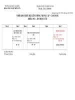
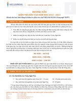
examination and simple bedside tests (Fig.
1.1 and Box 1.1).
WHAT’S IN A DIAGNOSIS?
Treatment before diagnosis
Table 1.1 Common functional syndromes1
Syndrome
Symptoms
Chronic fatigue
syndrome
Irritable bowel
syndrome
Persistent fatigue2
Sometimes, accurate diagnosis depends
upon the patient’s response to treatment.
In a few speciic situations this may be
life-saving as well as diagnostic. In any
patient with altered consciousness or acute
neurological dysfunction without a clearly
identiiable cause, two conditions need to
be excluded and treated immediately.
Abdominal pain, altered
bowel habit (diarrhoea or
constipation) and abdominal
bloating
Persistent pain in one or
more parts of the body,
sometimes following injury
but which outlasts the
original trauma2
Pain in the axial skeleton
with trigger points (tender
areas in the muscles)2
Pain, muscle tension or
stiffness localised below
the costal margin and
above the inferior gluteal
folds, with or without leg
pain2
Chronic pain
syndrome
Fibromyalgia
Chronic back
pain
1
Box 1.1 Symptoms and their relationship to
physical disease
More commonly
• Chest pain
• Breathlessness
• Syncope
• Abdominal pain
Less commonly
• Fatigue
• Back pain
• Headache
1
In all cases, physical examination and investigation fail to reveal an
underlying physical cause.
2
Symptoms must have lasted more than 3 months.
• Dizziness
10
Organic cause
3-year incidence (%)
8
6
4
2
s
es
Nu
m
bn
in
a
pa
ni
a
Ab
do
m
in
al
m
oe
so
In
in
pa
Dy
s
ck
pn
a
em
Ba
he
O
ed
s
ac
es
ad
He
in
tig
zz
Di
Fa
Ch
e
st
pa
in
ue
0
Fig. 1.1 Common symptoms and disease. From Douglas JG, Nicol F, Robertson C. Macleod’s Clinical
Examination 12E Edinburgh: Churchill Livingstone; 2009.
5
1
WHAT’S IN A DIAGNOSIS?
Hypoglycaemia can mimic conditions such
as epilepsy and hemiplegia. Check the
blood glucose level using a Stix test on a
inger-prick sample. If the value is low, take
a formal blood sample for laboratory blood
glucose determination, but do not wait
for this result before giving treatment –
give glucose or glucagon immediately. If
hypoglycaemia is the cause of the patient’s
symptoms, response will normally occur
within 5–10 minutes (rarely, in cases where
there has been severe, prolonged hypoglycaemia, this may take longer and residual
neurological deicit may persist).
Opioid intoxication is usually associated
with altered consciousness, reduced respiratory rate and depth, and small pupils. The
diagnosis is rarely dificult in younger
patients with a history or other features
of illicit drug use. However, these features
are not always present, and chronic opioid
toxicity may develop over hours/
days, particularly in elderly patients or
those with renal impairment. Naloxone
is a highly speciic opioid antagonist with
no agonist activity. Give 0.8 mg naloxone
(SC, IM or IV) immediately. If there is any
response, further doses of naloxone will
be required until no further reversal is
achieved. Remember that the half-life of
naloxone is much shorter than that of the
opioid that has been taken, so repeated stat.
doses or an infusion are likely to be needed.
There is one other situation where treatment is necessary before, or to achieve,
diagnosis. It is unnecessary, unhelpful and
inhumane to leave a patient in pain from
whatever cause. Put yourself in the patient’s
place. There is never any indication to
withhold analgesia from a patient in pain.
Concerns that you will ‘mask’ clinical signs
e.g. by giving opioids to a patient with an
‘acute’ abdomen, encourage opioid tolerance or addiction, and impair informed
consent are completely unfounded. In fact,
diagnostic accuracy is improved by making
the patient more cooperative, aiding the
performance of investigations such as
ultrasound, and pain relief brings additional beneits in reducing catecholamine
stimulation, improving respiratory and
6
cardiovascular function. The ‘pain ladder’
approach is useful, but for patients in acute
or severe pain, IV opioids titrated to the
clinical response are usually needed.
The patient who comes with
a diagnosis
Many patients have an idea of their own condition and, indeed, may begin the consultation by telling you their perceived diagnosis.
In part this relates to improved education,
greater exposure to medical conditions
through the media and the Internet. A
patient who has previously had a condition
that has recurred e.g. asthma or urinary tract
infection, or who has a lare-up of a chronic
condition e.g. inlammatory bowel disease,
will often present in this way. Remember
that many patients will be worrying about a
speciic diagnosis causing their presenting
complaint. This is particularly the case for
breast lumps, rectal bleeding and chronic
headache, where the perception may be that
the only possible diagnosis is cancer.
Self-diagnosis may also cause a delay in
seeking medical help because the patient
does not appreciate the signiicance of a
symptom or subconsciously may not want
to consider the possibility of serious disease.
Common examples include attributing
ischaemic chest pain to ‘indigestion’ and
assuming that rectal bleeding is due to
haemorrhoids.
One way of initially handling patients
who come with a diagnosis is to let them
express this openly and then to acknowledge their concerns. You must respect
these (indeed, the patient may well be right)
while taking care not to miss a more likely
diagnosis. In particular, do not take shortcuts with any of the components of history
taking, examination and investigation that
may be required.
Patients with rare or unusual diseases
often know much more about their condition than you. Use this golden opportunity.
There is no loss of face in admitting your
ignorance. Patients will respect you for
your honesty and you can learn much from
them about the disease and its treatment
and effects.
ASSESSING PATIENTS: A PRACTICAL GUIDE
Introduction
Before you can diagnose patients you must
irst obtain the necessary clinical and investigative information. Diagnostic success
depends upon the accuracy and completeness of this initial data gathering so your
history-taking and examination skills are
crucial. During clinical training, you may
have been taught a fairly idealised and rigid
‘method’ of patient assessment. However,
in everyday practice, a more lexible, luid
approach is preferable; this will allow you
to adapt to the clinical situation and acquire
the essential information in the most eficient manner.
Traditionally, the assessment of patients
has been divided into two distinct phases:
• the clinical assessment: history and
physical examination
• diagnostic investigations.
At least in the hospital setting, this distinction is highly artiicial due to the easy
and real-time availability of basic tests such
as ECG, CXR, glucose meter reading and
ABG, and routine laboratory blood tests
such as FBC, U+E and LFTs. Wherever
appropriate, these simple tests should be
carried out in tandem with the clinical
assessment to form a ‘routine patient workup’. The information from all of these
sources is combined to form a working or
differential diagnosis. Where necessary,
further targeted investigation can then be
undertaken to conirm the suspected diagnosis, narrow the differential diagnosis e.g.
exclude high-risk conditions, inform prognosis and guide management.
Thus, in this book, we advocate the following system for patient evaluation:
2
• the routine work-up: history, physical
examination and basic tests
• targeted, supplementary investigations.
The optimal approach to the routine workup varies, depending on the stability and
illness severity of the patient:
• Acutely unwell patients require a
rapid, targeted evaluation (‘ABCDE
assessment’) for life-threatening
disorders and major derangements of
physiology.
• Patients who are stable, including
those initially assessed by the ABCDE
approach, should have a full history and
clinical examination, as outlined below
(‘full clinical assessment’), alongside basic
tests relevant to the speciic presentation.
• The approach to frail, elderly patients
may need to be modiied to take account
of differences in the nature of illness
presentation, e.g. multi- versus singleorgan pathology; signiicant functional
decline secondary to minor illness
(p. 15).
Rapid assessment of
the sick patient
The ABCDE assessment (see Clinical Tool:
the ABCDE assessment) combines prompt
identiication of life-threatening pathology
with immediate management of any abnormalities detected, prioritising those that
are most rapidly fatal. Take an ABCDE
approach if the patient:
• appears unwell or is unresponsive
• exhibits evidence of acute physiological
derangement on basic observations (HR,
RR, BP, SpO2, temperature)
• has features of a serious acute problem
in any organ system.
7
2
ASSESSING PATIENTS: A PRACTICAL GUIDE
Clinical tool: The ABCDE assessment
What to look for
A. AIRWAY
A1 Ask: ‘How are you feeling?’
If patient can speak normally, airway is
patent – move straight to B.
A2 Assess for airway obstruction
• Lack of airlow at the mouth (complete
obstruction)
• Throat or tongue swelling
• Gurgling, snoring, choking, stridor
• Parodoxical, ‘see-saw’ breathing
(indrawing of chest with expansion of
abdomen on inspiration; vice-versa on
expiration)
Only move to B once airway patent
B. BREATHING
B1 Give high concentration O2
B2 Assess rate, depth and symmetry
of breathing, noting:
• poor respiratory effort: ↓↓ RR, feeble,
shallow breaths
• high respiratory effort: RR > 20/min, use
of accessory muscles, visibly tiring
• asymmetrical chest expansion
B3 Check for tracheal deviation
B4 Percuss and auscultate chest, noting:
• Hyperresonance – suggesting
pneumothorax
• Dullness – suggesting pleural effusion,
collapse, consolidation
• ↓ breath sounds – suggesting collapse
pneumothorax, pleural effusion,
• Wheeze – suggesting bronchospasm
• Crackles – suggesting pulmonary
oedema, ibrosis, consolidation
• Bronchial breathing suggesting
consolidation
B5 Record SpO2 on high FiO2
8
How to respond
If signs of obstruction:
• Get help!
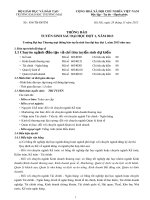
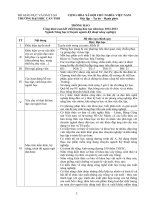
• Try simple airway manoeuvres: head tilt/chin
lift; jaw thrust (Fig. 2.1)
• Remove foreign bodies/secretions from
pharynx under direct vision
• Insert Guedel or nasopharygeal airway
If obstruction persists:
• Get expert assistance immediately
• Consider laryngeal mask airway or
endotracheal intubation
• If throat or tongue swelling, give IM adrenaline
(0.5 mg).
If respiratory effort inadequate:
• Get help!
• Manually ventilate via bag-valve-mask
• Consider a trial of naloxone.
If severe respiratory distress and signs of tension
pneumothorax (p 246), perform immediate
needle aspiration.
If widespread wheeze, check for signs of
anaphylaxis (see below). If present, manage
as described; otherwise, give nebulised
bronchodilator.
If chronic type 2 respiratory failure or severe
COPD, titrate FiO2 to patient’s baseline SpO2
(if known) or 90-92%. In all other critically ill
patients, continue high FiO2.
ASSESSING PATIENTS: A PRACTICAL GUIDE
2
Clinical tool: The ABCDE assessment—cont’d
What to look for
C. CIRCULATION
C1 Check colour & temperature of hands
• Cold, clammy, blue, mottled?
• Pink and warm?
C2 Measure capillary reill time (CRT)
Press irmly over ingertip for 5s, release
pressure then record time taken for skin to
return to normal colour
• ≤2 s is normal
• ≥2 s suggests ↓peripheral perfusion
C3 Palpate radial and carotid pulse:
• tachycardia: >100 bpm
• bradycardia: <60 bpm (or inappropriately
slow for context)
• Thready, weak – suggesting ↓cardiac
output, e.g. hypovolaemia
• Bounding – suggesting hyperdynamic
circulation, e.g. early sepsis
C4 Measure blood pressure
C5 Assess height of JVP at 45°
C6 Auscultate the heart for:
• Murmurs
• 3rd heart sound/gallop rhythm
C7 Attach ECG monitor and review
rhythm:
• Regular broad complex tachycardia
– likely VT (p. 227)
• Regular narrow complex tachycardia,
e.g. sinus tachy, SVT, A lutter (p. 225)
• Irregular tachycardia – likely atrial
ibrillation (p. 226)
• Bradycardia ≤40 bpm, e.g. 2nd or 3rd
degree AV block (p. 261)
C8 Perform a 12-lead ECG if chest pain
(p. 52) or arrhythmia
D. DISABILITY
D1 Check blood glucose meter reading
(‘BM’)
D2 Record Glasgow Coma Scale (p. 71)
How to respond
In patients with evidence of shock, e.g. ↑CRT,
cold peripheries, thready pulse, ↑HR, ↓BP:
Secure IV access (large bore if possible)
If VT, attempt deibrillation with a synchronised
DC shock (ask anaesthetist to sedate if
conscious)
If bradycardia:
• give atropine 0.5–3 mg
• if no response or HR < 40/min, get expert
help and consider IV adrenaline or
transcutaneous pacing
If tongue/throat swelling, severe respiratory
distress, widespread wheeze and/or a new
rash, assume anaphylaxis:
• Stop any potential trigger
• Give 0.5 mg IM adrenaline (anterolateral
aspect of middle 1/3 of the thigh)
• Give fast IV luids
• Get immediate anaesthetic help
Otherwise, give luid challenge unless evidence
of pulmonary oedema
If BM < 3 mmol/L:
• Send blood for formal lab glucose measurement
• Give immediate IV dextrose
If ↓GCS
• Perform an ABG if any suspicion of
hypercapnia, e.g. chronic lung disease,
depressed ventilation
• Give 0.8–2 mg IV naloxone if ↓pupil size or no
obvious cause
• Assess response after 1 minute and consider
further doses if partial response
9
2
ASSESSING PATIENTS: A PRACTICAL GUIDE
Clinical tool: The ABCDE assessment—cont’d
What to look for
How to respond
D3 Take a ‘3D’ history
Description of symptoms
Drugs and allergies
Disorders/Disability prior to this illness
D4 Examine pupils with a pen torch:
• Bilateral pinpoint – suggesting opioid
intoxication or pontine lesion
• Bilateral dilated – suggesting cocaine/
amphetamine intoxication or atropine
• Unilateral ixed, dilated suggesting ↑
intracranial pressure or 3rd nerve palsy
E. EXPOSURE
E1 Record body temperature
E2 Fully expose the body (preserve dignity),
looking for:
• Bleeding or injuries
• Rashes
• Jaundice
• Medic alert bracelet
E3 Examine the abdomen for distension,
tenderness, guarding, rigidity
If temperature < 34°C, conirm core temperature
with a low-reading thermometer and start
rewarming measures
Repeat the ABCDE assessment if a signiicant
abnormality was noted at any stage.
Refer to the relevant chapter for further
assessment of speciic presentations, e.g.
dyspnoea, shock, chest pain, ↓GCS,
abdominal pain, headache.
A
B
Fig. 2.1 Simple airway manoeuvres. A. Head-tilt, chin-lift method. B. Jaw-thrust method – preferred in
patients with suspected neck injury.
10
ASSESSING PATIENTS: A PRACTICAL GUIDE
Repeat the process to assess the effects of
interventions or in the event of any further
deterioration. Protect the spine at all times
if there is any suspicion of recent trauma.
Routine assessment of
the stable patient: The full
clinical assessment
Use Macleod’s Clinical Examination, for a
comprehensive guide to history taking and
systems-based clinical examination. The
following is an aide-mémoire with an emphasis on practical tips and avoidance of
common pitfalls.
2
record and medical case notes than by
simply asking the patient, particularly
with respect to the outcome of previous
investigations.
• If available, use a repeat prescription or
GP record to obtain the speciic names
and dosages of drugs, then ask patients
if they are actually taking the medicines
as prescribed. Ask about the use of any
additional over-the-counter or herbal
remedies. Also ask the patient about
side-effects of any current or previous
medications.
The history
Think of the history, not as a series of questions to be asked, but as a body of information that needs to be gathered using all
available sources (Table 2.1 and Boxes
2.1–2.3).
In particular, note:
Box 2.1 An alcohol history
• A supplementary account of the current
problem is essential in patients
presenting with confusion or transient
loss of consciousness.
• Details of the past medical history are
usually better established from the GP
• Alone or accompanied
• Quantity and type of drink
• Daily/weekly pattern (especially binge
drinking and morning drinking)
• Usual place of drinking
• Purpose of drinking
• Amount of money spent on alcohol
• Attitudes to alcohol
Table 2.1 Key information required from the history
Information to be established
Speciic details
Sources of information
Presenting complaint
Full details of recent symptoms and
events
Current and previous medical disorders
Previous investigations and results
Eficacy of previous treatments
All prescribed and over-the-counter
medications and doses; adherence to
prescription; recent changes to
medications; adverse drug reactions
(what drug? what happened?)
Smoking, alcohol (Box 2.1), drug
misuse* (Box 2.2), travel, pets, sexual
history* (Box 2.3)
Ability to mobilise, self-care, undertake
activities (work, driving, hobbies)
Effects on employment, family,
inance, conidence
Patient, relatives, carers,
witnesses, GP
Medical case notes, GP
record, patient
Past history
Drugs and allergies
Environmental risk factors
Impact and consequences
of illness (if relevant)
Repeat prescription, GP
record, patient, relatives,
carers
Patient, relatives
Patient, relatives, friends,
carers, GP
*Only if appropriate.
11
2
ASSESSING PATIENTS: A PRACTICAL GUIDE
Box 2.2 Non-prescribed drug history
Box 2.3 Sexual history questions
• What drugs are you taking?
• How often and how much?
• Do you have a regular sexual partner at the
moment?
• How long have you been taking drugs?
• Is your partner male or female?
• Any periods of abstinence? If so, when and
why did you start using drugs again?
• Have you had any (other) sexual partners in
the last 12 months?
• What symptoms do you have if you cannot
obtain drugs?
• How many were male? How many female?
• Do you ever inject?
• Do you use barrier contraception –
sometimes, always or never?
• Do you ever share needles, syringes or
other drug paraphernalia?
• Have you ever had a sexually transmitted
infection?
• Do you see your drug use as a problem?
• Do you want to make changes in your life
or change the way you use drugs?
The clinical examination
A routine ‘screening’ clinical examination
(see Clinical tool: The 20-step clinical examination) is required in most patients. Some
elements of the clinical examination that
have traditionally been considered routine
are only required in speciic circumstances.
These include examination of the fundi,
rectum, genitalia, breasts and individual
joints.
Clinical tool: The 20-step clinical examination
1. Assess general demeanour, appearance,
movements, odour, nutrition and
hydration.
2. Record routine observations, including
temperature, pulse, BP, RR and SpO2.
3. Examine the hands: temperature, capillary
reill, colour, nails, tremor, asterixis and
joints.
4. Feel the radial and brachial pulses.
5. Inspect the face and eyes (Tables 2.2 and
2.3).
6. Examine the mouth: dental hygiene,
cyanosis, tonsillar inlammation, ulcers,
blisters, candidiasis.
Position the patient at 45°
7. Assess the height and waveform of the
JVP and feel the carotid pulse.
8. Inspect and palpate the trachea: check
centrality and cricosternal distance.
12
9. Inspect and palpate the praecordium.
10. Auscultate the heart.
11. Examine the lung ields from the front.
Sit the patient up at 90°
12. Inspect the trunk (front and back) for
rashes, moles, spider naevi, scars etc.
13. Palpate for lymphadenopathy and goitre;
check for bony/renal angle tenderness,
sacral oedema.
14. Examine the lung ields from the back.
Lay the patient lat
15. Examine the abdomen and hernial
oriices.
16. Examine the legs:
• Inspect for swelling, colour, rashes,
skin changes.
• Feel for pitting, temperature, pulses,
capillary reill.