Neonatal respiratory disorders 2nd
Bạn đang xem bản rút gọn của tài liệu. Xem và tải ngay bản đầy đủ của tài liệu tại đây (10.92 MB, 569 trang )
Neonatal respiratory
disorders
This page intentionally left blank
Neonatal respiratory
disorders
Second edition
Edited by
Anne Greenough MD, FRCP, DCH
Professor of Clinical Respiratory Physiology and
Honorary Consultant Paediatrician
King’s College School of Medicine and Dentistry
London, UK
Anthony D Milner MD, FRCP, DCH
Professor of Neonatology
Department of Child Health
United Medical and Dental School of Guy’s and
St Thomas’ Hospitals
London, UK
A member of the Hodder Headline Group
LONDON
First published in Great Britain in 2003 by
Arnold, a member of the Hodder Headline Group,
338 Euston Road, London NW1 3BH
Distributed in the United States of America by
Oxford University Press Inc.,
198 Madison Avenue, New York, NY10016
Oxford is a registered trademark of Oxford University Press
© 2003 Arnold
All rights reserved. No part of this publication may be reproduced
or transmitted in any form or by any means, electronically or
mechanically, including photocopying, recording or any
information storage or retrieval system, without either prior
permission in writing from the publisher or a licence permitting
restricted copying. In the United Kingdom such licences are issued
by the Copyright Licensing Agency: 90 Tottenham Court Road,
London W1T 4LP.
Whilst the advice and information in this book are believed to be
true and accurate at the date of going to press, neither the
author[s] nor the publisher can accept any legal responsibility
or liability for any errors or omissions that may be made.
In particular (but without limiting the generality of the preceding
disclaimer) every effort has been made to check drug dosages;
however it is still possible that errors have been missed.
Furthermore, dosage schedules are constantly being revised and
new side-effects recognized. For these reasons the reader is
strongly urged to consult the drug companies’ printed
instructions before administering any of the drugs recommended
in this book.
British Library Cataloguing in Publication Data
A catalogue record for this book is available from the
British Library
Library of Congress Cataloging-in-Publication Data
A catalog record for this book is available from the
Library of Congress
ISBN 0 340 80813 6
1 2 3 4 5 6 7 8 9 10
Commissioning Editor: Joanna Koster
Development Editor: Sarah Burrows
Project Editor: Anke Ueberberg
Production Controller: Deborah Smith
Cover Design: Stewart Larking
Typeset in 10/12 Minion by Charon Tec Pvt Ltd., Chennai, India
Printed and bound in Italy
What do you think about this book? Or any other Arnold title?
Please send your comments to
To our very much loved daughter Antonia, for her unfailing good humor and
patience while Mummy and Daddy ‘wrote the book’.
This page intentionally left blank
Contents
List of contributors
xi
Preface
xiii
Acknowledgments
xiv
List of abbreviations
xv
PART 1
DEVELOPMENT AND PHYSIOLOGY OF THE RESPIRATORY SYSTEM
1
1
Fetal and postnatal anatomical lung development
Alison A Hislop
3
2
Surfactant
Bengt Robertson and Jan Johansson
12
3
Lung liquid
Dafydd V Walters
26
4
Control of breathing
Anthony D Milner, Hugo Lagercrantz and Ronny Wickstrom
37
5
Development of the immune system
Susan Leech
50
6
Adaptation at birth
Anthony D Milner
59
PART 2
ANTENATAL AND POSTNATAL INVESTIGATION
67
7
Clinical assessment
Anne Greenough, with contributions from NRC Roberton
69
8
Microbiology
Amanda Fife
72
9
Immunology
Susan Leech
77
10
Histopathology
DI Rushton, with contributions from S Gould
83
11A
Antenatal imaging and therapy
Kypros H Nicolaides, with contributions by Anne Greenough
92
11B
Neonatal imaging
John Karani
99
12
Neonatal bronchoscopy
Jacques de Blic
106
viii Contents
13
Measurement of lung function
Anthony D Milner and Gerrard F Rafferty
112
PART 3
CLINICAL MANAGEMENT OF THE NEONATE WITH RESPIRATORY PROBLEMS
131
14
Resuscitation at birth
Anthony D Milner
133
15
Respiratory support
Anne Greenough, including ‘Proportional Assist Ventilation’ by Andreas Schulze and
‘Liquid Ventilation’ by Thomas H Shaffer and Marla R Wolfson
149
16A
Intensive care
Neena Modi
205
16B
Feeding
Sean P Devane
216
17
Monitoring
Andrew Lyon and Ben Stenson
224
18
Physiotherapy
Annette Parker and Anne Greenough
236
PART 4
NEONATAL RESPIRATORY PROBLEMS
245
19
Respiratory distress syndrome
Henry L Halliday
247
20
Transient tachypnea of the newborn (TTN)
Anne Greenough
272
21
Pneumonia
Peter RF Dear, with contributions from Amanda Fife
278
22
Air leaks
Anne Greenough
311
23
Aspiration syndromes
Thomas E Wiswell and Pinchi Srinivasan, with contributions by NRC Roberton
334
24
Pleural effusions
Anne Greenough
355
25
Pulmonary hemorrhage
Grenville F Fox
365
26
Persistent pulmonary hypertension of the newborn
Steven H Abman and John P Kinsella
373
27
Respiratory presentation of cardiac disease
Edward Baker
387
28
Acute respiratory distress syndrome
Anne Greenough
396
29
Bronchopulmonary dysplasia
Ilene RS Sosenko and Eduardo Bancalari, with contributions from Anne Greenough
399
30
Apnea and bradycardia of prematurity
Jalal M Abu-Shaweesh, Terry M Baird and Richard J Martin
423
31
Neonatal upper airway obstruction
David Albert
437
Contents ix
32
Pulmonary agenesis and hypoplasia
Anne Greenough
449
33
Abnormalities of lung development
Anne Greenough and Mark Davenport
463
34
Abnormalities of the diaphragm
Anne Greenough and Mark Davenport
486
35
Abnormalities of the skeleton
Anne Greenough
505
36
Respiratory problems of infants with neurological disease
Janet M Rennie
519
APPENDICES
529
Appendix 1 Normal data for lung function in term infants during the neonatal period
Simon Hannam
531
Appendix 2 Normal blood gas values
Simon Hannam
532
Appendix 3 Pharmacopeia
Simon Hannam
533
Index
539
This page intentionally left blank
List of contributors
Steven H Abman MD
Professor of Pulmonary and Critical Care, Director,
Pediatric Heart Lung Center, Department of Pediatrics,
University of Colorado School of Medicine and
The Children’s Hospital, Denver, CO, USA
Jalal M Abu-Shaweesh MD
Assistant Professor of Pediatrics, Case Western Reserve
University, Cleveland, OH, USA
David Albert
Consultant Paediatric Otolaryngologist, Portland Hospital
Consulting Suite, London, UK
Terry M Baird MD
Assistant Professor of Pediatrics, Case Western Reserve
University, Cleveland, OH, USA
Edward Baker
Senior Lecturer and Honorary Consultant, Paediatric
Cardiology, Department of Congenital Heart Disease,
Guy’s Hospital, London, UK
Eduardo Bancalari MD
Professor of Pediatrics, Director, Division of Neonatology,
University of Miami School of Medicine, Miami, FL, USA
Jacques de Blic MD
Service de Pneumologie et d’Allergologie Pédiatriques,
Necker Enfants Malades Hospital, Paris, France
Mark Davenport
Consultant Paediatric Surgeon, Paediatric Surgery, King’s
College Hospital, London, UK
Peter RF Dear MD FRCP FRCPCH DCH
Consultant in Neonatal Medicine, Regional Neonatal
Intensive Care Unit, St James’s University Hospital,
Leeds, UK
Sean P Devane
Consultant Neonatologist, Children Nationwide Regional
Neonatal Intensive Care Centre, King’s College Hospital,
London, UK
Steven M Donn MD
Professor of Pediatrics, Director, Neonatal-Perinatal
Medicine, University of Michigan Health System, Mott
Children’s Hospital, Ann Arbor, MI, USA
Amanda Fife
Consultant Microbiologist, South London Public Health
Laboratory and Department of Infection, Guy’s, King’s
and St Thomas’ School of Medicine, Department of
Microbiology, King’s College Hospital, London, UK
Grenville F Fox MBChB MRCP FRCPCH
Consultant Neonatologist, Guy’s and St Thomas’ Hospital
Trust, Guy’s Hospital, London, UK
Anne Greenough
Professor of Clinical Respiratory Physiology,
Department of Child Health, King’s College Hospital,
London, UK
Henry L Halliday MD FRCPE FRCP FRCPCH
Professor, The Nuffield Department of Child Health,
Queen’s University of Belfast, Institute of Clinical Science,
Belfast, Northern Ireland
Simon Hannam
Consultant and Honorary Senior Lecturer in Neonatal
Medicine, Department of Child Health, King’s College
Hospital, London, UK
Alison A Hislop PhD
Reader in Developmental Vascular Biology, Vascular
Biology and Pharmacology Unit, Institute of Child Health,
Great Ormond Street Hospital, London, UK
Jan Johansson MD PhD
Professor, Department of Veterinary Medical Chemistry,
Swedish University of Agricultural Sciences, Uppsala,
Sweden, Karolinska Institutet, Stockholm, Sweden
John Karani
Consultant Radiologist, Department of Diagnostic
Radiology, King’s College Hospital, London, UK
John P Kinsella MD
Professor of Neonatology, Department of Pediatrics,
University of Colorado School of Medicine and
The Children’s Hospital, Denver, CO, USA
Hugo Lagercrantz
Professor, Karolinska Institutet, Astrid Lindgren Children’s
Hospital, Department of Woman and Child Health,
Neonatal Unit, Karolinska Hospital, Stockholm, Sweden
Susan Leech
Consultant Paediatrician, Department of Child Health,
King’s College Hospital, London, UK
Andrew Lyon MA MB FRCP FRCPCH
Consultant Neonatologist, Neonatal Unit, Simpson Centre
for Reproductive Health, Edinburgh, UK
xii List of contributors
Richard J Martin MD
Professor of Pediatrics, Case Western Reserve University;
and Director, Division of Neonatology, Rainbow Babies
and Children’s Hospital, Cleveland, OH, USA
Andreas Schulze MD
Professor of Pediatrics, Head, Division of Neonatology,
Department of Obstetrics and Gynecology, Division of
Neonatology, Klinikum Grosshadern, München, Germany
Anthony D Milner
Professor of Neonatology, Department of Child Health,
United Medical and Dental School of Guy’s and St Thomas’
Hospitals, London, UK
Thomas H Shaffer
Director, Nemours Lung Center, Department of Research,
Alfred I. duPoint Hospital for Children, Wilmington,
DE, USA; and Professor of Physiology and Pediatrics
and Director, Respiratory Physiology Section, Temple
University School of Medicine, Departments of Physiology
and Pediatrics, Temple University Children’s Hospital,
Philadelphia, PA, USA
Neena Modi MBChB MD FRCP FRCPCH
Reader and Consultant in Neonatal Medicine, Division of
Paediatrics, Obstetrics & Gynaecology, Faculty of
Medicine, Imperial College, Hammersmith Hospital and
Chelsea and Westminster Hospital, London, UK
Kypros H Nicolaides
Director, Harris Birthright Research Centre for Fetal
Medicine, King’s College Hospital, London, UK
Annette Parker MCSP
Superintendent Physiotherapist, Physiotherapy
Department, Taunton and Somerset Hospital,
Taunton, Somerset, UK
Gerrard F Rafferty
Lecturer in non-Clinical Respiratory Physiology,
Department of Child Health, Guy’s, King’s & St Thomas’
School of Medicine, London, UK
Janet M Rennie MA MD FRCP FRCPCH DCH
Consultant and Honorary Senior Lecturer in
Neonatal Medicine, King’s College Hospital,
London, UK
N R Clifford Roberton MA MB FRCP
Emeritus Consultant Paediatrician, Addenbrooke’s
Hospital, Cambridge, UK
Bengt Robertson MD PhD
Professor, Laboratory for Surfactant Research,
Department of Surgical Sciences, Karolinska Institute,
Stockholm, Sweden
David I Rushton MB chB FRCPCH FRCPath
Consultant Perinatal and Paediatric Pathologist,
Birmingham Women’s Hospital, UK
Ilene RS Sosenko MD
Professor of Pediatrics, Associate Director for Clinical
Development and Outreach, Division of Neonatology,
University of Miami School of Medicine, Miami, FL, USA
Pinchi Srinivasan MD
Fellow in Neonatology, Department of Pediatrics,
State University of New York, Stony Brook, NY, USA
Ben Stenson MB MD FRCP FRCPCH
Consultant Neonatologist, Simpson Centre for
Reproductive Health Pavilion, Edinburgh, UK
Dafydd V Walters
Professor of Child Health, St George’s Hospital Medical
School, University of London, London, UK
Ronny Wickstrom
Karolinska Institutet, Astrid Lindgren Children’s Hospital,
Department of Woman and Child Health, Neonatal Unit,
Karolinska Hospital, Stockholm, Sweden
Marla R Wolfson
Associate Professor of Physiology and Pediatrics,
Temple University School of Medicine, Department of
Physiology and Pediatrics,Temple University Children’s
Hospital, Philadelphia, PA, USA
Thomas E Wiswell MD
Attending Neonatologist and Professor of Pediatrics,
Health Sciences Center, State University of New York,
Stony Brook, NY, USA
Preface
Respiratory disorders remain a major problem in neonatal intensive care. As a consequence, this is an area of
intensive research. In writing the second edition we,
therefore, felt it important to involve leading researchers
from all over the world to contribute in their specialist
areas. Our aim is for this book to provide a comprehensive
and up-to-date statement on the physiology, pathology,
management and outcome of respiratory problems
facing neonatal clinicians on a daily basis.
Anne Greenough MD, FRCP, DCH
Anthony D Milner MD, FRCP, DCH
Acknowledgments
We would like to acknowledge the enormous debt we
owe to all who contributed chapters to this book and to
their secretaries. We are particularly grateful to Sue
Williams in the Department of Child Health at Guy’s,
King’s and St Thomas’ School of Medicine whose excellent secretarial, administrative and interpersonal relationship skills, patience and goodwill enabled this second
edition to be completed. We are enormously grateful to
Dr Paul Cheeseman who spent many hours meticulously
scanning in all the figures, to Dr John Karani who additionally provided the legends to all the chest radiographs
and Dr Johan Smith who provided key imaging pictures.
We also acknowledge the help received from all Edward
Arnold staff, particularly Sarah Burrows and Dr Joanna
Koster.
Anne Greenough MD, FRCP, DCH
Anthony D Milner MD, FRCP, DCH
Abbreviations
⌿L
heavy chain of IgM molecule
surrogate light chain of the
immunoglobulin molecule, formed
during B-cell development
arterial/alveolar oxygen
a/AO2
A/C
assist control
AaDO2
alveolar-arterial oxygen difference
AC
alternating current
AchR
acetylcholine receptor
ACT
activated clotting time
ADH
antidiuretic hormone
AFI
amniotic fluid index
AHA/AAP
American Heart Association/American
Academy of Pediatrics
ALE
acquired lobar emphysema
ALTE
acute life-threatening event
AMPA
alpha-amino-3-hydroxyl-5-methyl-4isoxazole-propionate
AP
anteroposterior
ARDS
acute respiratory distress syndrome;
adult respiratory distress syndrome
AREC
assistence respiratoire extracorporelle
ARF
acute renal failure
ATD
asphyxiating thoracic dystrophy
ATP
adenosine triphosphate
ATS
American Thoracic Society
AVP
arginine vasopressin
AWD
abdominal wall defects
B-1 cells
atypical, self-renewing B cells with a
less diverse receptor repertoire than
conventional B cells, secreting mainly
IgM
BAL
bronchoalveolar lavage
BCG
bacille Calmette–Guérin
bFGF
basic fibroblast growth factor
BLES
bovine liquid extract surfactant
BPD
bronchopulmonary dysplasia
bpm
beats per minute; breaths per minute
BUN
blood urea nitrogen
C
gene segment coding for the constant
region of the IgM heavy chain
C1 esterase
inhibitor of the classical pathway of
complement activation
C3, C4, C8, C9 complement component
CAM
cystic adenomatoid malformation of the
lung
cAMP
cyclic adenosine monophosphate
CBFV
cerebral blood flow velocity
CBS
captive bubble system
CC10
Clara cell 10 kDa protein
CCAM
congenital cystic adenomatoid
malformation of the lung
CD
clusters of differentiation representing cell
surface molecules
CDH
congenital diaphragmatic hernia
CDR3
complementarity determining region
3 – hypervariable loop at the end of variable
domain of antibodies or T-cell receptors
CFTR
cystic fibrosis transmembrane regulator
cGMP
cyclic guanosine monophosphate
CHAOS
congenital high airway obstruction
CHARGE coloboma of the iris and retina, heart
disease, atresia choanae, retarded growth,
genital hypoplasia, ear defects
CI
confidence interval
CK
creatine kinase
CLD
chronic lung disease
CMV
conventional mechanical ventilation;
CMV
cytomegalovirus
CNEP
continuous negative extrathoracic pressure
CNS
central nervous system
CO2
carbon dioxide
CoNS
coagulase-negative staphylococci
CPAP
continuous positive airways pressure
CPL
congenital pulmonary lymphangiectasis
CR3
complement receptor 3
CRD
carbohydrate recognition domain
CRP
C-reactive protein
CRT
capillary refill time
CSF
cerebrospinal fluid
CT
computerized tomography
CVP
central venous pressure
CVS
chorion villus sampling
CXR
chest X-ray
DC
direct current
DIC
disseminated intravascular coagulation
DNA
deoxyribonucleic acid
DPPC
dipalmitoylphosphatidylcholine
xvi Abbreviations
DTPA
EA
EBV
ECG
ECHO
ECMO
EDHF
EEG
EFA
EGF
ELBW
ELSO
EMG
ENaC
ENG
ENT
EOG
EPI
EPIMRI
EPO
ERS
ET
ET-1
EXIT
Factor B
FB
FBM
Fc
Fc␥receptor
FDLE
FEV0.5
FEV0.75
FG
FiO2
FPEFVL
FRC
FSP
FVC
GA
GABA
GBS
G-CSF
GER
GI
GM-CSF
GMP
HBIR
diethylene triamine pentaacetic acid
early amniocentesis
Epstein–Barr virus
electrocardiograph
echocardiograph
extracorporeal membrane oxygenation
endothelium-derived hyperpolarizing
factor
electroencephalograph
essential fatty acid
epidermal growth factor
extremely low birthweight
Extracorporeal Life Support
Organization
electromyograph
epithelial sodium channel
electroneurogram
ear, nose and throat
electro-oculograph
echoplanar imaging
echoplanar magnetic resonance imaging
erythropoietin
European Respiratory Society
endotracheal tube
endothelin-1
ex utero intrapartum treatment
component of the alternative pathway of
complement activation
fiberoptic bronchoscope
fetal breathing movements
fragment crystallizable – contains the
majority of the constant regions of the
IgG molecule
receptor for the constant arm of the IgG
molecule
fetal distal lung epithelial
forced expiratory volume in half a second
forced expiratory volume in threequarters of a second
French gauge
inspired oxygen concentration
forced partial expiratory flow–volume
loop
functional residual capacity
familial spontaneous pneumothorax
forced vital capacity
gestational age
gamma-aminobutyric acid
group B streptococcus
granulocyte-colony stimulating factor
gastroesophageal reflux
gastrointestinal
granulocyte-macrophage colonystimulating factor
guanosine 3,5-monophosphate
Hering–Breuer inflation reflex
HFFI
HFJV
HFO
HFOV
HFPPV
high-frequency flow interrupter
high-frequency jet ventilation
high-frequency oscillation
high-frequency oscillatory ventilation
high-frequency positive pressure
ventilation
HIV
human immunodeficiency virus
HLA
human leukocyte antigen – genetic
designation for MHC
HMD
hyaline membrane disease
HNF-3
hepatocyte nuclear factor-3
HRCT
high-resolution computed tomography
HSV
herpes simplex virus
I:E
inspiratory:expiratory
ICH
intracranial hemorrhage
ID
internal diameter
IFD
infant flow driver
IFN
interferon
Ig
immunoglobulin
IL
interleukin
ILCOR
International Liaison Committee on
Resuscitation
IMV
intermittent mandatory ventilation
iNO
inhaled nitric oxide
IPPV
intermittent positive pressure ventilation
IQ
intelligence quotient
IRDS
idiopathic RDS
ITPV
intratracheal pulmonary ventilation
IVC
inferior vena cava
IVH
intraventricular hemorrhage
IVIG
intravenous immunoglobulin
JCT
J chest tube
KGF
keratinocyte growth factor
L:S
lecithin:sphingomyelin (ratio)
LFT
lung function test
LPC
lysophosphatidylcholine
LPEP/LVET left ventricular pre-ejection period to
ejection time
L/T
lung–thorax
LV
liquid ventilation
M3G
morphine-3-glucuronide
M6G
morphine-6-glucuronide
MAP
mean airway pressure
MAS
meconium aspiration syndrome
MCT
medium chain triglyceride
MDI
metered drug inhaler
MHC
major histocompatibility complex
MRA
magnetic resonance angiography
MRI
magnetic resonance imaging
mRNA
messenger ribonucleic acid
MRSA
methicillin-resistant Staphylococcus
aureus
MUPG
3-methoxy-4-hydroxyphenylethylene
glycol
MV
mechanical ventilation
sodium ion
Naϩ
Abbreviations xvii
NADH
NANCi
nCPAP
NEC
nHFOV
NICHD
nicotinamide adenine dinucleotide
inhibitory non-adrenergic, non-cholinergic
nasal continuous positive airways pressure
necrotizing enterocolitis
nasal high-frequency oscillatory ventilation
National Institute of Child Health and
Human Development
NICU
neonatal intensive care unit
NIDCAP Neonatal Individualized Developmental
Care and Assessment Program
NIH
National Institutes of Health
nIPPV
nasal intermittent positive pressure
ventilation
NK cell
natural killer cell
NMDA
N-methyl-D-aspartate
NO
nitric oxide
NO2
nitrogen dioxide
NOS
nitric oxide synthase
nSIMV
nasal synchronized intermittent mandatory
ventilation
nSIPPV
nasal synchronized intermittent positive
pressure ventilation
NTB
necrotizing tracheobronchitis
OD
outside diameter
OH
oligohydramnios
OR
odds ratio
arterial carbon dioxide tension
PaCO2
PAF
platelet activating factor
Pao
pressure at the airway opening
PaO2
arterial oxygen tension
PAP
pulmonary alveolar proteinosis
PAV
proportional assist ventilation
PBS
pulsating bubble surfactometer
PC
phosphatidylcholine
PCA
postconceptional age
partial pressure of CO2
PCO2
PCR
polymerase chain reaction
PCV
packed cell volume; patient-controlled
ventilation
PDA
patent ductus arteriosus
PDE
phosphodiesterase
PDGF-AA platelet-derived growth factor
Pdi
diaphragmatic pressure
PE
expiratory pressure
PEEP
positive end expiratory pressure
maximal static expiratory pressure
PEMAX
PETCO2
endotracheal carbon dioxide pressure
PFC
perfluorochemical
PG
phosphatidylglycerol; prostaglandin
PHA
phytohemagglutinin
PI
inspiratory pressure
PIA
L-N-phenylisopropyladenosine
PIE
pulmonary interstitial emphysema
maximal static inspiratory pressure
PIMAX
PIP
peak inflating pressure; peak inspiratory
pressure
PKA
PLV
PMA
PO2
PPHN
protein kinase A
partial liquid ventilation
postmenstrual age
partial pressure of oxygen
persistent pulmonary hypertension of the
newborn
ppm
parts per million
PPROM
preterm premature rupture of the
membranes; prolonged preterm rupture
of the membranes
PPT
partial prothrombin time
PROM
premature rupture of the membranes
PSV
pressure support ventilation
PT
prothrombin time
PTV
patient-triggered ventilation
PVL
periventricular leukomalacia
PVR
pulmonary vascular resistance
RDS
respiratory distress syndrome
REM
rapid eye movement
RIP
respiratory inductive plethysmography
RIS
respiratory insufficiency syndrome
RMU
respiratory mechanical unloading
ROP
retinopathy of prematurity
RPR
rapid plasma reagent
RQ
respiratory quotient
RR
relative risk; respiratory rate
RSV
respiratory syncytial virus
SaO2
oxygen saturation
SAVI
synchronized assisted ventilation for
infants
SCID
severe combined immunodeficiency
SD
standard deviation
sGC
smooth muscle guanylate cyclase
SIADH
syndrome of inappropriate antidiuretic
hormone secretion
SIDS
sudden infant death syndrome
sIg
secretory immunoglobulin
SIMV
synchronized intermittent mandatory
ventilation
SIPPV
synchronized intermittent positive
pressure ventilation
SLN
superior laryngeal nerve
SMA
spinal muscular atrophy
SOD
superoxide dismutase
SP
substance P
SP-A, B, C surfactant proteins A, B, C
STOP-ROP supplemental therapeutic oxygen for
prethreshold retinopathy of prematurity
SVC
superior vena cava
SVD
spontaneous vaginal delivery
thyroxine
T4
TcCO2
transcutaneous CO2
TcO2
transcutaneous oxygen
expiratory time
TE
TGF-
transforming growth factor-beta
TGV
thoracic gas volume
xviii Abbreviations
THAM
TI
TLC
TLV
TNF
TOF
tPTEF/tE
TRH
tTdT
TTF
TTN
TwPdi
UAC
UKOS
URTI
US
UVC
tris(hydroxymethyl)aminomethane
inspiratory time
total lung capacity
total liquid ventilation
tumour necrosis factor
tracheoesophageal fistula
relationship between the time to reach peak
expiratory flow and total expiratory flow
time
thyrotropin-releasing hormone
Terminal deoxynucleotidyl transferase –
enzyme inserting nucleotides into the gene
segments in T-cell receptor and
immunoglobulin variable regions
thyroid transcription factor
transient tachypnea of the newborn
twitch transdiaphragmatic pressure
umbilical artery catheter
United Kingdom Oscillation Study
unspecific respiratory tract infection
ultrasound
umbilical venous catheter(ization)
V
VA
VCV
V-D-J
VDRL
VEGF
VG
VI
Vmax
VILI
VLBW
VLM
V/Q
V-region
VT
VV
volume
venoarterial
volume-controlled ventilation
Variable-diversity-joining gene segments
which recombine during development of the
T-cell receptor and immunoglobulin
molecule
Venereal Disease Research Laboratories
vascular epidermal growth factor; vascular
endothelial growth factor
volume guarantee
inspiratory volume
maximum flow
ventilator-induced lung injury
very low birthweight
ventrolateral medulla
ventilation–perfusion
variable region gene segments of the
immunoglobulin molecule
tidal volume
venovenous
PART
Development and physiology
of the respiratory system
Fetal and postnatal anatomical lung development
Surfactant
Lung liquid
Control of breathing
Development of the immune system
Adaptation at birth
3
12
26
37
50
59
1
This page intentionally left blank
1
Fetal and postnatal anatomical
lung development
ALISON A HISLOP
Fetal stages of lung development
Development of alveoli
Development of the pulmonary vasculature
3
7
7
There are major changes in the function of the lung at
the moment of birth. The lung has to be ready to function efficiently at this time, although it has grown while
not fulfilling its postnatal function. The lung at birth is
not a miniature version of the adult lung, but has grown
sufficiently to support the respiratory needs of the infant.
The primary function of the lung is gas exchange and the
airways and blood vessels are arranged to produce a distribution system for the air and blood to a large surface
area within a relatively small chest volume.
During infancy and childhood, as the body surface
increases the lung grows in size, increasing the size of airways and the surface area for gas exchange in the alveolar
region with a concomitant increase in the size of blood
vessels and number of capillaries. The structure of the components also mature.
FETAL STAGES OF LUNG DEVELOPMENT
The classic descriptions of lung growth have divided fetal
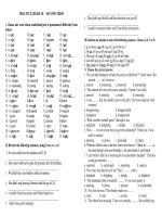
development into four major stages based on the appearance of the lung tissue. These are embryonic, pseudoglandular, canalicular and alveolar; the last is sometimes
divided into an earlier saccular or terminal sac phase and
a later alveolar stage (Table 1.1, Figure 1.1). The alveolar
stage continues after birth and in some species is entirely
postnatal. There is also considerable individual variation
and one stage gradually merges into the next. Within each
phase, development of specific structures is of major
importance. During the embryonic period the main hilar
The lung at birth
Factors affecting lung growth
References
9
9
10
connections of the airways and the pulmonary circulation are made. During the pseudoglandular phase the
pre-acinar airways with their accompanying arteries and
veins develop. During the canalicular phase the blood–gas
barrier thins and the maturation of the surfactant system
begins. In the alveolar phase alveoli multiply and by birth
up to half the adult number are present.
Development of the airways
The lung appears as a ventral diverticulum from the endodermal foregut in the fourth week after ovulation. The
complete lining epithelium of the lung is derived from the
endoderm. This bud is formed within the splanchnic mesoderm surrounding the gut and the dorsal aorta; it is from
this mesenchyme that the airway walls and blood vessels
are derived. A division produces the left and right bronchi by 26–28 days of gestational age and segmental airways are present by 6 weeks. Further division of airways
into the surrounding mesenchyme continues until the
end of the pseudoglandular stage (17 weeks of gestation)
by which time all pre-acinar airways to the level of the terminal bronchiolus are present. The majority of divisions
occur during the tenth to fourteenth weeks of gestation5
(Figures 1.1 and 1.2).
During the canalicular period (16–27 weeks of gestation) the pre-acinar airways increase in diameter and
length. The peripheral airways continue to divide to form
the prospective respiratory bronchioli (two to three
generations in humans) and beyond these the prospective alveolar ducts. The mesenchymal region between the
4 Fetal and postnatal anatomical lung development
Table 1.1 Phases of lung development in man
Embryonic
0–7 weeks of gestation
Lung buds form. Blood vessels connect to the heart
Pseudoglandular
6–17 weeks of gestation
Pre-acinar airways and blood vessels develop
Canalicular
16–27 weeks of gestation
Respiratory (intra-acinar) region develops. Thinning of peripheral
epithelium and mesenchyme. Type I and II pneumonocytes
Alveolar
27 weeks to term
Development of saccules and then alveoli
Postnatal
Up to 18 months
Alveoli and small blood vessels multiply. All structures increase in size
(a)
(b)
(c)
Figure 1.1 Photomicrographs illustrating the classical stages of fetal lung development: (a) pseudoglandular,
6–17 weeks of gestation; (b) canalicular, 16–27 weeks of gestation; (c) alveolar, 27 weeks to term.
Stages of lung
development
Lung structure
Trachea
24 days
Extrapulmonary main bronchus
28 days
Embryonic
0–7 weeks
gestation
Bronchi
8–13 generations
Bronchioli
3–10 generations
Pseudoglandular
7–17 weeks
gestation
Terminal bronchiolus
1 generation
Canalicular
17–27 weeks
gestation
Respiratory bronchioli
3–5 generations
Alveolar ducts
2–3 generations
Alveolar
28 weeks gestation
–2–3 years postnatal
Alveoli
300–600 million
10 000/acinus
Pleura
Acinus
Figure 1.2 Diagram representing the
number of airway generations in the
human lung and the stage and
gestational age at which they appear.
Fetal stages of lung development 5
airways thins and capillaries come to lie beneath the
epithelium of the peripheral airways, apparently causing
the epithelium to become thinner. The larger airways
(prospective bronchi) are lined by columnar epithelium,
but the distal bronchioli are lined by cuboidal cells. At the
level of the prospective respiratory bronchioli, part of the
wall is lined by flattened cells, as are the prospective alveolar ducts which at this stage are sac shaped (saccules).
By 20–22 weeks of gestation, type I and II alveolar epithelial cells can be identified lining all saccular air spaces.
The type I cells are flat and elongated and cover the majority of the surface. The type II cells maintain a cuboidal
shape and develop lamellar bodies around 24 weeks of
gestation, which is 4–5 weeks before surfactant can be
detected in the amniotic fluid. By the end of the canalicular
stage, the air to blood barrier is thin enough to support
gas exchange (about 0.6 m) but the gas exchange units
are the large thin-walled saccules. True alveoli develop
later (p. 7).
Increase in airway size in the prenatal period is linear
and continuous with antenatal growth. After the first
year of life, there is a slowing in growth, there being an
approximately twofold increase between 22 weeks of gestation and 8 months postnatal age and a two- to threefold increase between birth and adulthood.20 A previous
study, measuring airway length and diameter in children
from birth to adulthood, had reported symmetrical
growth throughout the lung.17 Tracheal size does not differ between sexes during early life,12 but adult males have
a larger trachea than females. Girls have wider and/or
shorter airways than boys during early childhood and
this may explain their lesser tendency to wheeze, but by
adulthood males have relatively large airways. This may
be a factor in the relative decline in reversible obstructive
airways disease in teenage boys.
Airway wall structure
As successive airways form, their walls first develop airway wall smooth muscle closely followed by cartilage,
submucosal glands and connective tissue. These structural elements of the airway wall appear from the hilum
towards the periphery and by 24 weeks of gestation the
airways have the same structure as they do in the adult.5,6
Smooth muscle cells are present in human trachea and
lobar bronchi by 6 weeks of gestation and extend along
the airway pathway as the peripheral airways divide
(Figure 1.3). Only the ultimate lung buds do not have any
airway smooth muscle.13,35 As in adult lungs, fetal airway
smooth muscle expresses contractile smooth muscle specific myofilaments such as smooth muscle ␣-actin and
smooth muscle myosin. In vitro studies of peripheral
explants of first trimester human lung have shown that
fetal airway smooth muscle cells have spontaneous
tone and peristalsis-like contractions which cause active
(a)
(b)
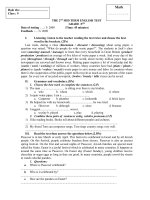
Figure 1.3 Photomicrograph of peripheral airway bud in
a 44-day-old fetus immunostained for (a) ␣ smooth muscle
actin and (b) CD31 (endothelial marker). Airway smooth
muscle (asm) ␣ smooth muscle positive, is seen to the
penultimate branch. Capillaries (arrowheads), positive for
CD31 are seen as far as the peripheral bud and coalesce
alongside the airway (arrowed). A muscle wall, one cell
thick and ␣ smooth muscle positive which is derived from
adjacent bronchial smooth muscle cells surrounds the
pulmonary artery (pa).
movement of intraluminal fluid.35 The movements are
sensitive to acetylcholine and isoproterenol, suggesting
that neurohumoral factors modulate smooth muscle
activity.35 Postnatally there is reactivity to methacholine
and subsequent bronchodilation after addition of
metaproterenol in healthy infants less than 15 months of
age.47 During fetal life and in the newborn, the amount
of muscle within a given sized airway is generally less
than in the adult (Figure 1.4).20 During the first year of
life, particularly in the first few weeks after birth, there is
6 Fetal and postnatal anatomical lung development
a rapid increase in bronchial smooth muscle mass relative to airway size. This rapid increase is probably related
to the change to air breathing since it occurs at a similar
postnatal age and therefore an earlier gestational age in
babies that are born prematurely (Figure 1.4). Airway
smooth muscle mass increases above normal in artificially ventilated babies.20
Cartilage first appears in the sixth gestational week in
the trachea, the tenth week in the main bronchi and
the twelfth week in segmental bronchi and by 24 weeks
of gestation extends as far as in the adult. There is a
progressive increase in the total cartilage mass through
infancy and childhood as airways increase in size. The submucosal glands are responsible for producing most of the
mucus found in the airways. They appear in the trachea
of the human at 10 weeks of gestation and gradually
extend towards the periphery of the lung, reaching their
adult position by the canalicular stage, but it is only at
13 years of age that the glands have the adult appearance.
During childhood there is relatively more submucosal
gland mass within the airway wall than in the adult.34
Airway epithelium
The epithelial cells develop by differentiation and maturation of the primitive endodermal cells. As well as producing the fluid and mucus for the ciliary escalator to
remove particles from inside the lung, the epithelium is
a source of smooth muscle inhibitory factor(s), and can
also generate endothelin, which is a contractile agonist as
well as a smooth muscle cell mitogen.46
Ciliated cells are found from the trachea to the respiratory bronchioli and at all levels are the most numerous
cells. They first appear at 11 weeks of gestation.25 They
do not divide, but originate from basal or secretory cells.
Mucus-secreting or goblet cells are found from the
trachea to the end of the bronchioli. The presence of
intracellular mucus has been demonstrated in the human
fetal lung at 13 weeks of gestation but at this age the cells
are sparse. At birth, there is still a relatively low number
of goblet cells, less than 10 percent of the total number of
epithelial cells. After birth, there is a rapid increase in the
number of goblet cells, reaching up to 40 percent of the
total in the bronchi by 3 months of age.20 Basal cells are
found in the larger airways and can be identified from 12
to 14 weeks of gestation and have been considered to be
a stem cell. In the terminal bronchioli a further cell type,
the Clara cell, is found. They are progenitors for the ciliated cells in peripheral airways.16 They produce Clara cell
10 kDa protein (CC10) which has immunomodulatory
and anti-inflammatory activity and may play a role in
controlling airway inflammation. Clara cells also produce
surfactant apoprotein, under -adrenergic control.
(a)
Bronchial smooth muscle (mm2/mm)
(b)
(c)
0.015
0.010
0.005
0
Bronchial blood vessels
0
20
40
60
80
Postconceptional age (weeks)
Figure 1.4 Photomicrograph of bronchiolar wall (a) at
term and (b) at 8 months of age (ϫ800). The bronchial
smooth muscle at arrows increases with age. (c) The area
of bronchial smooth muscle/mm of airway perimeter
in small bronchi related to postconceptional age. ᭹ ,
Normal fetus and infant; ᮀ , premature infant.
100
The airways are supplied with oxygenated blood via the
bronchial arteries, which appear from 8 weeks of gestation. They arise from the descending aorta as small
branches, probably by angiogenesis, and supply the extrapulmonary bronchi and extend down the intrapulmonary
airway wall alongside the cartilage plates. They divide to
form a subepithelial plexus and an adventitial plexus on
either side of the bronchial smooth muscle and cartilage.
By birth they extend to the end of the bronchioli. True