Annual update in intensive care and emergency medicine 2016
Bạn đang xem bản rút gọn của tài liệu. Xem và tải ngay bản đầy đủ của tài liệu tại đây (10.02 MB, 488 trang )
2016
Annual Update
in Intensive Care
and Emergency
Medicine 2016
Edited by J.-L.Vincent
123
Annual Update in Intensive Care and
Emergency Medicine 2016
The series Annual Update in Intensive Care and Emergency Medicine is the continuation of the series entitled Yearbook of Intensive Care Medicine in Europe and
Intensive Care Medicine: Annual Update in the United States.
Jean-Louis Vincent
Editor
Annual Update in
Intensive Care and
Emergency Medicine 2016
Editor
Prof. Jean-Louis Vincent
Université libre de Bruxelles
Dept. of Intensive Care
Erasme Hospital
Brussels, Belgium
ISSN 2191-5709
ISSN 2191-5717 (electronic)
Annual Update in Intensive Care and Emergency Medicine
ISBN 978-3-319-27348-8
ISBN 978-3-319-27349-5 (eBook)
DOI 10.1007/978-3-319-27349-5
© Springer International Publishing Switzerland 2016
This work is subject to copyright. All rights are reserved by the Publisher, whether the whole or part
of the material is concerned, specifically the rights of translation, reprinting, reuse of illustrations,
recitation, broadcasting, reproduction on microfilms or in any other physical way, and transmission or
information storage and retrieval, electronic adaptation, computer software, or by similar or dissimilar
methodology now known or hereafter developed.
The use of general descriptive names, registered names, trademarks, service marks, etc. in this publication does not imply, even in the absence of a specific statement, that such names are exempt from the
relevant protective laws and regulations and therefore free for general use.
The publisher, the authors and the editors are safe to assume that the advice and information in this book
are believed to be true and accurate at the date of publication. Neither the publisher nor the authors or
the editors give a warranty, express or implied, with respect to the material contained herein or for any
errors or omissions that may have been made.
Cover design: WMXDesign GmbH, Heidelberg
Printed on acid-free paper
This Springer imprint is published by Springer Nature
The registered company is Springer International Publishing AG Switzerland.
Contents
Common Abbreviations . . . . . . . . . . . . . . . . . . . . . . . . . . . . . . . . .
Part I
Infections and Antibiotics
Interpreting Procalcitonin at the Bedside . . . . . . . . . . . . . . . . . . . . .
J. Fazakas, D. Trásy, and Z. Molnár
Reducing Antibiotic Use in the ICU: A Time-Based Approach
to Rational Antimicrobial Use . . . . . . . . . . . . . . . . . . . . . . . . .
P. O. Depuydt, L. De Bus, and J. J. De Waele
Plasmacytoid Dendritic Cells in Severe Influenza Infection . . . . . . . . . .
B. M. Tang, M. Shojaei, and A. S. McLean
Critically Ill Patients with Middle East Respiratory Syndrome
Coronavirus Infection . . . . . . . . . . . . . . . . . . . . . . . . . . . . . .
H. M. Al-Dorzi, S. Alsolamy, and Y. M. Arabi
Part II
xi
3
15
25
35
Sepsis
Immunomodulation: The Future for Sepsis? . . . . . . . . . . . . . . . . . . .
T. Girardot, F. Venet, and T. Rimmelé
49
Norepinephrine in Septic Shock: Five Reasons to Initiate it Early . . . . .
M. Jozwiak, X. Monnet, and J.-L. Teboul
61
Myths and Facts Regarding Lactate in Sepsis . . . . . . . . . . . . . . . . . .
M. Nalos, A. S. McLean, and B. Tang
69
v
vi
Part III
Contents
Renal Issues
Creatinine-Based Definitions: From Baseline Creatinine
to Serum Creatinine Adjustment in Intensive Care . . . . . . . . . . .
S. De Rosa, S. Samoni, and C. Ronco
Detrimental Cross-Talk Between Sepsis and Acute Kidney Injury:
New Pathogenic Mechanisms, Early Biomarkers
and Targeted Therapies . . . . . . . . . . . . . . . . . . . . . . . . . . . . .
S. Dellepiane, M. Marengo, and V. Cantaluppi
81
91
Timing of Acute Renal Replacement Therapy . . . . . . . . . . . . . . . . . . 111
A. Jörres
(Multiple) Organ Support Therapy Beyond AKI . . . . . . . . . . . . . . . . 117
Z. Ricci, S. Romagnoli, and C. Ronco
Part IV
Fluid Therapy
Crystalloid Fluid Therapy . . . . . . . . . . . . . . . . . . . . . . . . . . . . . . . 133
S. Reddy, L. Weinberg, and P. Young
Part V
Bleeding
Emergency Reversal Strategies for Anticoagulants
and Antiplatelet Agents . . . . . . . . . . . . . . . . . . . . . . . . . . . . . 151
M. Levi
Part VI
Cardiovascular System
Bedside Myocardial Perfusion Assessment
with Contrast Echocardiography . . . . . . . . . . . . . . . . . . . . . . . 165
S. Orde and A. McLean
Pathophysiological Determinants of Cardiovascular Dysfunction
in Septic Shock . . . . . . . . . . . . . . . . . . . . . . . . . . . . . . . . . . . 177
F. Guarracino, R. Baldassarri, and M. R. Pinsky
Cardiovascular Response to ECMO . . . . . . . . . . . . . . . . . . . . . . . . . 185
S. Akin, C. Ince, and D. dos Reis Miranda
Mechanical Circulatory Support in the New Era: An Overview . . . . . . 195
K. Shekar, S. D. Gregory, and J. F. Fraser
Contents
Part VII
vii
Cardiac Arrest
Cardiac Arrest in the Elderly: Epidemiology and Outcome . . . . . . . . . 219
C. Sandroni, S. D’Arrigo, and M. Antonelli
Regional Systems of Care: The Final Link in the “Chain of Survival”
Concept for Out-of-Hospital Cardiac Arrest . . . . . . . . . . . . . . . . 231
T. Tagami, H. Yasunaga, and H. Yokota
Cardiac Arrest Centers . . . . . . . . . . . . . . . . . . . . . . . . . . . . . . . . . 241
E. L. Riley, M. Thomas, and J. P. Nolan
Part VIII
Oxygenation and Respiratory Failure
High-Flow Nasal Cannula Oxygen Therapy: Physiological Effects
and Clinical Data . . . . . . . . . . . . . . . . . . . . . . . . . . . . . . . . . 257
D. Chiumello, M. Gotti, and C. Chiurazzi
The Potential Value of Monitoring the Oxygen Reserve Index
in Patients Receiving Oxygen . . . . . . . . . . . . . . . . . . . . . . . . . 271
A. Perel
Variable Ventilation from Bench to Bedside . . . . . . . . . . . . . . . . . . . . 281
R. Huhle, P. Pelosi, and M. G. de Abreu
Monitoring Respiratory Effort by Means of the Electrical Activity
of the Diaphragm . . . . . . . . . . . . . . . . . . . . . . . . . . . . . . . . . 299
G. Grasselli, M. Pozzi, and G. Bellani
Dissipated Energy is a Key Mediator of VILI: Rationale for Using
Low Driving Pressures . . . . . . . . . . . . . . . . . . . . . . . . . . . . . . 311
A. Serpa Neto, M. B. P. Amato, and M. J. Schultz
Corticosteroids as Adjunctive Therapy in Severe CommunityAcquired Pneumonia . . . . . . . . . . . . . . . . . . . . . . . . . . . . . . . 323
C. Cillóniz, A. San José, and A. Torres
Part IX
Abdominal Issues
The Neglected Role of Abdominal Compliance
in Organ-Organ Interactions . . . . . . . . . . . . . . . . . . . . . . . . . . 331
M. L. N. G. Malbrain, Y. Peeters, and R. Wise
viii
Part X
Contents
Metabolic Support
Metabonomics and Intensive Care . . . . . . . . . . . . . . . . . . . . . . . . . . 353
D. Antcliffe and A. C. Gordon
The Rationale for Permissive Hyperglycemia in Critically Ill Patients
with Diabetes . . . . . . . . . . . . . . . . . . . . . . . . . . . . . . . . . . . . 365
J. Mårtensson and R. Bellomo
Indirect Calorimetry in Critically Ill Patients: Concept, Current Use,
and Future Challenges . . . . . . . . . . . . . . . . . . . . . . . . . . . . . . 373
E. De Waele, P. M. Honoré, and H. D. Spapen
Part XI
Ethical Issues
Managing Intensive Care Supply-Demand Imbalance . . . . . . . . . . . . . 385
C. C. H. Leung, W. T. Wong, and C. D. Gomersall
Advances in the Management of the Potential Organ Donor
After Neurologic Determination of Death . . . . . . . . . . . . . . . . . . 393
A. Confalonieri, M. Smith, and G. Citerio
Humanizing Intensive Care: Theory, Evidence, and Possibilities . . . . . . 405
S. M. Brown, S. J. Beesley, and R. O. Hopkins
Part XII
Applying New Technology
Ultrasound Simulation Education for Intensive Care
and Emergency Medicine . . . . . . . . . . . . . . . . . . . . . . . . . . . . 423
F. Clau-Terré, A. Vegas, and N. Fletcher
Virtual Patients and Virtual Cohorts: A New Way to Think About
the Design and Implementation of Personalized ICU Treatments . . 435
J. G. Chase, T. Desaive, and J.-C. Preiser
Part XIII
Intensive Care Unit Trajectories: The Bigger Picture
Predicting Cardiorespiratory Instability . . . . . . . . . . . . . . . . . . . . . . 451
M. R. Pinsky, G. Clermont, and M. Hravnak
Long-Term Outcomes After Critical Illness Relevant
to Randomized Clinical Trials . . . . . . . . . . . . . . . . . . . . . . . . . 465
C. L. Hodgson, N. R. Watts, and T. J. Iwashyna
Contents
ix
Long-Term Consequences of Acute Inflammation in the Surgical Patient:
New Findings and Perspectives . . . . . . . . . . . . . . . . . . . . . . . . 475
P. Forget
Kairotropy: Discovering Critical Illness Trajectories
Using Clinical Phenotypes with Big Data . . . . . . . . . . . . . . . . . . 483
G. E. Weissman and S. D. Halpern
Index . . . . . . . . . . . . . . . . . . . . . . . . . . . . . . . . . . . . . . . . . . . . . 497
Common Abbreviations
AKI
ARDS
BMI
CAP
CI
COPD
CPB
CPR
CRP
CRRT
CT
CVP
ECMO
EKG
ICU
IL
LPS
LV
MAP
MRI
OR
PEEP
PCR
RCT
RRT
RV
SvO2
SOFA
TNF
VILI
Acute kidney injury
Acute respiratory distress syndrome
Body mass index
Community-acquired pneumonia
Confidence interval/cardiac index
Chronic obstructive pulmonary disease
Cardiopulmonary bypass
Cardiopulmonary resuscitation
C-reactive protein
Continuous renal replacement therapy
Computed tomography
Central venous pressure
Extracorporeal membrane oxygenation
Electrocardiogram
Intensive care unit
Interleukin
Lipopolysaccharide
Left ventricular
Mean arterial pressure
Magnetic resonance imaging
Odds ratio
Positive end-expiratory pressure
Polymerase chain reaction
Randomized controlled trial
Renal replacement therapy
Right ventricular
Mixed venous oxygen saturation
Sequential organ failure assessment
Tumor necrosis factor
Ventilator-induced lung injury
xi
Part I
Infections and Antibiotics
Interpreting Procalcitonin at the Bedside
J. Fazakas, D. Trásy, and Z. Molnár
Introduction
One of the most challenging tasks in critical care medicine is the treatment of serious infection-related multiple organ dysfunction. Early detection of infection and
the immediate start of resuscitation paralleled with adequate antimicrobial therapy
undoubtedly give the best possible chance for survival and are strongly recommended in the Surviving Sepsis Campaign guidelines [1]. However, although recognizing organ failure via objective signs is relatively easy, diagnosing infection as
a possible underlying cause remains a challenge. Because of the non-specific properties of conventional signs of infection, such as body temperature and white blood
cell (WBC) count, biomarkers to aid diagnosis have been looked for for decades.
One of the most studied biomarkers is procalcitonin (PCT) [2]. Its role in assisting
antibiotic therapy has been studied extensively, with contradicting results. There are
positive studies [3, 4] showing that PCT-guided patient management reduced antibiotic exposure and length of antibiotic therapy without affecting patient outcomes.
There are also negative studies, which did not show this benefit [5–7]. However, to
understand the values and limitations of inflammatory biomarkers it is necessary to
understand the immunological background of critical illness determined mainly by
the host response. Putting the results of these studies into context, based on new
insights of the pathomechanisms of sepsis and systemic inflammation generated
mainly by the individual’s host response, may help explain the differences between
the reported results and help the clinician to interpret PCT data with more confidence at the bedside.
J. Fazakas
Department of Transplantation and Surgery, Semmelweis University
Budapest, Hungary
D. Trásy Z. Molnár ( )
Department of Anesthesiology and Intensive Therapy, University of Szeged
Budapest, Hungary
email:
© Springer International Publishing Switzerland 2016
J.-L. Vincent (ed.), Annual Update in Intensive Care and Emergency Medicine 2016,
DOI 10.1007/978-3-319-27349-5_1
3
4
J. Fazakas et al.
Sepsis Is not a ‘Definitive’ Disease
In classical medicine, for example, in most fields of surgery and internal medicine,
after taking a medical history, performing physical examination and diagnostic tests,
the diagnosis is often straightforward, and patients can receive treatment, which is
by-and-large well defined around the world. As examples, in stroke, myocardial infarction, bone fractures, intracranial hemorrhage, etc., we have diagnostic tests with
very high sensitivity and specificity. However, defining sepsis is not that simple.
The term “sepsis syndrome” was conceived in a hotel in Las Vegas in 1980, during the designing of the protocol of one of the first prospective randomized trials in
sepsis, performed by a group of scientists led by the late Roger Bone [8]. The study
ended with non-significant results, but a statement paper was later published by the
same authors entitled “Sepsis syndrome: a valid clinical entity” [9], after which
medical society started to deal with sepsis as a definitive disease. As a definitive
disease, physicians wanted a single test with high sensitivity and specificity to diagnose sepsis, and there was an urge to find an ‘anti-sepsis magic bullet’. Neither
of these wishes has or will ever come true.
Regarding the definition and diagnosis of sepsis, the classical signs of the “sepsis
syndrome”, such as fever/hypothermia, leukocytosis/leukopenia, tachycardia and
hypotension, were met by a very large and non-specific cohort of patients. For this
reason, a consensus conference was convened and defined ‘consensus criteria’ of
sepsis, which have been used for decades in research and clinical practice [10]. In
the most recent Surviving Sepsis Campaign guidelines more robust, more detailed
criteria were used as definition [1], but these were almost immediately questioned
by experts who had also taken part in the Surviving Sepsis Campaign process [11].
These efforts clearly show that finding the appropriate definition of sepsis has
been a continuous challenge for more than 30 years. The difficulty in defining
sepsis originates from its complex pathophysiology, which is affected by numerous individual variations of the host response. Furthermore, in most specialties,
diagnostic laboratory or radiological tests have very high sensitivity and specificity,
often reaching almost 95–100% [12]. However, in the case of sepsis, as we will
see in the subsequent paragraphs, the situation is different, which makes not just
diagnosis, but also interpretation of the results of clinical trials and epidemiological
data very difficult.
From Localized Insult to Cytokine Storm
The immune system is a complex network and the immune response to pathogens
relies on both innate and adaptive components, dynamically defined as the proand anti-inflammatory forces. The innate immune system (including the complement system, sentinel phagocytic and natural killer cells), is responsible for the
eradication of the invaders, whereas the adaptive immune system’s role is to control the process and keep it localized to the site of the insult [13]. Under normal
circumstances, these mechanisms remain in balance. The innate system acts by
Interpreting Procalcitonin at the Bedside
5
broad recognition of antigens, mainly by triggering pathogen-associated molecular
patterns (PAMPs) of lipopolysaccharide (LPS) elements of the surfaces of invading pathogens. When there is an imbalance due to dysregulation of the pro- and
anti-inflammatory forces, the local response escalates into a systemic host response
also termed a “cytokine storm” [14]. It was a surprising finding that after trauma,
burns, ischemia-reperfusion, pancreatitis, major surgery, etc., the same or similar
molecules that are found in PAMPs are released, mainly from the mitochondria of
the injured or stressed cells, and can also cause a cytokine storm. This process accompanying tissue injury is called damage-associated molecular patterns (DAMPs).
This similarity is due to the fact that the bacteria and the mitochondria (which are
more-or-less encapsulated bacteria) share very similar genetic backgrounds, and explains why tissue injury-induced DAMPs and bacterial infection-induced PAMPs
manifest as similar host responses and clinical manifestations [15].
The Role of PCT in Diagnosing Infection
The question “Is this patient septic?” is frequently asked on intensive care unit
(ICU) rounds. However, this may be an irrelevant issue. Why? Because, first,
we should recognize a critically ill patient via objective signs of organ dysfunction,
which determines the immediate start of basic and organ-specific resuscitation, regardless of the actual diagnosis. And, second, what is of pivotal importance is not
that the patient is septic or not, but whether the onset of critical illness is due to infection or not? Because, if it is due to infection, then we should start antibiotics or
perform another form of source control. But if there is no infection, then antibiotic
therapy should not be commenced, because of its undesired effects. Therefore, it is
not ‘sepsis’ that we treat, but organ dysfunction and infection.
Diagnosing infection on the ICU is not easy and requires a multimodal approach. Clinical signs are obviously the most important in recognizing critical
illness and suspecting infection and even the source of infection, but they cannot
prove it on their own. Conventional indicators, such as fever/hypothermia, leukocytosis/leukopenia, tachypnea, tachycardia, hypotension, taken from the classical sepsis-syndrome criteria are non-specific, and in fact poor indicators of infection. To
fill this gap, inflammatory biomarker measurements have been developed [2]. Every biomarker has its own merits and limitations, but there is no ‘ideal’ biomarker,
and there may never be one. Biomarkers can support decision-making but they will
never be able to differentiate between the inflammatory response to infection and
the host response to non-infectious insults with 100% sensitivity and specificity because of the complex, overlapping pathomechanism of PAMPs and DAMPs. This
situation is in sharp contrast with the diagnostic power of certain biomarkers used
in the world of ‘definitive’ diseases, where several laboratory parameters have this
ability. Furthermore, learning how to use biomarkers is not easy either.
The two markers most commonly used in infection/sepsis diagnostics and for
guiding therapeutic interventions are PCT and C-reactive protein (CRP) [2]. One
of the main limitations of CRP is that it moves ‘slowly’, and after a certain insult
6
J. Fazakas et al.
reaches its maximum value usually 48 h later. This is in general unacceptable on the
ICU, as every hour delay in starting, for example, appropriate antibiotic treatment
can affect mortality as indicated in the study of Kumar et al. [16]. Furthermore,
levels are generally elevated in most ICU patients, making interpretation of CRP
very difficult [17].
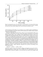
PCT is detectable in the serum within a few (4–6) h after its induction, which
is most often by bacterial infection. During the ‘normal’ course of an infection it
reaches its peak within 24 h and then starts to decline, if treatment is adequate, with
levels reducing by roughly 50% daily according to its half-life [18]. PCT differentiates bacterial infections from a systemic inflammatory response of other etiologies
with higher sensitivity and specificity than CRP [19], and also has good prognostic value [20]. However, interpreting PCT concentrations on admission or after the
onset of an acute insult, whether infectious or not, is not simple. There are many
studies reporting that PCT concentrations correlate with severity and differ significantly in patients with SIRS, sepsis, severe sepsis and septic shock [21]. Clec’h
et al. found that patients with septic shock had more than 10 times higher median
PCT concentrations as compared to those admitted with shock of non-septic origin
[22]. However, looking at the data carefully reveals that although there was a remarkable and statistically significant difference, there was also a huge scatter and
overlap of the PCT data between the groups (septic shock: 14 (0.3–767) vs. nonseptic shock: 1 (0.15–36) ng/ml), which makes individual interpretation of a single
measurement very difficult – a finding, which is generally true for every biomarker
of inflammation. This observation was reinforced by the same group in a subsequent study, in which they found that the median PCT concentrations in medical
vs. surgical patients differed in SIRS (0.3 (0.1–1.0) vs. 5.7 (2.7–8.3)) and in septic
shock (8.4 (3.6–76.0) vs. 34.0 (7.1–76.0) ng/ml) [23]. These differences and the
large overlap can be explained by the PAMP- and DAMP-based host response. In
certain cases, there is a single PAMP or DAMP, but they can also occur in combination as PAMP + DAMP. The latter is bound to have a pronounced inflammatory
response reflected in higher PCT concentrations. Therefore, it has become clear
that the same PCT concentration, in other words a given ‘normal’ concentration,
cannot be used in every condition. Medical patients with infection should, in general, have lower PCT concentrations (single PAMP insult) as compared to surgical
patients with infection, in which DAMP and PAMP are present at the same time.
Moreover, it is also important to acknowledge that any cellular injury, whether direct tissue or ischemia-reperfusion injury without infection, can result in elevation
of PCT induced by a single DAMP type insult.
In recent large multicenter trials some authors did not show benefit of a PCTbased approach to antibiotic management in ICU patients [5–7]. However, in these
studies, fixed PCT values were applied in the protocols with a low (1 ng/ml) cut-off
value for intervention. In the studies by Jensen et al. [6] and Layios et al. [5], 40%
of the patients were surgical, in whom, as we have shown before, this threshold of
PCT is too low; hence these patients may have received antibiotics unnecessarily,
which may in part explain the negative results.
Interpreting Procalcitonin at the Bedside
7
Although PCT absolute values have the above mentioned limitations, there is
overwhelming evidence that in most cases, high PCT concentrations indicate bacterial infection. The shortcomings of absolute PCT values may be compensated when
the kinetics of PCT are taken into account to indicate infection. In a recent study
by Tsangaris et al., daily measurements of PCT were performed and kinetics were
evaluated in patients who had already been treated on the ICU for > 10 days and
had a sudden onset of fever [24]. These authors found a two-fold increase in PCT
levels from the day before to the day when there was a sudden onset of fever in
patients with proven infection, but there was no change in PCT concentrations in
patients with fever but no infection. They concluded that in patients treated longerterm on the ICU, PCT values on the day of fever onset must be compared to values
measured on the previous day in order to define whether this rise in temperature is
due to infection or not. It is also important to note that these were medical patients,
in whom median PCT concentrations varied between 0.1–0.75 ng/ml. Nevertheless,
despite the low absolute values, a two-fold increase was able to detect those with
proven infection.
In a recent observational study, we also found that an increase in PCT from the
day before (t 1 ) to the day when infection was suspected (t0 ) predicted infection.
The best cut-off for the absolute PCT concentration was 0.84 ng/ml, with a sensitivity of 61% (95% CI 50–72) and specificity 72% (95% CI 53–87), which shows that
the absolute value was a poor indicator. However, the percentage change in PCT
concentration, with an increase of > 88% from t 1 to t0, had a sensitivity of 75%
(95% CI 65–84) and specificity of 79% (95% CI 60–92) and a PCT delta change
of > 0.76 ng/ml had a sensitivity of 80% (95% CI 70–88) and specificity of 86%
(95% CI 68–96) to indicate infection. Furthermore, neither the absolute values of
conventional indicators of infection, such as WBC, body temperature and CRP, nor
their change from t 1 to t0 , could predict infection [25].
Despite the discussed limitations, elevated PCT concentration has so far been
shown to have the best sensitivity and specificity to indicate infection compared to
other markers, and in the case of high levels one must at least suspect infection,
whereas low PCT concentrations may help to exclude infection and suggest that
antibiotics not be started. However, careful multimodal evaluation of the clinical
picture together with PCT results and consideration of all the issues discussed earlier is necessary to correctly interpret PCT results and make the best decisions for
our ICU patients.
PCT-assisted Antibiotic Therapy
There are three fundamental questions to be answered during our ward rounds
when treating patients with suspected or proven infections on the ICU: 1) is there
infection, in other words should we start empirical antibiotic therapy?; 2) is the
commenced antibiotic effective?; and finally, 3) when should we stop antibiotic
treatment?
8
J. Fazakas et al.
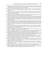
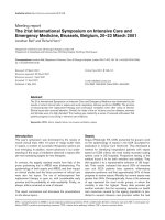
Fig. 1 Starting antibiotic
(AB) therapy. PCT: procalcitonin; *: patient requires
clinically significant dose of
vasopressors after initial resuscitation. For explanation
see text
Suspected infection
Hemodynamic instability
Yes*
No
PCT ↑
AB
Yes
No
AB
No AB:
- Observe
- Reassess later
Diagnosing Infection and Starting Antibiotics
The role of PCT in diagnosing infection has been discussed in detail earlier. Several studies have investigated the effects of PCT-guided antibiotic therapy on patient
outcomes. In a landmark study by Christ-Crain et al., antibiotic exposure was reduced by 50% (44% versus 83%) in patients admitted to emergency wards with
acute respiratory complaints when antibiotic therapy was guided by admission PCT
levels as compared to using conventional signs of infection only [3]. Two subsequent multicenter trials also found a decrease in antibiotic exposure in patients
treated on the ICU with infections [4, 26]. The possible reasons why other studies
[5, 7], could not find positive results were discussed earlier.
Patients treated on the ICU for a longer period of time may develop an imbalance
between pro- and anti-inflammatory forces such that the latter become prominent.
These patients will become immunoparalyzed, making them prone to a series of
recurrent infections. Detecting infection in these patients may prove even more
difficult. Rau et al. [27] found that in patients with secondary peritonitis, PCT
levels increased and indicated infection, but the peak values decreased significantly
with each new insult. This observation was also supported by Charles et al. [28].
They found that during the first infectious insult, the mean PCT concentration was
55 ng/ml, but during the second infectious insult, despite the same clinical gravity,
it was several fold less at 6.4 ng/ml. These data indicate that with time, patients
become immunoparalyzed and lower PCT concentrations should then be taken just
as seriously as higher levels in the early course of the disease. Furthermore, this
Organ function
Microbiology
Procalcitonin
PCT:
- No change
- Small ↓
-↑
2
PCT ↓
1
PCT:
- No change
- Small ↓
-↑
2
Micro: positive
+ABs appropriate
B
- Change ABs
- Reassess organ
support
Micro: positive
+ABs inappropriate
C
Continue Reconsider: Continue Reconsider:
- Source
- Source
- ABs
- Reassess
organ support
PCT ↓
1
Micro: negative
A
A
PCT:
- No change
- Small ↓
-↑
Continue
+ exclude
other source
STOP
ABs
2
PCT ↓
1
Micro: negative
Clinical improvement
Continue
PCT ↓
1
Exclude
other
sources
PCT:
- No change
- Small ↓
-↑
2
Micro: positive
+ABs appropriate
B
Yes
PCT:
- No change
- Small ↓
-↑
2
STOP ABs - Change ABs
Reassess - Consult
micro
infect/micro
specialist
PCT ↓
1
Micro: positive
+ABs inappropriate
C
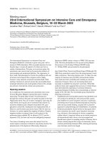
Fig. 2 Multimodal reassessment of antibiotic (AB) therapy on day 2–3. After empirical antibiotics have been commenced, a multimodal reassessment of the
situation when microbiological data are available, usually on day 2 or 3, is strongly recommended. No clinical improvement: Despite no clinical improvement,
a decrease in procalcitonin (PCT) (A-1) may still indicate that the infection is under control, but the patient needs more time to benefit from treatment; therefore,
antibiotics should be continued. By contrast (A-2), if PCT is not decreasing or even increasing, these can be important signs that infection is not under control,
hence source of infection and antibiotics (type, dose) should be reassessed. If antibiotics are appropriate, depending on PCT changes, antibiotics should either
be continued (B-1) or other sources of infection should be looked for (B-2). In case of inappropriate antibiotics and no clinical improvement, regardless of
the PCT concentration, therapy should be changed (C). Clinical improvement: If there is no proof of infection (micro: negative), based on PCT changes (#
or ") infection may be excluded and antibiotics stopped (A-1) or continued (A-2). Similar algorithms can be applied if antibiotics are appropriate (B-1, 2).
If antibiotics are inappropriate and PCT decreases, then one may consider the microbiology as a false positive and stop antibiotics (C-1), because it is highly
unlikely that there is clinical improvement and decreasing PCT if an infection is not under control due to inappropriate antibiotics. This scenario happens when
there are pathogens (colonization for example), but no infection. Finally, if antibiotics are inappropriate and PCT changes unfavorable (C-2), consultation
with infectious disease specialists/microbiologists is recommended. Micro: microbiology; ": increasing concentrations; #: decreasing concentrations; infect:
infectious disease; micro: microbiology
Intervention
No
Interpreting Procalcitonin at the Bedside
9
10
J. Fazakas et al.
provides further evidence that PCT will respond to the same insult differently during
the course of the disease, and hence values should be interpreted with care.
To put these considerations into a clinical context, two very common scenarios
will be discussed, which are summarized in two decision trees (Figs. 1 and 2).
Figure 1 shows how PCT can help in decision-making when infection is suspected.
If the patient is hemodynamically unstable and infection is likely, by definition
he/she has septic shock or at least one cannot exclude it, hence antibiotic therapy
should not be delayed but has to be commenced immediately, regardless of the PCT
or any biomarker value [16]. However, if the patient is stable hemodynamically,
and PCT is ‘low’ or decreasing (based on the problems with absolute PCT values,
discussed in detail above, we deliberately avoid giving exact numbers), then we can
wait, observe the patient and reassess later (Fig. 1; [3]).
The next scenario demonstrates the reevaluation of the situation when microbiological data become available, which usually takes place 2–3 days after specimens
were sent to the laboratory and antibiotics have been started (Fig. 2). There are several issues to consider. A multimodal approach considering the presence or absence
of clinical improvement, combined with microbiological and PCT results, can help
in decisions as to whether to continue, reconsider or change antibiotics and/or reassess organ support and most importantly, to stop antibiotics if they are considered
unnecessary (for more explanation see Fig. 2).
This multimodal evaluation could be called the MET-concept: Measure, Evaluate and Treat, which may help to individualize suspected infection management in
the early course of sepsis on the ICU.
Evaluating Antibiotic Appropriateness
Once it has been decided that empirical antibiotic therapy should be started, it is of
utmost importance to confirm the appropriateness, type and dose, or change treatment if needed as soon as possible, because antibiotic therapy is a double-edged
sword: on the one hand, we have some evidence that in septic shock every hour
delay in starting adequate antibiotic therapy could have a serious effect on survival
[16]; on the other hand, unnecessary overuse of antibiotics can cause harm, such
as increased bacterial resistance, the occurrence of multi-resistant strains, invasive
fungal infections, adverse effects of the drug itself, and increased costs [29]. International guideline-based local protocols and antibiotic stewardship may help in
choosing the right medication. Unfortunately, it seems that inappropriate empirical antibiotic therapy is still a common feature on the ICU and in the hospital in
general, and can be as high as 25–30% [28, 30]. The gold standard for proving the
appropriateness of antibiotic therapy is microbiological confirmation of the bacteria
and its susceptibility. However, these results may come far too late, in reality days
after the specimen had been sent, and treatment cannot be delayed. At present there
is very little to help clinicians identify appropriate antibiotic treatment at the early
stage of patient care. In a recent pilot study, we measured PCT before initiating
antibiotics (t0 ), and then 8 hourly (t8 , t16 , t24 ) after commencing empirical antibiotic
Interpreting Procalcitonin at the Bedside
11
therapy, and found that there was a significant difference in PCT kinetics between
patients who received appropriate compared to those who received inappropriate
antibiotic therapy (Molnar et al., unpublished data). Receiver operating characteristic (ROC) curve analysis revealed that a PCT elevation 55% within the first 16 h
(i.e., from t0 –t16 ) had an area under the curve (AUC) for predicting inappropriate
antibiotic treatment of 0.78 (95% CI: 0.66–0.85), p < 0.001; from t0 –t24 a 70%
increase had an AUC of 0.85 (0.75–0.90), p < 0.001. These data suggest that early
response of PCT within the first 24 h of commencing empirical antibiotics in critically ill patients may help the clinician to evaluate the appropriateness of therapy,
a concept which certainly will have to be tested in the future. Hospital mortality
was 35% in the appropriate and 65% in the inappropriate group (p = 0.001), which
provides further evidence that choosing inappropriate antibiotic therapy seriously
affects survival.
Stopping Antibiotic Therapy
PCT, mainly due to its favorable kinetic profile, can potentially also be a useful
biomarker for stopping antibiotic treatment [31]. In the PRORATA study [4], PCTguided antibiotic management was tested in an ICU population. Similar to the
study by Christ-Crain et al. [3], antibiotics were encouraged when PCT concentrations were elevated, and discouraged when concentrations were low. The novelty
of this trial was that investigators were encouraged to discontinue antibiotics when
the PCT concentration was less than 80% of the peak value or when an absolute
concentration of less than 0.5 ng/ml was reached. This approach shortened antibiotic exposure by 23% and by almost 3 days in the PCT-group compared to
conventionally treated patients. It is very important, however, to acknowledge that
patients in the PCT-group also received antibiotics for a shorter period of time as
recommended by guidelines and local protocols. Despite the significantly shorter
antibiotic therapy, the researchers were unable to show any difference in outcome
between the groups; in other words, patients did not suffer harm from not receiving
antibiotics for the length of time recommended by guidelines. In two other studies,
on high-risk surgical patients with suspected infection, PCT-guided therapy during
the postoperative period in the ICU resulted in significant reduction of antibiotic
therapy and length of ICU stay [32, 33].
In a recent, large, multicenter study by Shehabi et al., the authors found no significant differences between PCT-guided and conventional groups, although PCTmanaged patients received a median of 2 days fewer (9 vs 11 day) antibiotics, which
in fact was the main outcome measure [7]. However, as was clearly stated by the
authors, the study was powered for a very ambitious 25% reduction in the length
of antibiotic treatment based on an estimated 9 days of antibiotic therapy, which in
fact translated after the final analysis into an almost 4 day reduction in the study
arm, with 11 antibiotic treatment days being the baseline in the control arm.
12
J. Fazakas et al.
Conclusion
In this deadly battle of fighting the burden of serious infections on the ICU, we often keep missing the point. Although sepsis exists, just like critical illness, precisely
defining it is probably impossible because of its diversity in etiology, pathomechanisms and clinical manifestation. Therefore, interpreting the results of sepsis studies
is a daunting task. PCT is definitely one of the most reliable inflammatory markers
in the critically ill to date, and there is also convincing evidence that its use to guide
antibiotic therapy can rationalize the starting, escalating and stopping of antibiotic
therapy. Furthermore, when the concept highlighted in this chapter is applied, PCT
may also become cost-effective, because of the effects of not starting antibiotic therapy or stopping it early. However, starting or stopping antibiotic treatment is more
complex than just treating a single value or even the kinetics of PCT concentrations.
A multimodal, individualized concept, consisting of recognizing organ dysfunction,
identifying the possible source, following the clinical picture, and taking PCT and
PCT-kinetics into account, is necessary in order to correctly interpret PCT concentrations and optimize everyday patient management.
References
1. Dellinger RP, Levy MM, Rhodes A et al (2013) Surviving sepsis campaign: international
guidelines for management of severe sepsis and septic shock, 2012. Intensive Care Med
39:165–228
2. Pierrakos C, Vincent JL (2010) Sepsis biomarkers: a review. Crit Care 14:R15
3. Christ-Crain M, Jaccard-Stolz D, Bingisser R et al (2004) Effect of procalcitonin-guided treatment on antibiotic use and outcome in lower respiratory tract infections: cluster-randomised,
single-blinded intervention trial. Lancet 21:600–607
4. Bouadma L, Luyt CE, Tubach F et al (2010) Use of procalcitonin to reduce patients’ exposure
to antibiotics in intensive care units (PRORATA trial): a multicentre randomised controlled
trial. Lancet 375:463–474
5. Layios N, Lambermont B, Canivet JL et al (2012) Procalcitonin usefulness for the initiation of
antibiotic treatment in intensive care unit patients. Crit Care Med 40:2304–2309
6. Jensen JU, Lundgren B, Hein L et al (2008) The Procalcitonin and Survival Study (PASS) – a
randomised multi-centre investigator initiated trial to investigate whether daily measurements
biomarker procalcitonin and pro-active diagnostic and therapeutic responses to abnormal procalcitonin levels, can improve survival in intensive care unit patients. Calculated sample size
(target population): 1000 patients. BMC Infect Dis 8:91–101
7. Shehabi Y, Sterba M, Garrett PM et al (2014) Procalcitonin algorithm in critically ill adults
with undifferentiated infection or suspected sepsis. A randomized controlled trial. Am J Respir
Crit Care Med 190:1102–1110
8. Bone RC, Fisher CJ Jr, Clemmer TP, Slotman GJ, Metz CA, Balk RA (1987) A controlled clinical trial of high-dose methylprednisolone in the treatment of severe sepsis and septic shock.
N Engl J Med 317:653–658
9. Bone RC, Fisher CJ Jr, Clemmer TP, Slotman GJ, Metz CA, Balk RA (1989) Sepsis syndrome:
a valid clinical entity. Methylprednisolone Severe Sepsis Study Group. Crit Care Med 17:389–
393
Interpreting Procalcitonin at the Bedside
13
10. American College of Chest Physicians, Society of Critical Care Medicine (1992) Consensus
Conference: definitions for sepsis and organ failure and guidelines for the use of innovative
therapies in sepsis. Crit Care Med 20:864–874
11. Vincent JL, Opal SM, Marshall JC, Tracey KJ (2013) Sepsis definitions: time for change.
Lancet 381:774–775
12. Sartori M, Cosmi B, Legnani CJ et al (2012) The Wells rule and D-dimer for the diagnosis of
isolated distal deep vein thrombosis. J Thromb Haemost 10:2264–2269
13. Cavaillon JM, Adrie C, Fitting C, Adib-Conqui M (2005) Reprogramming of circulatory cells
in sepsis and SIRS. J Endotoxin Res 11:311–320
14. Cavaillon JM, Adib-Conquy M (2006) Bench-to-bedside review: endotoxin tolerance as a
model of leukocyte reprogramming in sepsis. Crit Care 10:233
15. Zhang Q, Raoof M, Chen Y (2010) Circulating mitochondrial DAMPs cause inflammatory
responses to injury. Nature 464:104–107
16. Kumar A, Roberts D, Wood KE et al (2006) Duration of hypotension before initiation of effective antimicrobial therapy is the critical determinant of survival in human septic shock. Crit
Care Med 34:1589–1596
17. Dandona P, Nix D, Wilson MF et al (1994) Procalcitonin increase after endotoxin injection in
normal subjects. J Clin Endocrinol Metab 79:1605–1608
18. Meisner M (2010) Procalcitonin – Biochemistry and Clinical Diagnosis, 1st edn. UNI-MED
Science, Germany
19. Müller B, Becker KL, Schächinger H et al (2000) Calcitonin precursors are reliable markers
of sepsis in a medical intensive care unit. Crit Care Med 28:977–983
20. Jensen JU, Heslet L, Jensen TH, Espersen K, Steffensen P, Tvede M (2006) Procalcitonin
increase in early identification of critically ill patients at high risk of mortality. Crit Care Med
34:2596–2602
21. Pupelis G, Drozdova N, Mukans M, Malbrain ML (2014) Serum procalcitonin is a sensitive
marker for septic shock and mortality in secondary peritonitis. Anaesthesiol Intensive Ther
46:262–273
22. Clec’h C, Ferriere F, Karoubi P et al (2004) Diagnostic and prognostic value of procalcitonin
in patients with septic shock. Crit Care Med 32:1166–1169
23. Clec’h C, Fosse JP, Karoubi P et al (2006) Differential diagnostic value of procalcitonin in
surgical and medical patients with septic shock. Crit Care Med 34:102–107
24. Tsangaris I, Plachouras D, Kavatha D et al (2009) Diagnostic and prognostic value of procalcitonin among febrile critically ill patients with prolonged ICU stay. BMC Infect Dis 9:213
25. Nemeth M, Trasy D, Osztroluczki A et al (2013) Increase in procalcitonin kinetics may be a
good indicator of starting empirical antibiotic treatment in critically ill patients (a pilot study).
Intensive Care Med 39(S2):80
26. Schuetz P, Christ-Crain M, Thomann R et al (2009) Effect of procalcitonin-based guidelines
vs standard guidelines on antibiotic use in lower respiratory tract infections: the ProHOSP
randomized controlled trial. JAMA 302:1059–1066
27. Rau BM, Frigerio I, Büchler MW et al (2007) Evaluation of procalcitonin for predicting septic
multiorgan failure and overall prognosis in secondary peritonitis: a prospective, international
multicenter study. Arch Surg 142:134–142
28. Charles PE, Tinel C, Barbar S et al (2009) Procalcitonin kinetics within the first days of sepsis:
relationship with the appropriateness of antibiotic therapy and the outcome. Crit Care 13:R38
29. Ohl CA, Luther VP (2011) Antimicrobial stewardship for inpatient facilities. J Hosp Med
1:S4–S15
30. Mettler J, Simcock M, Sendi P et al (2007) Empirical use of antibiotics and adjustment of
empirical antibiotic therapies in a university hospital: a prospective observational study. BMC
Infect Dis 7:21
31. Gogos CA, Drosou E, Bassaris HP, Skoutelis A (2000) Pro- versus anti-inflammatory cytokine
profile in patients with severe sepsis: a marker for prognosis and future therapeutic options. J
Infect Dis 181:176–180
14
J. Fazakas et al.
32. Hochreiter M, Kohler T, Schweiger AM et al (2009) Procalcitonin to guide duration of antibiotic therapy in intensive care patients: a randomized prospective controlled trial. Crit Care
13:R83
33. Schroeder S, Hochreiter M, Koehler T et al (2009) Procalcitonin (PCT)-guided algorithm
reduces length of antibiotic treatment in surgical intensive care patients with severe sepsis:
results of a prospective randomized study. Langenbecks Arch Surg 394:221–226
Reducing Antibiotic Use in the ICU:
A Time-Based Approach to Rational
Antimicrobial Use
P. O. Depuydt, L. De Bus, and J. J. De Waele
Introduction
Antibiotics are life-saving drugs and among the most important therapeutic
weapons of the intensive care unit (ICU) physician. In severe bacterial infection,
community-acquired, healthcare-associated and hospital-acquired, timely administration of antibiotic therapy active against the causal pathogen is one of the main
determinants of a favorable patient outcome. On the other hand, it is an undeniable
fact that antibiotics need to be used judiciously, as the induction and rapid spread of
resistance threatens to reduce their lifespan. Trying to spare our current antibiotic
armamentarium has become urgent, as development of new antibiotics has lagged
completely behind the emergence of resistance [1]. As such, the ICU physician
faces a daily dilemma: Using antibiotics may improve individual patient outcome
(inasmuch as clinical deterioration is due to bacterial infection), but will induce
selection pressure and potential harm to future patients or to the same patient in the
future, whereas withholding antibiotics will avoid selection pressure but may put
the individual patient at increased risk of harm caused by an untreated infection.
This dilemma is made worse by the fact that, in critically ill patients, clinical presentation of hospital-acquired infection may be subtle or atypical at the time when
the decision of whether or not to start antibiotics has to be made. Moreover, at that
time, the causative pathogen is usually not identified but assumed to be potentially
resistant to multiple antibiotics. These uncertainties lead ICU physicians to err on
the side of caution for the immediate benefit of the individual patient and to accept
a certain overuse of broad-spectrum antibiotics. However, as the ecological impact
of antibiotic consumption is escalating, efforts have been made to reconcile maximum short-term patient safety (the least number of ‘missed’ infections or ‘missed’
pathogens) with a reduction in overall antibiotic use.
P. O. Depuydt L. De Bus J. J. De Waele ( )
Department of Critical Care Medicine, Ghent University Hospital
Ghent, Belgium
email:
© Springer International Publishing Switzerland 2016
J.-L. Vincent (ed.), Annual Update in Intensive Care and Emergency Medicine 2016,
DOI 10.1007/978-3-319-27349-5_2
15