Application of chitin and chitosanbased materials for enzyme immobilizations: a review
Bạn đang xem bản rút gọn của tài liệu. Xem và tải ngay bản đầy đủ của tài liệu tại đây (168.29 KB, 14 trang )
Enzyme and Microbial Technology 35 (2004) 126–139
Application of chitin- and chitosan-based materials
for enzyme immobilizations: a review
Barbara Krajewska∗
Jagiellonian University, Faculty of Chemistry, 30-060 Kraków, Ingardena 3, Poland
Received 11 September 2003; received in revised form 24 December 2003; accepted 24 December 2003
Abstract
As functional materials, chitin and chitosan offer a unique set of characteristics: biocompatibility, biodegradability to harmless products,
nontoxicity, physiological inertness, antibacterial properties, heavy metal ions chelation, gel forming properties and hydrophilicity, and
remarkable affinity to proteins. Owing to these characteristics, chitin- and chitosan-based materials, as yet underutilized, are predicted to be
widely exploited in the near future especially in environmentally benign applications in systems working in biological environments, among
others as enzyme immobilization supports. This paper is a review of the literature on enzymes immobilized on chitin- and chitosan-based
materials, covering the last decade. One hundred fifty-eight papers on 63 immobilized enzymes for multiplicity of applications ranging
from wine, sugar and fish industry, through organic compounds removal from wastewaters to sophisticated biosensors for both in situ
measurements of environmental pollutants and metabolite control in artificial organs, are reviewed.
© 2004 Elsevier Inc. All rights reserved.
Keywords: Chitin; Chitosan; Enzyme immobilization; Applications; Review
1. Why enzymes?
While conventional methodologies of chemical processes
have been developed in the past decades to a level allowing production, separation and analytical determination of
an enormous range of sophisticated products, alternative
methodologies that are not only efficient and safe but also
environmentally benign and resource- and energy-saving,
are being increasingly sought. One of the most promising
strategies to achieve these goals is the utilization of enzymes
[1–5]. Enzymes exhibit a number of features that make their
use advantageous as compared to conventional chemical
catalysts. Foremost among them are a high level of catalytic
efficiency, often far superior to chemical catalysts, and a
high degree of specificity that allows them to discriminate
not only between reactions but also between substrates (substrate specificity), similar parts of molecules (regiospecificity) and between optical isomers (stereospecificity).
These specificities warrant that the catalyzed reaction is not
perturbated by side-reactions, resulting in the production
of one wanted end-product, whereas production of undesirable by-products is eliminated. This provides substantially
higher reaction yields reducing material costs. In addition,
∗
Tel.: +48 12 6336377; fax: +48 12 6340515.
E-mail address: (B. Krajewska).
0141-0229/$ – see front matter © 2004 Elsevier Inc. All rights reserved.
doi:10.1016/j.enzmictec.2003.12.013
enzymes generally operate at mild conditions of temperature, pressure and pH with reaction rates of the order of
those achieved by chemical catalysts at more extreme conditions. This makes for substantial process energy savings and
reduced manufacturing costs. Also, enzymes practically do
not present disposal problems since, being mostly proteins
and peptides, they are biodegradable and easily removed
from contaminated streams. This unique set of advantageous
features of enzymes as catalysts has been exploited since the
1960s and several enzyme-catalyzed processes have been
successfully introduced to industry, e.g. in the production
of certain foodstuffs, pharmaceuticals and agrochemicals,
but now also increasingly to organic chemical synthesis.
2. Why immobilize enzymes?
In addition to the unquestionable advantages, there exists
a number of practical problems in the use of enzymes. To
these belong: the high cost of isolation and purification of
enzymes, the instability of their structures once they are isolated from their natural environments, and their sensitivity
both to process conditions other than the optimal ones, normally narrow-ranged, and to trace levels of substances that
can act as inhibitors. The latter two result in enzymes’ short
operational lifetimes. Also, unlike conventional heteroge-
B. Krajewska / Enzyme and Microbial Technology 35 (2004) 126–139
neous chemical catalysts, most enzymes operate dissolved
in water in homogeneous catalysis systems, which is why
they contaminate the product and as a rule cannot be recovered in the active form from reaction mixtures for reuse.
Several methods have been proposed to overcome these
limitations, one of the most successful being enzyme immobilization [1–6]. Immobilization is achieved by fixing
enzymes to or within solid supports, as a result of which
heterogeneous immobilized enzyme systems are obtained.
By mimicking the natural mode of occurence in living cells,
where enzymes for the most cases are attached to cellular
membranes, the systems stabilize the structure of enzymes,
hence their activities. Thus, as compared to free enzymes
in solution immobilized enzymes are more robust and more
resistant to environmental changes. More importantly, the
heterogeneity of the immobilized enzyme systems allows
easy recovery of both enzyme and product, multiple reuse
of enzymes, continuous operation of enzymatic processes,
rapid termination of reactions and greater variety of bioreactor designs.
Enzymes may be immobilized by a variety of methods,
which may be broadly classified as physical, where weak interactions between support and enzyme exist, and chemical,
where covalent bonds are formed with the enzyme [1–4,6,7].
To the physical methods belong: (i) containment of an enzyme within a membrane reactor, (ii) adsorption (physical, ionic) on a water-insoluble matrix, (iii) inclusion (or
gel entrapment), (iv) microencapsulation with a solid membrane, (v) microencapsulation with a liquid membrane, and
(vi) formation of enzymatic Langmuir-Blodgett films. The
chemical immobilization methods include: (i) covalent attachment to a water-insoluble matrix, (ii) crosslinking with
use of a multifunctional, low molecular weight reagent, and
(iii) co-crosslinking with other neutral substances, e.g. proteins. Numerous other methods which are combinations of
the ones listed or original and specific of a given support
or enzyme have been devised. However, no single method
and support is best for all enzymes and their applications.
This is because of the widely different chemical characteristics and composition of enzymes, the different properties of
substrates and products, and the different uses to which the
product can be applied. Besides, all of the methods present
advantages and drawbacks. Adsorption is simple, cheap and
effective but frequently reversible, covalent attachment and
crosslinking are effective and durable, but expensive and
easily worsening the enzyme performance, and in membrane reactor-confinment, entrapment and microencapsulations diffusional problems are inherent. Consequently, as a
rule the optimal immobilization conditions for a chosen enzyme and its application are found empirically by a process
of trial and error in a way to ensure the highest possible
retention of activity of the enzyme, its operational stability
and durability.
Advantageous though it is, the immobilization involves a
number of effects worsening the performance of enzymes
[1–4,6,7]. Compared with the free enzyme, most commonly
127
the immobilized enzyme has its activity lowered and the
Michaelis constant increased. These alterations result from
structural changes introduced to the enzyme by the applied immobilization procedure and from the creation of
a microenvironment in which the enzyme works, different
from the bulk solution. The latter is strongly dependent on
the reaction taking place, the nature of the support and on
the design of the reactor. Furthermore, being two phase
systems, the immobilized enzyme systems suffer from inevitable mass transfer limitations, producing unfavourable
effects on their overall catalytic performances. These,
however, may be reduced by applying appropriate reactor
designs.
For the implementation in a commercial process all beneficial and detrimental effects of whether a chemical catalyst
or an enzyme is chosen, and whether a free or immobilized
enzyme is used, have to be weighed taking into account all
relevant aspects, health and environmental included, in addition to obvious economical viability. To date, several immobilized enzyme-based processes have proved economic
and have been implemented on a larger scale, mainly in
the food industry, where they replace free enzyme-catalyzed
processes, and in the manufacture of fine speciality chemicals and pharmaceuticals, particularly where asymmetric
synthesis or resolution of enantiomers to produce optically
pure products are involved [1–5,8]. A selection of currently
used immobilized-enzyme processes, in the approximate order of the decreasing scale of manufacture, is given in Table
1. The scale of the processes ranges from about 106 t per year
for high-fructose corn syrup, arguably one of the most commercially important immobilized enzyme-based process, to
about 102 t per year for enantiopure l-DOPA [5].
Areas of present and potential future applications of immobilized enzyme systems other than industrial (Table 1)
include: laboratory scale organic synthesis, and analytical
and medical applications [1–5,7]. Having been shown to be
able to catalyze reactions not only in aqueous solutions but
also in organic media, enzymes offer great potential for assisting organic synthesis [9]. They can simplify the chemical procedures by reducing the number of synthetic steps,
they can enhance the purity of the products, and most importantly, they can catalyze regio- and stereoselective synthesis
giving, otherwise unobtainable compounds with the desired
properties.
In analytical applications immobilized enzymes are used
chiefly in biosensors [3,10–12] and to a lesser extent, in diagnostic test strips. Biosensors are constructed by integrating
biological sensing systems, e.g. enzyme(s), with transducers. These obtain a chemical signal produced by the interaction of the biological system with an analyte and transduce
it into a measurable response. Different kinds of transducers have been employed in biosensors, viz potentiometric,
amperometric, conductometric, thermometric, optical and
piezo-electric, most of the current research being placed on
the first two. Enzymes for the most cases are immobilized either directly on a transducer’s working tip or in/on a polymer
128
B. Krajewska / Enzyme and Microbial Technology 35 (2004) 126–139
Table 1
Some of the more important industrial applications of immobilized enzyme systems [1–3,5]
Enzyme (EC number)
Substrate
Product
Glucose isomerase (5.3.1.5)
-Galactosidase (3.2.1.23)
Lipase (3.1.1.3)
Nitrile hydratase (4.2.1.84)
Aminoacylase (3.5.1.14)
Raffinase (3.2.1.22)
Invertase (3.2.1.26)
Aspartate ammonia-lyase (4.3.1.1)
Thermolysin (3.4.24.27)
Glucoamylase (3.2.1.3)
Papain (3.4.22.2)
Hydantoinase (3.5.2.2)
Penicillin amidase (3.5.1.11)
Glucose
Lactose
Triglycerides
Acrylonitrile
3-Cyanopyridine
Adiponitrile
d,l-Aminoacids
Raffinose
Sucrose
Ammonia + fumaric acid
Peptides
Starch
Proteins
d,l-Amino acid hydantoins
Penicillins G and V
-Tyrosinase (4.1.99.2)
Pyrocatechol
Fructose (high-fructose corn syrup)
Glucose and galactose (lactose-free milk and whey)
Cocoa butter substitutes
Acrylamide
Nicotinamide
5-Cyanovaleramide
l-Amino acids (methionine, alanine, phenylalanine, tryptophan, valine)
Galactose and sucrose (raffinose-free solutions)
Glucose/fructose mixture (invert sugar)
l-Aspartic acid (used for production of synthetic sweetener aspartame)
Aspartame
d-Glucose
Removal of “chill haze” in beers
d,l-Amino acids
6-Aminopenicillanic acid (precursor of semi-synthetic penicillins,
e.g. ampicillin)
l-DOPA
membrane tightly wrapping it up. In principle, due to enzyme specificity and sensitivity biosensors can be tailored
for nearly any target analyte, and these can be both enzyme
substrates and enzyme inhibitors. Advantageously, their determination is performed without special preparation of the
sample. Meeting the demand for practical, cost-effective and
portable analytical devices, enzyme-based biosensors have
enormous potential as useful tools in medicine, environmental in situ and real time monitoring, bioprocess and food control, and in biomedical and pharmaceutical analysis. Their
use, impaired as yet by not quite satisfactory reliability, is
predicted to become widely accepted once their storage and
operational stabilities have been improved. The most extensively studied enzymes for the application in enzyme-based
biosensors are presented in Table 2. Of these, glucose sensors are the most widely studied constituting ca. 1/3 of the
enzyme-biosensors literature, the subsequent ten sensors occupy another 1/3 of the literature and the other sensors the
remaining 1/3 [11]. From a practical and commercial point
of view, four of the sensors listed, namely glucose, lactate,
urea and glutamate have been widely used [12].
Medical applications of immobilized enzymes include
[1,4,13] diagnosis and treatment of diseases, among those
enzyme replacement therapies, as well as artificial cells and
organs, and coating of artificial materials for better biocompatibility. Offering a great potential in this area, real
application of immobilized enzymes has as yet suffered
from serious problems from their toxicity to the human organism, allergenic and immunological reactions as well as
from their limited stability in vivo. Examples of potential
medical uses of immobilized enzyme systems are listed in
Table 3.
Table 2
Some of the most frequently studied enzymes for enzyme-based biosensors [3,10–12]
Enzyme (EC number)
Substrate
Application
Glucose oxidase (1.1.3.4)
Horseradish peroxidase (1.11.1.7)
Glucose
H 2 O2
Lactate oxidase (1.13.12.4)
Tyrosinase (1.14.18.1)
Lactate
Phenols, polyphenols
Glutamate oxidase (1.4.3.11)
Urease (3.5.1.5)
Alcohol dehydrogenase (1.1.1.1)
Acetylcholinesterase (3.1.1.7)
Glutamate
Urea
Ethanol
Acetylcholine, acetylthiocholine
Choline oxidase (1.1.3.17)
Lactate dehydrogenase (1.1.1.27)
Cholesterol oxidase (1.1.3.6)
Penicillinase (3.5.2.6)
Alliinase (4.4.1.4)
Choline
lactate
Cholesterol
Penicillins
Cysteine sulfoxides
Diagnosis and treatment of diabetes, food science, biotechnology
Biological and industrial applications, inhibition-based
determination of heavy metal ions and pesticides
Sports medicine, critical care, food science, biotechnology
Determination of phenolic compounds in foods, inhibition-based
determination of carbamate pesticides
Food science, biotechnology
Medical diagnosis, artificial kidney, environmental monitoring
Food science, biotechnology
Inhibition-based determination of organophosphorus and carbamate
pesticides
Enzyme used in conjunction with acetycholinesterase
Sports medicine, critical care, food science, biotechnology
Medical applications
Pharmaceutical applications
Food industry (garlic-, onions- and leek-derived products)
B. Krajewska / Enzyme and Microbial Technology 35 (2004) 126–139
Table 3
Selected potential medical uses of immobilized enzymes [1,4,13]
Enzyme (EC number)
Condition
Asparaginase (3.5.1.1)
Arginase (3.5.3.1)
Urease (3.5.1.5)
Glucose oxidase (1.1.3.4)
Carbonate dehydratase (4.2.1.1)
+ catalase (1.11.1.6)
Catalase (1.11.1.6)
Glucoamylase (3.2.1.3)
Glucose-6-phosphate
dehydrogenase (1.1.1.49)
Xanthine oxidase (1.1.3.22)
Phenylalanine ammonia lyase
(4.3.1.5)
Urate oxidase (1.7.3.3)
Heparinase (4.2.2.7)
Leukemia
Cancer
Artificial kidney, uraemic disorders
Artificial pancreas
Artificial lungs
Acatalasemia
Glycogen storage disease
Glucose-6-phosphate dehydrogenase
deficiency
Lesch–Nyhan disease
Phenylketonuria
Hyperuricemia
Extracorporeal therapy procedures
OH
HO
O
NH
C=O
CH3
OH
HO
NH2
OH
HO
O
O
OH
129
OH
O
HO
Chitin
O
NH
C=O
CH3
OH
O
HO
O
NH2
OH
O
HO
OH
O
OH
O
HO
OH
O
O
NH2
OH
O
HO
O
NH
C=O
CH3
Chitosan
O
HO
O
O
O
OH
Cellulose
3. Why immobilize enzymes on chitin- and
chitosan-based materials?
The properties of immobilized enzymes are governed by
the properties of both the enzyme and the support material
[4,6]. The interaction between the two lends an immobilized
enzyme specific physico-chemical and kinetic properties that
may be decisive for its practical application, and thus, a support judiciously chosen can significantly enhance the operational performance of the immobilized system. Although it is
recognized that there is no universal support for all enzymes
and their applications, a number of desirable characteristics
should be common to any material considered for immobilizing enzymes. These include: high affinity to proteins,
availability of reactive functional groups for direct reactions
with enzymes and for chemical modifications, hydrophilicity, mechanical stability and rigidity, regenerability, and ease
of preparation in different geometrical configurations that
provide the system with permeability and surface area suitable for a chosen biotransformation. Understandably, for
food, pharmaceutical, medical and agricultural applications,
nontoxicity and biocompatibility of the materials are also
required. Furthermore, to respond to the growing public
health and environmental awareness, the materials should be
biodegradable, and to prove economical, inexpensive.
Of the many carriers that have been considered and studied for immobilizing enzymes, organic or inorganic, natural
or synthetic, chitin and chitosan are of interest in that they
offer most of the above characteristics.
Chitin and chitosan are natural polyaminosaccharides
[14–28], chitin being one of the world’s most plentiful, renewable organic resources. A major constituent
of the shells of crustaceans, the exoskeletons of insects
and the cell walls of fungi where it provides strength
and stability, chitin is estimated to be synthesized and
degraded in the biosphere in the vast amount of at
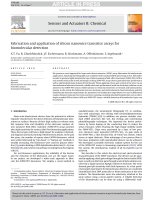
least 10 Gt each year. Chemically, chitin is composed of
(1 → 4) linked 2-acetamido-2-deoxy--d-glucose units
Fig. 1. Structure of chitin, chitosan and cellulose.
(or N-acetyl-d-glucosamine) [14], forming a long chain
linear polymer (Fig. 1). It is insoluble in most solvents.
Chitosan, the principal derivative of chitin, is obtained by
N-deacetylation to a varying extent that is characterized by
the degree of deacetylation, and is consequently a copolymer of N-acetyl-d-glucosamine and d-glucosamine. Chitin
and chitosan can be chemically considered as analogues
of cellulose, in which the hydroxyl at carbon-2 has been
replaced by acetamido and amino groups, respectively. Chitosan is insoluble in water, but the presence of amino groups
renders it soluble in acidic solutions below pH about 6.5. It
is important to note that chitin and chitosan are not single
chemical entities, but vary in composition depending on the
origin and manufacture process. Chitosan can be defined as
chitin sufficiently deacetylated to form soluble amine salts,
the degree of deacetylation necessary to obtain a soluble
product being 80–85% or higher.
Commercially, chitin and chitosan are obtained at a relatively low cost from shells of shellfish (mainly crabs,
shrimps, lobsters and krills), wastes of the seafood processing industry [15,18,20,22–24]. Basically, the process consits
of deproteinization of the raw shell material with a dilute
NaOH solution and decalcification with a dilute HCl solution. To result in chitosan, the obtained chitin is subjected
to N-deacetylation by treatment with a 40–45% NaOH solution, followed by purification procedures. Thus, production
and utilization of chitosan constitutes an economically attractive means of crustacean shell wastes disposal sought
worldwide.
Chitosan possesses distinct chemical and biological
properties [14–28a]. In its linear polyglucosamine chains
of high molecular weight, chitosan has reactive amino
and hydroxyl groups, amenable to chemical modifications
[14,18,19,23]. Additionally, amino groups make chitosan a
cationic polyelectrolyte (pKa ≈ 6.5), one of the few found
130
B. Krajewska / Enzyme and Microbial Technology 35 (2004) 126–139
in nature. This basicity gives chitosan singular properties: chitosan is soluble in aqueous acidic media at pH <
6.5 and when dissolved possesses high positive charge on
–NH3 + groups, it adheres to negatively charged surfaces,
it aggregates with polyanionic compounds, and chelates
heavy metal ions. Both the solubility in acidic solutions and
aggregation with polyanions impart chitosan with excellent gel-forming properties. Along with unique biological
properties that include biocompatibility, biodegradability to
harmless products, nontoxicity, physiological inertness, remarkable affinity to proteins, hemostatic, fungistatic, antitumoral and anticholesteremic properties, chitin and chitosan,
as yet underutilized, offer an extraordinary potential in a
broad spectrum of applications which are predicted to grow
rapidly once the standardized chitinous materials become
available. Crucially, as bio- and biodegradable polymers
chitin/chitosan materials are eco-friendly, safe for humans
and the natural environment.
Increasingly over the last decade chitin- and chitosanbased materials have been examined and a number of potential products have been developed for areas such as
[14,17,19,23,24,27,28b] wastewater treatment (removal of
heavy metal ions, flocculation/coagulation of dyes and proteins, membrane purification processes), the food industry
(anticholesterol and fat binding, preservative, packaging material, animal feed additive), agriculture (seed and fertilizer
coating, controlled agrochemical release), pulp and paper
industry (surface treatment, photographic paper), cosmetics
and toiletries (moisturizer, body creams, bath lotion).
But owing to the unparalleled biological properties,
the most exciting uses of chitin/chitosan-based materials are those in the area of medicine and biotechnology
[16,20–22,28a]. In medicine they may be employed as bacteriostatic and fungistatic agents, drug delivery vehicles,
drug controlled release systems, artificial cells, wound healing ointments/dressings, haemodialysis membranes, contact
lenses, artificial skin, surgical sutures and for tissue engineering. In biotechnology on the other hand, they may find
application as chromatographic matrices, membranes for
membrane separations, and notably as enzyme/cell immobilization supports.
As enzyme immobilization supports chitin- and chitosanbased materials are used in the form of powders, flakes and
gels of different geometrical configurations. Chitin/chitosan
powders and flakes are available as commercial products among others from Sigma-Aldrich and chitosan gel
beads (Chitopearl) from Fuji Spinning Co. Ltd. (Tokyo,
Japan). Otherwise the chitinous supports are laboratorymanufactured. Preparation of chitosan gels is promoted by
the fact that chitosan dissolves readily in dilute solutions
of most organic acids, including formic, acetic, tartaric
and citric acids, to form viscous solutions that precipitate
upon an increase in pH and by formation of water-insoluble
ionotropic complexes with anionic polyelectrolytes. In this
way chitosan gels in the form of beads, membranes, coatings,
capsules, fibres, hollow fibers and sponges can be manufac-
tured. Commonly, different follow-up treatments and modifications are applied to improve gel stability and durability.
The methods of chitosan gel preparation described in the
literature can be broadly divided into four groups: solvent
evaporation method, neutralization method, crosslinking
method and ionotropic gelation method [15,20,21,23–27].
3.1. Solvent evaporation method
The method is mainly used for the preparation of membranes and films, the latter being especially useful in preparing minute enzymatically active surfaces deposited on tips
of electrodes. A solution of chitosan in organic acid is cast
onto a plate or an electrode tip and allowed to dry, if possible at elevated temperature (ca. 65 ◦ C). Upon drying the
membrane/film is normally neutralized with a dilute NaOH
solution and crosslinked to avoid disintegration in solutions
of pH < 6.5. A crosslinking agent may also be mixed with
the initial chitosan solution before drying. Enzymes may be
immobilized on such prepared membranes either on their
surfaces by adsorption, frequently followed by crosslinking
(reticulation), or covalent binding, commonly preceded by
chemical activation of the surface, or included into chitosan
solution to achieve inclusion.
Spray drying is a variant of the solvent evaporation
method allowing the preparation of beads smaller in size
than those prepared with the other methods [44].
3.2. Neutralization method
If an acidic chitosan solution is mixed with alkali, an increase in pH results in precipitation of solid chitosan. This
method is exploited to produce chitosan precipitates, membranes, fibers, but foremost spherical beads of different sizes
and porosities. These are obtained by adding a chitosan
solution dropwise to a solution of NaOH, most frequently
prepared in water-ethanol mixtures, where ethanol, being
a non-solvent for chitosan, facilitates the solidification of
chitosan beads. Following the preparation, the beads are
commonly subjected to crosslinking. Enzyme immobilization, similar to the solvent evaporation method, is achieved
by binding onto the gel surface by adsorption, reticulation or covalent binding, or by inclusion if the enzyme is
dissolved in the initial chitosan solution.
3.3. Crosslinking method
In this method an acidic chitosan solution is subjected to
straightforward crosslinking by mixing with a crosslinking
agent, which results in gelling. Gels obtained in bulk solution are later crushed into particles. To obtain gel membranes, the chitosan solution cast on a plate is immersed
in a crosslinking bath, and to obtain beads the solution is
added dropwise therein. In the case of electrodes, crosslinking treatment is frequently done upon covering the tip of the
electrode with chitosan solution. Clearly, immobilization of
Table 4
Enzymes immobilized on chitin- and chitosan-based materials
Application
Support (preparation method)
Immobilization
Reference
Acid phosphatase (3.1.3.2)
F. Hydrophobic interaction chromatography; I.
Mercapto-chitin powder
Chitosan beads (b)
Chitosan precipitate (b)
I
III, IV
III
[29]
[30,31]
[32]
Alanine dehydrogenase (1.4.1.1)
E. Determination of l-alanine (medicine)
Chitosan beads
III
[33]
Alkaline phosphatase (3.1.3.1)
F. Hydrophobic interaction chromatography
C. Molecular cloning
Chitosan precipitate (b)
Chitosan beads (c)
III
III
[32]
[34]
Alkaline protease (3.4.21.62)
B. Production of laundry detergents
Chitin powder
Chitosan powder
III (78%)a
I (15%), III
[35]
[35]
H. Ester and peptide synthesis; transesterification
Chitosan beads
I
[36]
Alcohol dehydrogenase (1.1.1.1)
I.
Chitosan beads
Chitosan membrane (a)
III (25%)
III, IV
[37]
[38]
Alcohol oxidase (1.1.3.13)
Aminoacylase (3.5.1.14)
E. Determination of ethanol
B. Production of l-phenylalanine
Chitosan beads
Chitosan-coated alginate beads (d)
III
V (>100%)
[39]
[40]
␣-Amylase (3.2.1.1)
A. Hydrolysis of starch for glucose syrup and
E. for BOD analysis in waters
F. Hydrophobic interaction chromatography
Chitin powder
Chitosan beads
Chitosan precipitate (b)
Chitosan microbeads (a)
III (38%) [41]
I
III
V
[41,42]
[43]
[32]
[44]
-Amylase (3.2.1.2)
A. Production of high maltose syrup from starch
Chitosan beads
I
[45]
␣-l-Arabinofuranosidase (3.2.1.55)
A. Aromatization of musts, alcoholic beverages and
fruit juices
Bromelain (3.4.22.32)
Carbonic anhydrase (4.2.1.1)
I.
I.
Chitosan powder
Glyceryl-chitosan powder
Chitosan particles (c)
Chitosan beads
Chitosan-coated alginate beads (d)
I, II (3.2%) [47], III
II
V
IV
V
[46–48]
[46]
[49]
[50,51]
[52]
Catalase (1.11.1.6)
A. Removal of H2 O2 from food
C. Treatment of hyperoxaluria
I.
Chitosan powder
Chitosan film (a)
Chitosan membrane (a)
Chitosan-organosilane particles (c)
Chitosan beads (c)
I, IV, II
V
III (4%)
I
V
[53a,b]
[54]
[55]
[56]
[57]
Cellulase (3.2.1.4)
A. Decrease in viscosity of fruit/vegetable juices
F. Affinity chromatography
Chitin powder
Chitosan beads (b)
Chitosan solution
IV (15%)
I
protective additive
[58]
[59]
[60]
Chitosanase (3.2.1.132)
I.
Chitin powder
III
[61]
␣-Chymotrypsin (3.4.21.1)
H. Ester and peptide synthesis
F. Preparation of trypsin-free chymotrypsin
Chitin film
Chitosan beads
Chitosan-magnetite beads
II
I
I
[62]
[36]
[63]
Creatinine deaminase (3.5.4.21)
D. Creatinine biosensor (medical diagnosis)
Chitosan membrane (a)
I, III
[64]
B. Krajewska / Enzyme and Microbial Technology 35 (2004) 126–139
Enzyme (EC number)
131
132
I.
Chitosan powder
I (3.5%), IV(5.2%)
[65]
Dextranase (3.2.1.11)
C. Partial hydrolysis of dextran for preparation of
blood substitutes and B. of dentifrices
Chitin powder and colloidal chitin
Chitosan powder
I, III
I, III (63%)
[66]
[66]
endo-1,4--Xylanase (3.2.1.8)
C. Conversion of hemicelluloses (pulp industry)
Chitosan powder
Chitosan beads
Chitosan-xanthan beads (d)
I (24%)
III (20%)
V (180%) [70]
[67]
[67]
[68–70]
Ficin (3.4.22.3)
Galactose oxidase (1.1.3.9)
I.
D. Galactose biosensor
Chitosan beads
Chitosan membrane (a)
IV
III
[50]
[71a]
␣-Galactosidase (3.2.1.22)
A. Raffinose hydrolysis in beet molasses
C. Blood group specificity; Fabry disease
Chitin powder
IV (67%)
[71b,c]
-Galactosidase (3.2.1.23)
A. Hydrolysis of lactose (lactose-free dairy products)
Chitin powder
Chitosan powder
Chitosan beads (b)
Chitosan beads
Chitosan-polyphosphate beads (d)
Chitosan precipitate (b)
III
III
III (100%) [75]
I, III
V
II
[72,73]
[74]
[75,76]
[77–79]
[80]
[81]
Glucoamylase (3.2.1.3)
A. Hydrolysis of starch (ethanol production)
Chitin powder
Chitosan magnetite beads (c)
Chitosan powder
Chitosan beads
III
I
I
I, III
[42]
[63]
[82]
[83]
Glucose oxidase (1.1.3.4)
D. Glucose biosensors
E. Determination of glucose
Chitin powder
-Chitin membrane (coagulation)
Chitin film (coagulation)
Chitosan beads
Chitosan membrane (a)
Chitosan membrane (a, c, d)
Sol–gel/chitosan membrane (c)
Chitosan-organosilane particles (c)
Chitosan beads-liposomes
I
V
I
III
III
V
V
I
III
[84]
[85,86]
[87]
[88]
[71a,89a,89b]
[90–93a]
[93b]
[56,93c]
[94]
␣-Glucosidase (3.2.1.20)
A. Hydrolysis of maltose (food/feed additives)
Chitosan beads
III
[95]
-Glucosidase (3.2.1.21)
A. Wine making and juice processing
F. Hydrophobic interaction chromatography
Chitosan
Chitosan
Chitosan
Chitosan
Chitosan
Chitosan
Chitosan
III, II (29%) [98]
V
III (60%), IV
I (90%)
III, IV
III
I
[48,96–98]
[49,99]
[100]
[101]
[31]
[32]
[63]
Glutamate dehydrogenase (1.4.1.2)
E. Glutamate determination (food industry and
medicine)
Chitosan membrane (a)
Succinyl-, glutaryl-, phtalyl-chitosan
membranes (a)
III
IV
[102]
[102]
Glutamate oxidase (1.4.3.11)
D. Glutamate biosensor
Chitosan membrane (a)
III
[71a]
powder
particles (c)
flakes
solution
beads (b)
precipitate (b)
magnetite beads (c)
B. Krajewska / Enzyme and Microbial Technology 35 (2004) 126–139
Cyclodextrin glycosyltransferase (2.4.1.19)
Table 4 (Continued )
Enzyme (EC number)
Application
Support (preparation method)
Immobilization
Reference
-Glycosidase (3.2.1.group)
A. Cellobiose hydrolysis for glucose production
Chitosan powder
Chitosan precipitate (b)
II
II
[103]
[104,105]
Horseradish peroxidase (1.11.1.7)
D. H2 O2 biosensor; E. determination of H2 O2
B. Oxidative polymerization of aniline
G. Removal of phenols from petroleum refinery
wastewaters
E. Inhibition-based determination of Hg(II)
Chitosan powder
Chitosan beads
Chitosan membrane (c)
III, IV (62%) [106b]
III
III
[106a,b]
[39,88,107]
[108]
Chitosan film (a)
Chitosan solution
Silica sol–gel chitosan film (c)
Chitosan-carbon film (a)
V
Protective additive
I
I
[54,109,110]
[111]
[112–114]
[115]
Chitosan powder
Chitosan solution
Chitosan microbeads (a)
Chitosan-organosilane particles (c)
Chitosan-magnetite beads (c)
I (91%), III (44%), IV (70%)
Protective additive
V
I
I
[116]
[117]
[44]
[56]
[63]
Isoamylase (3.2.1.68)
A. Hydrolysis of starch (glucose and maltose)
Chitin powder
III (46%)
[118,119]
Laccase (1.10.3.2)
B. Pulp and paper industry
G. Removal of phenols from effluents
Chitosan precipitate (b)
V, II (45%) [121]
[120,121]
Lactate oxidase (1.13.12.4)
Leucine dehydrogenase (1.4.1.9)
D. Lactate biosensor
E. Determination of l-leucine (medicine)
Chitosan-enzyme beads (d)
Chitosan beads
V
III
[122]
[33]
Limonoid glucosyltransferase (2.4.1.210)
A. Debittering of citrus juice
Chitosan powder
Chitosan precipitate (b)
III
III
[123]
[123]
Lipase (3.1.1.3)
H. Esterifications and transesterifications
B. Hydrolysis of olive oil
Chitosan flakes
Chitosan beads
Chitosan beads
Chitosan-polyphosphate beads (d)
Chitosan membrane (a)
Chitosan-PVA membrane (a)
Chitosan-xanthan beads (d)
I (7.1%)
I (14.7%) [124], IV
IV + II (91.5%)
V (42–50%)
V, III (47%) [130]
V
V (90–99%)
[124]
[124–126a,c]
[126b]
[127,128]
[129,130]
[129]
[131–133]
Lysozyme (3.2.1.17)
F. Affinity membrane chromatography
A. Cheesemaking
Microporous chitin membrane (a)
Chitosan powder
PHEMA-chitosan membranes
microporous chitin membrane (a)
I
I (10%)
I
[134]
[135]
[136–138]
Neutral proteinase (3.4.24.28)
Nucleoside phosphorylase (2.4.2.1)
5 -Nucleotidase (3.1.3.5)
Octopine dehydrogenase (1.5.1.11)
Oxalate oxidase (1.2.3.4)
A. Hydrolysis of soybean protein
E. Determination of fish and shellfish freshness
E. Determination of fish and shellfish freshness
E. Determination of shellfish freshness
C. Treatment of hyperoxaluria
Chitosan
Chitosan
Chitosan
Chitosan
Chitosan
II
III
III
III
II
[139]
[140–142]
[140,142]
[143]
[53b]
Papain (3.4.22.2)
A. Removal of “chill haze” in beers; I.
B. Hydrolysis of collagen/keratin (cosmetics)
Chitin powder
Chitosan beads
Chitosan precipitate (b)
II
IV
II (82%)
[144]
[50,145,146]
[147]
Pectin lyase (4.2.2.10)
A. Reduction of fruit/vegetable juices’ viscosity
Chitin powder
III (26%)
[58]
precipitate (b)
beads
beads
beads
powder
133
A. Hydrolysis of sucrose (production of invert sugar)
B. Krajewska / Enzyme and Microbial Technology 35 (2004) 126–139
Invertase (3.2.1.26)
Chitosan beads
I (15%)
[148]
Chitosan precipitate (b)
III
[32]
Pepsin (3.4.23.1)
Phospholipase A2 (3.1.1.4)
I.
C. Lowering plasma cholesterol level
Succinylated chitosan powder
Chitosan beads
IV (80%)
IV (50%)
[149]
[150]
Proteases (3.4.groups)
A. Casein hydrolysate debittering; I.
Chitin powder
Chitin film
Chitosan-xanthan beads (d)
III
II
V
[151]
[62]
[68,133]
Pullulanase (3.2.1.41)
A. Hydrolysis of starch (glucose/maltose syrup)
Chitin powder
Chitosan-magnetite particles (c)
Chitosan powder
Chitosan beads
III
IV
I, III
I
[152]
[153]
[152]
[45]
Putrescine oxidase (1.4.3.10)
E. Determination of meat freshness
Chitosan beads
III
[154]
␣-l-Rhamnopyranosidase (3.2.1.40)
A. Aromatization of musts, alcoholic beverages and fruit juices
Chitin powder
Chitosan powder
Chitosan particles (c)
II
III, II
V
[155]
[48,155]
[49]
Sulfite oxidase (1.8.3.1)
D. Sulfite biosensor
Chitosan-PHEMA membrane (b)
I
[156,157]
Tannase (3.1.1.20)
A. Hydrolysis of tea tannins
Chitin powder and colloidal chitin
Chitosan precipitate (b)
Chitosan-triphosphate beads (d)
III, I
III
V
[158]
[158]
[159]
Transglutaminase (2.3.2.13)
A. Deamidation of food proteins
Chitosan beads
III
[160]
Trypsin (3.4.21.4)
F. Affinity purification
A. Hydrolysis of proteins
Chitin flakes
Chitosan-magnetite particles (c)
II, IV (67%)
I
[161]
[162]
Tyrosinase (1.14.18.1)
C. Production of l-DOPA
G. Detection and removal of phenols
Chitin flakes
Chitin powder
Chitosan flakes
Chitosan beads (b)
Chitosan-organosilane film (c)
Chitosan membrane (a, b)
III
I (95%)
III
V (15%) [163], III
IV
I
[163]
[164]
[163,165]
[163,165]
[166]
[167,168]
Urease (3.5.1.5)
C.
D.
G.
A.
Chitosan-triphosphate beads (d)
Chitosan beads
Chitosan membrane (a)
Chitosan-PVA capsules (d)
Chitosan-PGMA precipitate (d)
Chitosan-coated alginate beads (d)
Chitosan-organosilane particles (c)
III (64%)
III (100%)
I, II, III (94%) [172]
V
I (82%)
V
I
[169]
[170]
[171–173]
[174]
[175]
[176]
[56]
Uricase (1.7.3.3)
Xanthine oxidase (1.1.3.22)
E. Determination of uric acid (medicine)
E. Determination of fish freshness
Chitosan membrane (a)
Chitosan beads
IV
III
[177]
[140–142]
-Xylolidase (3.2.1.37)
B. Production of lignocellulosic fibers
Chitosan powder
Chitosan beads
I (25%)
III (33%)
[67]
[67]
Artificial kidney
Urea biosensor
Treatment of fertilizer effluents
Removal of urea from beverages and food
Applications are presented in nine cathegories: (A) food industry; (B) industries other than food; (C) medicine; (D) biosensors; (E) enzyme reactors for biosensing; (F) separation, purification and
recovery of enzymes; (G) environmental; (H) chemical synthesis; (I) immobilization studies. Support preparation methods are presented as: (a) solvent evaporation method; (b) neutralization method; (c)
crosslinking method; (d) ionotropic gelation method. Commercial powders, flakes or gel beads are not marked. Immobilizations are presented as five techniques: (I) adsorption of enzyme on support; (II)
adsorption of enzyme on support followed by cross-linking with glutaraldehyde (reticulation); (III) covalent binding of enzyme to glutaraldehyde-activated support; (IV) covalent binding of enzyme to
support activated with agents other than glutaraldehyde; (V) gel inclusion.
a In brackets activity retention is given, if reported.
B. Krajewska / Enzyme and Microbial Technology 35 (2004) 126–139
C. Production of pectate oligosaccharides (inducers of flowering and
antibacterial agents)
F. Hydrophobic interaction chromatography
134
Pectinase (3.2.1.15)
B. Krajewska / Enzyme and Microbial Technology 35 (2004) 126–139
enzymes on such prepared gels does not require chemical
activation, as the crosslinker, normally a bifunctional agent,
fullfils two functions, crosslinking and activation. The enzyme may also be entrapped in the gel if mixed with chitosan prior to crosslinking.
Overwhelmingly, as a crosslinking and surface activating
agent glutaraldehyde is used. This is due to its reliability
and ease of use, but more importantly, due to the availability of amino groups for the reaction with glutaraldehyde not
only on enzymes but also on chitosan. Other less frequently
employed difunctional agents include glyoxal [30,31,57],
tris(hydroxymethyl)phosphine P(CH2 OH)3 [38,100], hexamethylenediamine [65,153], ethylenediamine [116], carbodiimides [102,106b,126b,149], epichlorohydrin [129] and
N-hydroxysuccinimide [50,51].
A comparatively newly developed method of chitosan
gelling is by use of sol–gel processes resulting in chitosanorganosilane hybrid gels. The method employs silylating agents, such as (CH3 O)3 Si–R–NH2 [56], (CH3 O)2 CH3 Si–R–O–CO–CH=CH2 [113,166], (C2 H5 O)3 Si–O–
C2 H5 [114], however, often regarded simply as crosslinkers.
3.4. Ionotropic gelation method (or coacervation)
By virtue of the attraction of oppositely-charged
molecules, chitosan, owing to its cationic polyelectrolyte
nature, spontaneously forms water-insoluble complexes
with anionic polyelectrolytes [22,27,69]. The anionic polyelectrolytes used include alginate, carrageenan, xanthan,
various polyphosphates and organic sulfates or enzymes
themselves [122]. The method is utilized chiefly for the
preparation of gel beads, which is achieved by adding an
anionic polyelectrolyte solution dropwise into an acidic
chitosan solution. Enzyme immobilization is achieved here
by preparing an enzyme-containing anionic polyelectrolyte
solution prior to gelation. The enzyme is immobilized by
inclusion in the interior of the beads/capsules.
An overview of enzymes immobilized on chitin- and
chitosan-based materials, reported in the literature over the
last decade, is presented in Table 4. It implies that there continues to be vivid interest in utilizing chitin-based materials,
predominantly chitosan, as a promising enzyme immobilization support for a multiplicity of applications ranging
from the wine, sugar and fish industries, through organic
contaminants removal from wastewaters to sophisticated
biosensors for both in situ measurements of environmental
pollutants and metabolite control in artificial organs. Studies like those summarized in Table 4 can play a decisive
role in advancing this hitherto underutilized, renewable
biopolymer of great potential to the market of biomaterials.
Acknowledgments
This work was supported by the KBN grant no. PB
7/T09A/048/20.
135
References
[1] Bullock C. Immobilised enzymes. Sci Progress 1995;78:119–34.
[2] Woodley JM. Immobilized biocatalysts. Solid Supports Catal Org
Synth 1992;254–271.
[3] Chaplin MF, Bucke C. Enzyme technology. Cambridge University
Press; 1990.
[4] Kennedy JF, Cabral JMS. Immobilized enzymes. In: Scouten WH,
editor. Solid phase biochemistry. Analytical and synthetic aspects.
New York: John Wiley & Sons; 1983. p. 253–391.
[5] van de Velde F, Lourenço ND, Pinheiro HM, Bakker M.
Carrageenan: a food-grade and biocompatible support for
immobilisation techniques. Adv Synth Catal 2002;344:815–35.
[6] Tischer W, Wedekind F. Immobilized enzymes: methods and
applications. Top Curr Chem 1999;200:95–126.
[7] Scouten WH, Luong JHT, Brown RS. Enzyme or protein
immobilization techniques for applications in biosensor design.
TIBTECH 1995;13:178–85.
[8] Wiseman A. Designer enzyme and cell applications in industry and
in environmental monitoring. J Chem Tech Biotechnol 1993;56:3–
13.
[9] Carrea G, Riva S. Properties and synthetic applications of enzymes
in organic solvents. Angew Chem Int Ed 2000;39:2226–54.
[10] Guilbault GG. Immobilized enzyme electrode probes. In: Scouten
WH, editor. Solid phase biochemistry. Analytical and synthetic
aspects. New York: John Wiley & Sons; 1983. p. 479–505.
[11] Davis J, Vaughan DH, Cardosi MF. Elements of biosensor
construction. Enzyme Microb Tech 1995;17:1030–5.
[12] Wilson GS, Hu Y. Enzyme-based biosensors for in vivo
measurements. Chem Rev 2000;100:2693–704.
[13] Pi¸skin AK. Therapeutic potential of immobilized enzymes. NATO
ASI Series, Ser E 1993;252:191–200.
[14] Peter M. Applications and environmental aspects of chitin and
chitosan. J Macromol Sci Pure Appl Chem 1995;A32:629–40.
[15] Hudson SM, Smith C. Polysaccharides: chitin and chitosan:
chemistry and technology of their use as structural materials. In:
Kaplan DL, editor. Biopolymers from renewable resources. Berlin:
Springer; 1998. p. 96–118.
[16] Khor E. Chitin: a biomaterial in waiting. Curr Opin Solid State
Mater Sci 2002;6:313–7.
[17] Felse PA, Panda T. Studies on applications of chitin and its
dervatives. Bioprocess Eng 1999;20:505–12.
[18] Tharanathan RN, Kittur FS. Chitin—the undisputed biomolecule of
great potential. Crit Rev Food Sci Nutr 2003;43:61–87.
[19] Kurita K. Controlled functionalization of the polysaccharide chitin.
Prog Polym Sci 2001;26:1921–71.
[20] Paul W, Sharma CP. Chitosan, a drug carrier for the 21st century:
a review. STP Pharmaceut Sci 2000;10:5–22.
[21] Felt O, Buri P, Gurny R. Chitosan: a unique polysaccharide for
drug delivery. Drug Dev Ind Pharm 1998;24:979–93.
[22] Singla AK, Chawla M. Chitosan: some pharmaceutical and
biological aspects—an update. Pharm Pharmacol 2001;53:1047–67.
[23] Dutta PK, Ravikumar MNV, Dutta J. Chitin and chitosan for
versatile applications. J Macromol Sci 2002;C42:307–54.
[24] Shahidi F, Arachchi JKV, Jeon YJ. Food applications of chitin and
chitosan. Food Sci Technol 1999;10:37–51.
[25] Agboh OC, Qin Y. Chitin and chitosan fibers. Polym Adv Technol
1996;8:355–65.
[26] Kawamura Y, Yoshida H, Asai S, Kurahashi I, Tanibe H. Effects
of chitosan concentration and precipitation bath concentration on
the material properties of porous crosslinked chitosan beads. Sep
Sci Technol 1997;32:1959–74.
[27] Kubota N, Kikuchi Y. Macromolecular complexes of chitosan.
In: Dumitriu S, editor. Polysaccharides. New York: Dekker; 1998.
p. 595–628.
136
B. Krajewska / Enzyme and Microbial Technology 35 (2004) 126–139
[28] (a) Krajewska B. Chitin and its derivatives as supports for
immobilization of enzymes. Acta Biotechnol 1991;11:269–77;
(b) No HK, Meyers SP. Application of chitosan for treatment of
wastewaters. Rev Environ Contam Toxicol 2000;163:1–28.
[29] Kurita K, Yoshino H, Nishimura S-I, Ishii S, Mori T,
Nishiyama Y. Mercapto-chitins: a new type of supports for
effective immobilization of acid phosphatase. Carbohydr Polym
1997;32:171–5.
[30] Juang R-S, Wu F-C, Tseng R-L. Solute adsorption and enzyme
immobilization on chitosan beads prepared from shrimp shell
wastes. Bioresour Tech 2001;80:187–93.
[31] Juang R-S, Wu F-C, Tseng R-L. Use of chemically modified
chitosan beads for sorption and enzyme immobilization. Adv
Environ Res 2002;6:171–7.
[32] Agarval R, Gupta MN. Evaluation of glutaraldehyde-modified
chitosan as a matrix for hydrophobic interaction chromatography.
Anal Chim Acta 1995;313:253–7.
[33] Kiba N, Oyama Y, Furusawa M. Determination of aliphatic
amino acids in serum by HPLC with fluorimetric detection using
co-immobilized enzyme reactor. Talanta 1993;40:657–60.
[34] Zubriene A, Budriene S, Lubiene J, Dienys G. Immobilized
alkaline phosphatase for molecular cloning. Biocatal Biotransform
2002;20:423–7.
[35] Abdel-Naby MA, Ismail A-MS, Ahmed SA, Abdel-Fattah AF.
Production and immobilization of alkaline protease from Bacillus
mycoides. Bioresour Tech 1998;64:205–10.
[36] Kise H, Hayakawa A. Immobilization of proteases to porous
chitosan beads and their catalysis for ester and peptide synthesis in
organic solvents. Enzyme Microb Tech 1991;13:584–8.
[37] Soni S, Desai JD, Devi S. Immobilization of yeast alcohol
dehydrogenase and covalent binding to polymeric support. J Appl
Polym Sci 2001;82:1299–305.
[38] Cochrane FC, Petach HH, Henderson W. Application of tris(hydroxymethyl)phosphine as a coupling agent for alcohol dehydrogenase
immobilization. Enzyme Microb Tech 1996;18:373–8.
[39] Taniai T, Sukuragawa A, Okutani T. Fluorometric determination
of ethanol in liquor samples by flow-injection analysis using an
immobilized enzyme-reactor column with packing prepared by
coupling alcohol oxidase and peroxidase onto chitosan beads. J
AOAC Int 2001;84:1475–83.
[40] Lee KH, Lee PM, Siaw YS. Studies of l-phenyl-alanine production
by immobilized aminoacylase in stabilized calcium alginate beads.
J Chem Tech Biotechnol 1992;54:375–82.
[41] Abdel-Naby MA, Hashem AM, Esawy MA, Abdel-Fattah AF.
Immobilization of Bacillus subtilis ␣-amylase and characterization
of its enzymatic properties. Microbiol Res 1999;153:319–25.
[42] Reiss M, Heibges A, Metzger J, Hartmeier W. Determination of
BOD-values of starch-containing waste water by a BOD-biosensor.
Biosens Bioelectron 1998;13:1083–90.
[43] Morita T, Karube I. Enzymatic hydrolysis in water-imiscible organic
solvent, two-phase systems. Appl Biochem Biotechnol 1995;55:75–
86.
[44] Siso MIG, Lang E, Carrenõ-Gómez B, Becerra M, Espinar FO,
Méndez JB. Enzyme encapsulation on chitosan microbeads. Process
Biochem 1997;32:211–6.
[45] Noda T, Furuta S, Suda I. Sweet potato -amylase immobilized on
chitosan beads and its application in the semi-continuous production
of maltose. Carbohydr Polym 2001;44:189–95.
[46] Spagna G, Andreani F, Salatelli E, Romagnoli D, Casarini D, Pifferi
PG. Immobilization of the glycosidases: ␣-l-arabinofuranosidase
and ␣-d-glucopyranosidase from Aspergillus niger on chitosan
derivative to increase the aroma of wine. Part II. Enzyme Microb
Tech 1998;23:413–21.
[47] Spagna G, Andreani F, Salatelli E, Romagnoli D, Pifferi PG.
Immobilization of ␣-l-arabinofuranosidase on chitin and chitosan.
Process Biochem 1998;33:57–62.
[48] Martino A, Schiraldi C, Di Lazzaro A, Fiume I, Spagna G, Pifferi
PG, et al. Improvement of the flavour of Falanghina white wine
using a purified glycosidase preparation from Aspergillus niger.
Process Biochem 2000;36:93–102.
[49] Spagna G, Barbagallo RN, Greco E, Manenti I, Pifferi PG.
A mixture of purified glycosidases from Aspergillus niger for
oenological application immobilized by inclusion in chitosan gels.
Enzyme Microb Tech 2002;30:80–9.
[50] Hayashi T, Ikada Y. Protease immobilization onto porous chitosan
beads. J Appl Polym Sci 1991;42:85–92.
[51] Seo H, Itoyama K, Morimoto K, Takagishi T, Oka M, Hayashi
T. Spacer effects on enzymatic activity of bromelain immobilized
onto porous chitosan beads. Eur Polym J 1998;34:917–22.
[52] Simsek-Ege FA, Bond GM, Stringer J. Matrix molecular weight
cut-off for encapsulation of carbonic anhydrase in polyelectrolyte
beads. J Biomater Sci Polym Ed 2002;13:1175–87.
[53] (a) Pifferi PG, Bonora V, Spagna G, Tramontini M. Immobilization
of catalase on macromolecular supports activated with acid dyes.
Process Biochem 1993;28:29–38;
(b) Ramakrishnan V, Lathika KM, D’Souza SJ, Singh BB, Raghavan
KG. Investigation with chitosan-oxalate oxidase-catalase conjugate
for degrading oxalate from hyperoxaluric rat chyme. Ind J Biochem
Biophys 1997;34:373–8.
[54] Huang H, Hu N, Zeng Y, Zhou G. Electrochemistry and
electrocatalysis with heme proteins in chitosan biopolymer films.
Anal Biochem 2002;308:141–51.
[55] Çetinus SA,
¸
Öztop HN. Immobilization of catalase on chitosan
film. Enzyme Microb Tech 2000;26:497–501.
[56] Airoldi C, Monteiro OAC. Chitosan-organosilane hybrids—syntheses, characterization, copper adsorption and enzyme immobilization.
J Appl Polym Sci 2000;77:797–804.
[57] Çetinus SA,
¸ Öztop HN. Immobilization of catalase into chemically
crosslinked chitosan beads. Enzyme Microb Tech 2003;32:889–94.
[58] Vaillant F, Millan A, Millan P, Dormier M, Decloux M, Reynes M.
Co-immobilized pectinlyase and endocellulase on chitin and nylon
supports. Process Biochem 2000;35:989–96.
[59] Roy I, Sardar M, Gupta MN. Exploiting unusal polysaccharides
for separation of enzymes on fluidized bed. Enzyme Microb Tech
2000;27:53–65.
[60] Darias R, Villalonga R. Functional stabilization of cellulase by
covalent modification with chitosan. J Chem Tech Biotechnol
2001;76:489–93.
[61] Zeng J, Zheng L-Y. Studies on Penicillium sp. ZDZ1 chitosanase
immobilized on chitin by cross-linking reaction. Process Biochem
2002;38:531–5.
[62] Ge SJ, Zhang L-X. The immobilized porcine pancreatic
exopeptidase and its application in casein hydrolysates debittering.
Appl Biochem Biotechnol 1996;59:159–65.
[63] Ghosh M, Tyagi R, Gupta MN. Preparation of trypsin free
chymotrypsin. Biotechnol Tech 1995;9:149–52.
[64] Magalhães JMCS, Machado AASC. Array of potentiometric
sensors for the analysis of creatinine in urine samples. Analyst
2002;127:1069–75.
[65] Sobral KCA, Rodrigues RMO, De Oliveira RD, De Moraes
FF, Zanim GM. Immobilization of cyclodextringlycosyltransferase
(CGTase) from Bacillus firmus in commercial chitosan. J Incl Phen
Macrocycl Chem 2002;44:383–6.
[66] Abdel-Naby MA, Ismail A-MS, Ahmed SA, Abdel-Fattah AM,
Abdel-Fattah AF. Preparation and some properties of immobilized
Penicillinum funiculosum 258 dextranase. Process Biochem
1999;34:391–8.
[67] Abdel-Naby MA. Immobilization of Aspergillus niger NRC 107
xylanase and -xylolidase, and properties of the immobilized
enzymes. Appl Biochem Biotechnol 1993;38:69–81.
[68] Dumitriu S, Magny P, Montané D, Vidal PF, Cornet E. Polyionic
hydrogels obtained by complexation between xanthan and chitosan:
their properties as supports for enzyme immobilization. J Bioact
Compat Polym 1994;9:184–209.
B. Krajewska / Enzyme and Microbial Technology 35 (2004) 126–139
[69] Dumitriu S, Chornet E. Inclusion and release of proteins from
polysaccharide-based polyion complexes. Adv Drug Deliv Rev
1998;31:223–46.
[70] Dumitriu S, Chornet E. Immobilization of xylanase in
chitosan-xanthan hydrogels. Biotechnol Prog 1997;13:539–45.
[71] (a) Wang Y, Zhu J, Zhu R, Zhu Z, Lai Z, Chen Z. Chitosan/Prussian
blue-based biosensors. Meas Sci Technol 2003;14:831–6;
(b) Önal S, Telefoncu A. Comparison of chitin and Amberlite
IRA-938 for ␣-galactosidase immobilization. Artif Cells Blood
Subst Biotechnol 2003;31:19–33;
(c) Önal S, Telefoncu A. Preparation and properties of
␣-galactosidase chemically attached to activated chitin. Artif Cells
Blood Subst Biotechnol 2003;31:339–55.
[72] Illanes A, Altamirano C, Aillapán A, Tomasello G, Zuñiga ME.
Packed-bed reactor performance with immobilized lactase under
thermal inactivation. Enzyme Microb Tech 1998;23:3–9.
[73] Illanes A, Wilson L, Tomasello G. Effect of modulation of enzyme
inactivation on temperature optimization for reactor operation with
chitin-immobilized lactase. J Mol Catal B: Enzym 2001;11:531–40.
[74] Figueroa ARC, Talavera J, Colomina M. Flow optimization in a
class of enzymatic plug-flow reactor. Biotechnol Prog 1997;13:109–
12.
[75] Rejikumar S, Devi S. Immobilization of -galactosidase onto
polymeric supports. J Appl Polym Sci 1995;55:871–8.
[76] Rejikumar S, Devi S. Hydrolysis of lactose and milk whey using a
fixed-bed reactor containing -galactosidase covalently bound onto
chitosan and cross-linked poly(vinyl alcohol). Int J Food Sci Tech
2001;36:91–8.
[77] Shin H-J, Park J-M, Yang J-W. Continuous production of galactooligosaccharides from lactose by Bullera singularis -galactosidase
immobilized in chitosan beads. Process Biochem 1998;33:787–92.
[78] Sheu D-C, Li S-Y, Duan K-J, Chen CW. Production of galactooligosaccharides by -galactosidase immobilized on glutaraldehydetreated chitosan beads. Biotechnol Tech 1998;12:273–6.
[79] Carrara CR, Rubiolo AC. Immobilization of -galactosidase on
chitosan. Biotechnol Prog 1994;10:220–4.
[80] Ghanem A, Skonberg D. Effect of preparation method on the capture
and release of biologically active molecules in chitosan gel beads.
J Appl Polym Sci 2002;84:405–13.
[81] Portaccio M, Stellato S, Rossi S, Bencivenga U, Eldin MSM,
Gaeta FS, et al. Galactose competitive inhibition of -galactosidase
(Aspergillus oryzae) immobilized on chitosan and nylon supports.
Enzyme Microb Tech 1998;23:101–6.
[82] Takahashi T, Kayama N. Binding to chitosan of multiple forms
of glucoamylases from Aspergillus saitoi and Rhizopus sp. Chem
Pharm Bull 1992;40:2775–9.
[83] Dhar GM, Mitsutomi M, Ohtakara A. Immobilization of
glucoamylases on chitosan beads and application to the conversion
of starch to glucose. Bull Fac Agr Saga Univ 1993;74:59–68.
[84] Sugawara K, Takano T, Fukushi H, Hoshi S, Akatsuka K,
Kuramitz H, et al. Glucose sensing by a carbon-paste electrode
containing chitin modified with glucose oxidase. J Electroanal Chem
2000;482:81–6.
[85] Ohashi E, Koriyama T. Simple and mild preparation of an
enzyme-immobilized membrane for a biosensor using -type
crystalline chitin. Anal Chim Acta 1992;262:19–25.
[86] Ohashi E, Karube I. Development of a thin membrane glucose
sensor using -type crystalline chitin for implantable biosensor. J
Biotechnol 1995;40:13–9.
[87] Sugawara K, Fukushi H, Hoshi S, Akatsuka K. Electrochemical
sensing of glucose at a platinum electrode with a chitin/glucose
oxidase film. Anal Sci 2000;16:1139–43.
[88] Taniai T, Sakuragawa A, Okutani T. Fluorometric determination
of glucose by flow injection analysis using immobilized
enzyme-reactor columns prepared by coupling glucose oxidase and
peroxidase onto chitosan beads. Anal Sci 2000;16:517–21.
137
[89] (a) Zhu J, Zhu Z, Lai Z, Wang R, Guo X, Wu X, et al. Planar
amperometric glucose sensor based on glucose oxidase immobilized
by chitosan film on Prussian blue layer. Sensors 2002;2:127–36;
(b) Hsieh B-C, Cheng T-J, Wang T-Y, Chen RLC. Use of
chitosan membrane from the carapace of the soldier crab
Mictyris brevidactylus for biosensor construction. Mar Biotechnol
2003;5:119–25.
[90] Miao Y, Chia LS, Goh NK, Tan SN. Amperometric glucose
biosensor based on immobilization of glucose oxidase in
chitosan matrix cross-linked with glutaraldehyde. Electroanalysis
2001;13:347–9.
[91] Chen L, Gorski W. Bioinorganic composites for enzyme electrodes.
Anal Chem 2001;73:2862–8.
[92] Wei X, Cruz J, Gorski W. Integration of enzymes and electrodes:
spectroscopic and electrochemical studies of chitosan-enzyme films.
Anal Chem 2002;74:5039–46.
[93] (a) Xu XH, Han B, Fu YS, Han J, Shi HB, Wu B, et al. Preparation
of chitosan/glucose oxidase nanolayered films for electrode
modification by the technique of layer-by-layer self-assembly.
J Mater Sci Lett 2003;22:695–7;
(b) Chen X, Jia J, Dong S. Organically modified sol–gel/chitosan
composite based glucose biosensor. Electroanalysis 2003;15:608–
12;
(c) Yang YM, Wang JW, Tan RX. Immobilization of glucose oxidase
on chitosan-SiO2 gel. Enzyme Microb Tech 2004;34:126–31.
[94] Wang S, Yoshimoto M, Fukunaga K, Nakao K. Optimal covalent
immobilization of glucose oxidase-containing liposomes for highly
stable biocatalyst in bioreactor. Biotechnol Bioeng 2003;83:444–53.
[95] Sheu D-C, Huang C-I, Duan K-J. Production of isomaltooligosaccharides by a ␣-glucosidase immobilized in chitosan beads and
by polyethyleneimine-glutaraldehyde treated mycelia of Aspergillus
carbonarius. Biotechnol Tech 1997;11:287–91.
[96] Martino A, Durante M, Pifferi PG, Spagna G, Bianchi G.
Immobilization of -glucosidase from a commercial preparation.
Part 1. A comparative study of natural supports. Process Biochem
1996;31:281–5.
[97] Martino A, Pifferi PG, Spagna G. Immobilization of -glucosidase
from a commercial preparation. Part 2. Optimization of the immobilization process on chitosan. Process Biochem 1996;31:287–93.
[98] Gallifuoco A, D’Ercole L, Alfani F, Cantarella M, Spagna G,
Pifferi PG. On the use of chitosan-immobilized -glucosidase in
wine-making: kinetics and enzyme inhibition. Process Biochem
1998;33:163–8.
[99] Abdel-Fattah AF, Osman MY, Abdel-Naby MA. Production and
immobilization of cellobiase from Aspergillus niger A20. Chem
Eng J 1997;68:189–96.
[100] Oswald PR, Evans RA, Henderson W, Daniel RM, Fee CJ.
Properties of a thermostable -glucosidase immobilized using
tris(hydroxymethyl)phosphine as a highly effective coupling agent.
Enzyme Microb Tech 1998;23:14–9.
[101] Agarwal L, Gupta MN. Sequential precipitation with reversibly
soluble insoluble polymers as a bioseparation strategy: purification
of -glucosidase from Trichoderma longibrachiatum. Protein Exp
Purif 1996;7:294–8.
[102] Petach HH, Driscoll J. Transparent chitosan derivatives for the
immobilization of glutamate dehydrogenase. Biotechnol Bioeng
1994;44:1018–22.
[103] D’Auria S, Pellino F, La Cara F, Barone R, Rossi M, Nucci
R. Immobilization on chitosan of a thermophilic -glycosidase
expressed in Saccharomyces cerevisiae. Appl Biochem Biotechnol
1996;61:157–66.
[104] Briante R, La Cara F, Febbraio F, Patumi M, Nucci R. Bioactive
derivatives from oleuropein by a biotransformation on Olea
europaea leaf extracts. J Biotechnol 2002;93:109–19.
[105] Briante R, La Cara F, Febbraio F, Barone R, Picciali G, Carolla R,
et al. Hydrolysis of oleuropein by recombinant -glycosidase from
hyperthermophilic archaeon Sulfolobus solfataricus immobilized on
chitosan matrix. J Biotechnol 2000;77:275–86.
138
B. Krajewska / Enzyme and Microbial Technology 35 (2004) 126–139
[106] (a) Jin Z, Su Y, Duan Y. A novel method for polyaniline synthesis
with immobilized horseradish peroxidase enzyme. Synthet Met
2001;122:237–42;
(b) Bindhu LV, Abraham TE. Immobilization of horseradish
peroxidase on chitosan for use in nonaqueous media. J Appl Polym
Sci 2003;88:1456–64.
[107] Sakuragawa A, Taniai T, Okutani T. Fluorometric determination of
microamounts of hydrogen peroxide with an immobilized enzyme
prepared by coupling horseradish peroxidase to chitosan beads.
Anal Chim Acta 1998;374:191–200.
[108] Miao Y, Tan SN. Amperometric hydrogen peroxide biosensor based
on immobilization of peroxidase in chitosan matrix crosslinked with
glutaraldehyde. Analyst 2000;125:1591–4.
[109] Veselova IA, Shekhovtsova TN. Visual determination of mercury(II)
using horseradish peroxidase immobilized on polyurethane foam.
Anal Chim Acta 1999;392:151–8.
[110] Shekhovtsova TN, Muginova SV, Bagirova NA. Determination of
organomercury compounds using immobilized peroxidase. Anal
Chim Acta 1997;344:145–51.
[111] Wagner M, Nicell JA. Peroxidase-catalyzed removal of phenols from
a petroleum refinery wastewater. Water Sci Tech 2001;43:253–60.
[112] Miao Y, Tan SN. Amperometric hydrogen peroxide biosensor with
silica sol–gel/chitosan film as immobilization matrix. Anal Chim
Acta 2001;437:87–93.
[113] Wang G, Xu J-J, Chen H-Y, Lu Z-H. Amperometric hydrogen
peroxide biosensor with sol–gel/chitosan network-like film as
immobilization matrix. Biosens Bioelectronics 2003;18:335–43.
[114] Zhou G-J, Wang G, Xu J-J, Chen H-Y. Reagentless chemiluminescence biosensor for determination of hydrogen peroxide
based on the immobilization of horseradish peroxidase on biocompatible chitosan membrane. Sens Actuators B 2002;81:334–9.
[115] Lei C-X, Hu SQ, Shen G-L, Yu R-Q. Immobilization of horseradish
peroxidase to a nano-Au monolayer modified chitosan-entrapped
carbon paste electrode for the detection of hydrogen peroxide.
Talanta 2003;59:981–8.
[116] Hsieh H-J, Liu P-C, Liao W-J. Immobilization of invertase via
carbohydrate moiety on chitosan to enhance its thermal stability.
Biotechnol Lett 2000;22:1459–64.
[117] Gómez L, Ramirez HL, Vilallonga R. Stabilization of invertase
by modification of sugar chains with chitosan. Biotechnol Lett
2000;22:347–50.
[118] Liu H-S, Chen W-H, Lai J-T. Immobilization of isoamylase
on carboxymethyl-cellulose and chitin. Appl Biochem Biotechnol
1997;66:57–67.
[119] Chen J-P, Lee J-J, Liu H-S. Comparison of isoamylase
immobilization to insoluble and temperature-sensitive reversibly
soluble carriers. Biotechnol Lett 1997;11:109–12.
[120] Vasquez-Duhalt R, Tinoco R, D’Antonio P, Topoleski LDT,
Payne GF. Enzyme conjugation to the polysaccharide chitosan:
smart biocatalysts and biocatalytic hydrogels. Bioconjugate Chem
2001;12:301–6.
[121] D’Annibale A, Stazi SR, Vinciguerra V, Di Mattia E, Sermanni
GG. Characterization of immobilized laccase from Lentinula edodes
and its use in olive-mill wastewater treatment. Process Biochem
1999;34:697–706.
[122] Wei X, Zhang M, Gorski W. Coupling the lactate oxidase to
electrodes by ionotropic gelation of biopolymer. Anal Chem
2003;75:2060–4.
[123] Karim MR, Hashinaga F. Preparation and properties of
immobilized pummelo limonoid glucosyltransferase. Process
Biochem 2002;38:809–14.
[124] Pereira EB, De Castro HF, De Moraes FF, Zanin GM. Kinetic
studies of lipase from Candida rugosa. Appl Biochem Biotechnol
2001;91-93:739–52.
[125] Pereira EB, De Castro HF, De Moraes FF, Zanin GM. Esterification
activity and stability of Candida rugosa lipase immobilized into
chitosan. Appl Biochem Biotechnol 2002;98:977–86.
[126] (a) Itoyama K, Tokura S, Hayashi T. Lipoprotein lipase immobilization onto porous chitosan beads. Biotechnol Prog 1994;10:225–9;
(b) Hung T-C, Giridhar R, Chiou S-H, Wu W-T. Binary
immobilization of Candida rugosa lipase on chitosan. J Mol Catal
B: Enzym 2003;26:69–78;
(c) Chiou S-H, Wu W-T. Immobilization of Candida rugosa lipase
on chitosan with activation of the hydroxyl groups. Biomaterials
2004;25:197–204.
[127] Betigeri SS, Neau SH. Immobilization of lipase using hydrophilic polymers in the form of hydrogel beads. Biomaterials
2002;23:3627–36.
[128] Alsarra IA, Betigeri SS, Zhang H, Evans BA, Neau SH. Molecular
weight and degree of deacetylation effects on lipase-loaded chitosan
bead characteristics. Biomaterials 2002;23:3637–44.
[129] Tan T, Wang F, Zhang H. Preparation of PVA/chitosan lipase
membrane reactor and its application in synthesis of monoglyceride.
J Mol Catal B: Enzym 2002;18:325–31.
[130] Amorim RVS, Melo ES, Carneiro-da Cunha MG, Ledingham WM,
Campos-Takaki GM. Chitosan from Syncephalastrum racemosum
used as a film support for lipase immobilization. Bioresour Tech
2003;89:35–9.
[131] Magnin D, Dumitriu S, Magny P, Chornet E. Lipase immobilization
into porous chitoxan beads: activities in aqueous and organic media
and lipase characterization. Biotechnol Prog 2001;17:734–7.
[132] Dumitriu S, Chornet E, Vidal PF, Moresoli C. Polyionic hydrogels as
supports for immobilization of lipase. Biotechnol Tech 1995;9:833–
6.
[133] Dumitriu S, Chornet E. Inclusion and release of proteins from
polysaccharide-based polyion complexes. Adv Drug Del Rev
1998;31:223–46.
[134] Ruckenstein E, Zeng X. Macroporous chitin affinity membranes for
lysozyme separation. Biotechnol Bioeng 1997;56:610–7.
[135] Crapisi A, Lante A, Pasini G, Spettoli P. Enhanced microbial cell
lysis by the use of lysozyme immobilized on different carriers.
Process Biochem 1993;28:17–21.
[136] Bayramo˘glu G, Arica MY. Procion Green H-4G immobilized on
a new IPN hydrogel membrane composed of poly(2-hydroxyethylmethacrylate)/chitosan: preparation and its application to the
adsorption of lysozyme. Colloids Surf A: Phys Eng Asp 2002;
202:41–52.
[137] Bayramo˘glu G, Yilmaz M, Arica MY. Affinity dye-ligand
poly(2-hydroxyethyl methacrylate)/chitosan composite membrane
for adsorption lysozyme and kinetic properties. Biochem Eng J
2003;13:35–42.
[138] Bayramo˘glu G, Kaya B, Arica MY. Procion Brown MX-5BR
attached and Lewis metals ion-immobilized poly(hydroxyethyl
methacrylate)/chitosan IPNs membranes: their lysozyme adsorption
equilibria and kinetics characterization. Chem Eng Sci
2002;57:2323–34.
[139] Guo M-L, Jiang Y-M, Ma Z-L, Li Y-L. Hydrolytic characteristics of
chitosan-immobilized As 1.398 neutral proteinase (from B. subtilis)
to soybean protein. Food Chem 1996;55:373–7.
[140] Okuma H, Takahashi H, Yazawa S, Sekimukaki S. Development
of a system with double enzyme reactors for the determination of
fish freshness. Anal Chim Acta 1992;260:93–8.
[141] Okuma H, Watanabe E. Flow system for fish freshness determination
based on a double multi-enzyme reactor electrodes. Biosens
Bioelectron 2002;17:367–72.
[142] Park IS, Kim N. Simultaneous determination of hypoxanthine,
inosine and inosine 5 -monophosphate with serially connected three
enzyme reactors. Anal Chim Acta 1999;394:201–10.
[143] Shin SJ, Yamanaka H, Endo H, Watanabe E. Development of an
octopine biosensor and its application to the estimation of scallop
freshness. Enzyme Microb Tech 1998;23:10–3.
[144] Lin H, Wang H, Xue C, Ye M. Preparation of chitosan oligomers
by immobilized papain. Enzyme Microb Tech 2002;31:588–92.
B. Krajewska / Enzyme and Microbial Technology 35 (2004) 126–139
[145] Itoyama K, Tanibe H, Hayashi T, Ikada Y. Spacer effects on
enzymatic activity of papain immobilized onto porous chitosan
beads. Biomaterials 1994;15:107–12.
[146] Hayashi T. Polymer microspheres as carriers of the immobilized
enzymes. Makromol Chem Macromol Symp 1993;70/71:137–
45.
[147] Kilinç A, Önal S, Telefoncu A. Stabilization of papain by
modification with chitosan. Turk J Chem 2002;26:311–6.
[148] Iwasaki K, Inoue M, Matsubara Y. Continuous hydrolysis of pectate
by immobilized endo-polygalacturonase in a continuously stirred
tank reactor. Biosci Biotech Biochem 1998;62:262–7.
[149] González G, Alea J. Pepsin immobilized by covalent binding
to a chitosan derivative. J Chem Tech Biotechnol 1995;63:
247–8.
[150] Chen J-P, Chen J-Y. Preparation and characterization of immobilized
phospholipase A2 on chitosan beads for lowering serum cholesterol
concentration. J Mol Catal B: Enzym 1998;5:483–90.
[151] Han X-Q, Shahidi F. Extraction of harp seal gastric proteases and
their immobilization on chitin. Food Chem 1995;52:71–6.
[152] Monolov RJ, Kambourova MS, Emanuilova EJ. Immobilization and
properties of Bacillus stearothermophilus pullulanase. Biotechnol
Appl Biochem 1993;18:409–15.
[153] Hisamatsu M, Hirata M, Teranishi K, Yamada T. Magnetic support
from partially deacetylated chitin for enzyme immobilization. J
Ferment Bioeng 1993;76:342–3.
[154] Okuma H, Okazaki W, Usami R, Horikoshi K. Development of
the enzyme reactor system with an amperometric detection and
application to estimation of the incipient stage of spoilage of
chicken. Anal Chim Acta 2000;411:37–43.
[155] Spagna G, Barbagallo RN, Casarini D, Pifferi PG. A novel chitosan
derivative to immobilize ␣-l-rhamnopyranosidase from Aspergillus
niger for application in beverage technologies. Enzyme Microb
Tech 2001;28:427–38.
[156] Ng L-T, Yuan YJ, Zhao H. Natural polymer-based sulfite biosensor.
Electroanalysis 1998;10:1119–24.
[157] Ng L-T, Guthrie JT, Yuan YJ, Zhao H. UV-cured natural
polymer-based membrane for biosensor application. J Appl Polym
Sci 2001;79:466–72.
[158] Abdel-Naby MA, Sherif AA, El-Tnash AB, Mankarios AT.
Immobilization of Aspergillus oryzae tannase and properties
of the immobilized enzyme. J Appl Microbiol 1999;87:108–
14.
[159] Boadi DK, Neufeld RJ. Encapsulation of tannase for the hydrolysis
of tea tannins. Enzyme Microb Tech 2001;28:590–5.
[160] Nonaka M, Sawa A, Matsuura Y, Motoki M, Nio N. Determination of several food proteins using free and immobilized
Cu2+ -independent microbial transglutaminase. Biosci Biotechnol
Biochem 1996;60:532–3.
[161] Ge S-J, Bai H, Zhang L-X. Trypsin immobilization on shrimp chitin
with formaldehyde and its application to continuous hydrolysis of
casein. Biotechnol Appl Biochem 1996;24:1–5.
139
[162] An X, Su Z. Characterization and application of high magnetic
property chitosan particles. J Appl Polym Sci 2001;81:1175–
81.
[163] Carvalho GMJ, Alves TLM, Freire DMG. l-DOPA production by
immobilized tyrosinase. Appl Biochem Biotechnol 2000;84-86:791–
800.
[164] Batra R, Gupta MN. Non-covalent immobilization of potato
(Solanum tuberosum) polyphenol oxidase on chitin. Biotechnol Appl
Biochem 1994;19:209–15.
[165] Wu F-C, Tseng R-L, Juang R-S. Enhanced abilities of highly
swollen chitosan beads for color removal and tyrosinase
immobilization. J Hazard Mater B 2001;81:167–77.
[166] Wang G, Xu J-J, Ye L-H, Zhu J-J, Chen H-Y. Highly sensitive
sensors based on the immobilization of tyrosinase in chitosan.
Bioelectrochemistry 2002;57:33–8.
[167] Wu LQ, Chen TH, Wallace KK, Vazquez-Duhalt R, Payne GF.
Enzymatic coupling of phenol vapors onto chitosan. Biotechnol
Bioeng 2001;76:325–32.
[168] Edwards W, Leukes WD, Rose PD, Burton SG. Immobilization
of polyphenol oxidase on chitosan-coated polysulphone capillary
membranes for improved phenolic effluent bioremediation. Enzyme
Microb Tech 1999;25:769–73.
[169] Kayastha AM, Srivastava PK. Pigeonpea (Cajanus cajan L.) urease
immobilized on glutaraldehyde-activated chitosan beads and its
analytical applications. Appl Biochem Biotechnol 2001;96:41–53.
[170] Chen JP, Chiu SH. Preparation and characterization of urease
immobilized onto porous chitosan beads for urea hydrolysis.
Bioprocess Eng 1999;21:323–30.
[171] Magalhães JMCS, Machado AACS. Urea potentiometric biosensor
based on urease immobilized on chitosan membrane. Talanta
1998;47:183–91.
[172] Krajewska B, Leszko M, Zaborska W. Urease immobilized on
chitosan membrane: preparation and properties. J Chem Tech
Biotechnol 1990;48:337–50.
[173] Magalhães JMCS, Machado AACS. Enzyme immobilization on
chitin and chitosan for enzymatic sensors. NATO ASI Ser, Ser E
1993;252:191–200.
[174] Miguez MJB, Rodrigues BC, Sanchez MD, Laranjeira MCM.
Preparation and scanning electronic microscopy study of chitosan/
polyvinyl(alcohol)-encapsulated crude urease extract. J Microencapsulation 1997;14:639–46.
[175] Chellapandian M, Krishnan MVR. Chitosan-poly(glycidyl mathacryalte) copolymer for immobilization of urease. Process Biochem
1998;33:595–600.
[176] DeGroot AR, Neufeld RJ. Encapsulation of urease in alginate
beads and protection from ␣-chymotrypsin with chitosan membrane.
Enzyme Microb Tech 2001;29:321–7.
[177] Yao D, Vlessidis AG, Evmiridis NP. Microdialysis sampling and
monitoring of uric acid in vivo by a chemiluminescence reaction
and an enzyme on immobilized chitosan support membrane. Anal
Chim Acta 2003;478:23–30.