cell cycle regulation - Ebook USA ( biotechnology)
Bạn đang xem bản rút gọn của tài liệu. Xem và tải ngay bản đầy đủ của tài liệu tại đây (4.85 MB, 381 trang )
Results and Problems in Cell Differentiation
42
Series Editors
D. Richter, H. Tiedge
Philipp Kaldis (Ed.)
Cell Cycle Regulation
With 26 Figures, 1 in Color, and 9 Tables
123
Philipp Kaldis,PhD
National Cancer Institute, NCI-Frederick
1050 Boyles Street
Bldg. 560
Frederick, MD 21702-1201
USA
ISSN 0080-1844
ISBN-10 3-540-34552-3 Springer Berlin Heidelberg New York
ISBN-13 978-3-540-34552-7 Springer Berlin Heidelberg New York
Library of Congress Control Number: 2006925965
This work is subject to copyright. All rights are reserved, whether the whole or part of the material
is concerned, specifically the rights of translation, reprinting, reuse of illustrations, recitation, broad-
casting, reproduction on microfilm or in any other way, and storage in data banks. Duplication of
this publication or parts thereof is permitted only under the provisions of the German Copyright Law
of September 9, 1965, in its current version, and permission for use must always be obtained from
Springer. Violations are liable for prosecution under the German Copyright Law.
Springer is a part of Springer Science+Business Media
springer.com
c
Springer-Verlag Berlin Heidelberg 2006
Printed in Germany
The use of registered names, trademarks, etc. in this publication does not imply, even in the absence
of a specific statement, that such names are exempt from the relevant protective laws and regulations
and therefore free for general use.
Cover design: Design & Production GmbH, Heidelberg
Typesetting and Production: LE-T
E
XJelonek,Schmidt&VöcklerGbR,Leipzig
Printed on acid-free paper 31/3150/YL – 5 4 3 2 1 0
Preface
The cell cycle is tightly regulated on many different levels to ensure properly
controlled proliferation. In the last 20 years, through the contributions of
many laboratories, we have gained insight into many important aspects of
the regulation of the cell cycle and its relation to cancer, which culminated
in the 2001 Nobel Prize being awarded to Leland Hartwell, Tim Hunt, and
Paul Nurse. In the investigations of cell cycle regulation, it has been essential
to use different model systems from yeast to mouse, where the results from
one system have led to advances in another system. Recently, studies have been
done using more complex organisms like the mouse, which has taught us much
about redundancy and flexibility in the regulation of the cell cycle. Some of
the (even fundamental) results from yeast or mammalian cell lines had to be
revised since they were not completely applicable to complex animal systems.
Itisamajorchallengetokeepanopenmindwhennewresultsoverthrow
established dogmas, especially since some of the dogmas have never been
backed by convincing experiments. This book will provide an updated view of
some of the most exciting areas of cell cycle regulation.
The chapters of this book have been written by experts in the cell cycle
field and cover topics ranging from yeast to mouse and from Rb to sterility. In
the first chapter Moeller and Sheaff review recent results regarding G1 phase
control, which might suggest that depending on the context or cell type, the
G1 phase control could be different. The second chapter by Teer and Dutta
deals with the regulation of DNA replication during the S phase. They discuss
the origin of replication complex, MCMs, and how they are controlled by
different factors. The next chapter, by Yang and Zou, reviews checkpoints and
the response to DNA damage, followed by a chapter by Hoffmann, which deals
with protein kinases that are involved in the regulation of the mitotic spindle
checkpoint. The regulation of the centrosome cycle is discussed in the chapter
by Mattison and Winey. In the sixth chapter Reed reviews the regulation of the
cell cycle by ubiquitin-mediated degradation. The next chapter, by Dannenberg
and Te Riele, deals with the Rb family and its control of the cell cycle using
in vivo systems. Lili Yamasaki reviews the relations between cancer and the
Rb/E2F pathway in the eighth chapter and Hiroaki Kiyokawa then discusses
interactions of senescence and cell cycle control. Aleem and Kaldis follow with
new concepts obtained by studying mouse models of cell cycle regulators. In
VI Preface
the eleventh chapter Bernard and Eilers review the functions of Myc in the
control of cell growth and proliferation. The book concludes with a chapter
by Rajesh and Pittman, who discuss the relations of cell cycle regulators and
mammalian germ cells.
Thefuturechallengesincellcycleresearchwillbetointegrateourknowledge
coming from different systems, extend it to tumorigenesis in humans, and use
all this information to design clinically relevant studies. This cannot happen
in one step or overnight and will necessitate a lot of effort. It will continue
to require broad-based basic research, along with the development of relevant
animal models. These animal models need to recapitulate human diseases
as closely as possible. Currently, many questions remain regarding animals
being good models for human diseases. Nevertheless, more effort needs to
be expended in developing better animal models before conclusions can be
drawn. It is obvious that without appropriate animal models we will have to
continue to test newly developed drugs in clinical trials without knowing the
potential outcome. This is a time-consuming and risky procedure, which has
been going on for too long a time. The future of cell cycle research is bright and
the results of such studies will hopefully influence the battle against cancer.
This book could not have been completed without the outstanding contri-
butions from the authors and I would like to thank them all for their valuable
effort. In addition, I thank the members of the Kaldis lab as well as Michele
Pagano for encouragement and support. I also acknowledge the support of
Ursula Gramm, Sabine Schreck (Springer, Heidelberg), and Michael Reinfarth
(Le-TeX GbR, Leipzig) for editorial managing and production of this book.
March 2006 Philipp Kaldis
Contents
G1 Phase: Components, Conundrums, Context
Stephanie J. Moeller, Robert J. Sheaff ................ 1
1 Introduction .......................... 1
2 ArrivaloftheCycle....................... 2
2.1 DiscreteEventsduringDivision ................ 2
2.2 MaintainingOrder ....................... 3
2.3 CellCycleMachinery...................... 4
3 G1ProgressioninCulturedCells................ 5
3.1 CoordinatingCellGrowthandDivision............ 6
3.2 InformationIntegration .................... 7
3.3 TheCyclin-CdkEngine..................... 8
3.4 RemovingImpediments:InactivatingRb ........... 9
3.5 Removing Impediments: Inactivating p27
kip1
......... 10
3.6 PreparingfortheFuture .................... 11
4 AblatingG1RegulatorsinMice ................ 12
4.1 Cyclin D-Cdk4/6 ........................ 12
4.2 Cyclin E/Cdk2.......................... 14
4.3 G1Targets............................ 16
5 ImplicationsandFutureDirections .............. 19
5.1 Conundrums .......................... 19
5.2 G1inContext .......................... 20
6 Conclusions........................... 23
References.................................. 24
Regulation of S Phase
Jamie K. Teer, Anindya Dutta ..................... 31
1 Introduction .......................... 31
2 OriginsofReplication ..................... 32
2.1 GenomeReplicatorSequences................. 32
3 Pre-ReplicationComplex.................... 35
3.1 ORC ............................... 35
3.2 Cdt1 ............................... 37
3.3 Cdc6............................... 38
VIII Contents
3.4 MCM2-7............................. 40
3.5 Geminin............................. 41
3.6 Summary ............................ 42
4 Pre-InitiationComplex..................... 43
4.1 Mcm10.............................. 43
4.2 Cdc45 .............................. 44
4.3 Dbf4/Cdc7 ........................... 45
4.4 GINS............................... 46
4.5 DPB11.............................. 47
4.6 Summary ............................ 47
5 S-phaseRegulationandCancer ................ 49
6 Conclusion ........................... 50
References.................................. 52
Checkpoint and Coordinated Cellular Responses to DNA Damage
Xiaohong H. Yang, Lee Zou ....................... 65
1 Introduction .......................... 65
2 SensingDNADamageandDNAReplicationStress...... 66
2.1 RecruitmentofATRtoDNA .................. 66
2.2 DNA Damage Recognition
by the RFC- and PCNA-like Checkpoint Complexes . . . . . 69
2.3 ProcessingofDNALesions................... 71
2.4 MRNComplexandActivationofATMandATR ....... 73
3 TransductionofDNADamageSignals............. 74
4 Regulation of Downstream Cellular Processes . . . . . . . . 76
4.1 RegulationoftheCellCycle .................. 77
4.2 RegulationofDNAReplicationForks ............. 78
4.3 RegulationofDNARepair ................... 79
4.4 RegulationofTelomeres .................... 80
5 Interplay between Checkpoint Signaling and Chromatin . . . 81
6 Perspectives........................... 82
References.................................. 83
Protein Kinases Involved in Mitotic Spindle Checkpoint Regulation
Ingrid Hoffmann ............................. 93
1 Introduction .......................... 93
2 TheSpindleAssemblyCheckpoint............... 94
3 Regulation of the Spindle Checkpoint by Protein Kinases . . 95
3.1 Bub1............................... 95
3.2 BubR1.............................. 98
3.3 AuroraB............................. 99
3.4 Mps1............................... 101
3.5 Mitogen-activatedproteinkinase ............... 102
4 TheSpindleCheckpointandCancer.............. 102
Contents IX
5 Conclusions........................... 104
References.................................. 104
The Centrosome Cycle
Christopher P. Mattison, Mark Winey ................ 111
1 Introduction .......................... 111
1.1 History ............................. 111
1.2 MicrotubuleOrganizingCenters ............... 112
1.3 CentrosomeFunctions ..................... 112
1.4 Centrosome Dysfunction and Cancer/Disease ........ 113
1.5 CentrosomeStructure ..................... 113
2 TheCentrosomeCycle ..................... 114
2.1 Introduction .......................... 114
2.2 CentrosomeDuplication.................... 116
2.3 CentrosomeMaturation .................... 126
2.4 CentrosomeSeparation..................... 130
2.5 LicensingofCentrosomeDuplication............. 133
2.6 Post-MitosisReturntoG1 ................... 133
3 Conclusion ........................... 134
References.................................. 135
The Ubiquitin-Proteasome Pathway in Cell Cycle Control
Steven I. Reed ............................... 147
1 Introduction .......................... 147
2 TheUbiquitin-ProteasomePathway.............. 148
3 Protein-Ubiquitin Ligases in the Cell Cycle Core Machinery . 149
3.1 APC/CProtein-UbiquitinLigases ............... 151
3.2 APC/CSubstratesandBiology................. 154
3.3 APC/CandMeiosis....................... 156
3.4 SCFProtein-UbiquitinLigases................. 156
3.5 SCFSubstratesandBiology .................. 157
3.6 RegulationofSCFActivity................... 162
4 CheckpointControl....................... 163
5 Atypical Roles of Proteasomes and Ubiquitylation . . . . . . 166
6 DeubiquitylatingEnzymes................... 167
7 Conclusions........................... 167
References.................................. 169
The Retinoblastoma Gene Family
in Cell Cycle Regulation and Suppression of Tumorigenesis
Jan-Hermen Dannenberg, Hein P. J. te Riele ............. 183
1 CancerandGeneticAlterations ................ 183
2 The pRb Cell Cycle Control Pathway:
ComponentsandtheCancerConnection ........... 184
X Contents
3 RegulationofE2FResponsiveGenesbypRb ......... 185
4 TheRetinoblastomaGeneFamily ............... 187
4.1 RbGeneFamilyMembers ................... 187
4.2 pRbFamilyProteinStructure ................. 187
4.3 Similar and Distinct Functions of the pRb Protein Family . . 188
4.4 pRb Family Mediated Regulation of E2F
byCellularLocalization .................... 190
4.5 RegulationofE2FMediatedGeneExpression......... 190
4.6 The pRb Family and the Cellular Response
TowardsGrowth-InhibitorySignals .............. 192
5 The pRb and p53 Pathway
inSenescenceandTumorSurveillance ............ 193
5.1 ReplicativeSenescence ..................... 193
5.2 TumorSurveillance....................... 195
6 Interconnectivity between the pRb and p53 Pathway . . . . . 196
7TheRb Gene Family in Tumor Suppression in Mice . . . . . 199
7.1 Mechanistic Insights in the Tumor Suppressive Role
oftheRbGeneFamily ..................... 205
8Roleofp107 and p130 inHumanCancer ........... 207
9 The Retinoblastoma Gene Family
inDifferentiationandTumorigenesis ............. 208
9.1 A Link between Pax, bHLH and Pocket Proteins
inDifferentiationandTumorigenesis ............. 209
9.2 Pax and bHLH Proteins
in Retina and Pulmonary Epithelium Development . . . . . 209
10 Conclusion ........................... 210
References.................................. 211
Modeling Cell Cycle Control and Cancer with pRB Tumor Suppressor
Lili Yamasaki ............................... 227
1 IntroductionandBackground................. 227
1.1 Epidemiology.......................... 227
1.2 ModelingHumanCancerintheMouse ............ 228
2 TheUniversalityoftheCellCycle ............... 230
3 ThepRBTumorSuppressorPathway ............. 231
3.1 TheDiscoveryofpRB...................... 231
3.2 UpstreamRegulatorsofpRB.................. 232
3.3 Phenotype of Mice Lacking pRB Family Members . . . . . . 233
3.4 pRBRegulatesGrowthandDifferentiation .......... 236
4TheE2F/DPTranscriptionFactorFamily........... 237
4.1 E2FTargetGenesandRepression ............... 237
4.2 MiceDeficientinE2FFamilyMembers ............ 238
5 Cyclin-dependentKinasesandtheirInhibitors........ 240
5.1 Deregulation of Cyclins, Cdks and CKIs in Human Tumors . 240
Contents XI
5.2 MiceDeficientinCyclins,CdksandCKIs ........... 241
6 Links Between the pRB and p53 Tumor Suppressor Pathway . 243
7 MurineModelsofRetinoblastoma............... 245
8 RevisingCellCycleModels................... 246
References.................................. 248
Senescence and Cell Cycle Control
Hiroaki Kiyokawa ............................ 257
1 Senescence ........................... 257
2 Roleofthep53PathwayinSenescence ............ 258
3 RoleoftheRbPathwayinSenescence ............. 260
4TheRoleoftheINK4A/ARFLocusinSenescence ...... 262
5 Mouse Cells vs. Human Cells:
Roles of Reactive Oxygen Species and Telomere Attrition . . 263
6 Conclusions........................... 266
References.................................. 266
Mouse Models of Cell Cycle Regulators: New Paradigms
Eiman Aleem, Philipp Kaldis ...................... 271
1 Introduction .......................... 271
2 HistoryoftheCellCycleModel ................ 273
2.1 The Concept of Mammalian Cell Cycle Regulation . . . . . . 273
2.2 LessonsfromYeast ....................... 273
2.3 HumanCdc2,Cdk2andCyclinE ............... 275
2.4 G1PhaseinMammalianCulturedCells............ 276
3 MouseModelsofCellCycleRegulators ............ 279
3.1 Targeting of Individual Cell Cycle Regulators Results
inEmbryonicLethality..................... 279
3.2 Sterility ............................. 281
3.3 MouseModelswithHematopoieticDefects.......... 287
3.4 MouseModelswithPancreaticDefects ............ 289
3.5 PlacentalDefectsandEndoreduplication ........... 291
4 Tumorigenesis in Mouse Models of Cell Cycle Regulators . . 294
4.1 PituitaryTumors ........................ 294
4.2 SkinCancerandMelanoma .................. 298
4.3 BreastCancer.......................... 298
4.4 OvarianTumors......................... 300
5 NewFunctionsforOldPlayers................. 301
5.1 Cdc2RegulatesSPhaseEntry ................. 301
5.2 p27RegulatestheRhoPathway ................ 303
6 Genetic Interaction and Functional Complementation
ofCellCycleRegulators..................... 304
6.1 InteractionsofCyclinD1andp27 ............... 304
XII Contents
6.2 Functional Complementation of Cdc2 and Cdk2
inG1/SPhaseTransition.................... 305
6.3 Functional Cooperation Between Cdk2, Cdk4 and p27 . . . . 307
6.4 CompensationBetweentheD-typeCyclins.......... 308
6.5 InteractionsBetweenCdk4andCdk6 ............. 309
6.6 Cyclin E Can Functionally Compensate for Cyclin D1 . . . . 310
7 Implications of Data from Cell Cycle Mouse Models
toHumanCancer........................ 310
7.1 Cdk2 in Human Tumors and in Tumor Cell Lines . . . . . . . 311
8 Conclusions........................... 312
References.................................. 314
Control of Cell Proliferation and Growth by Myc Proteins
Sandra Bernard, Martin Eilers .................... 329
1 Introduction .......................... 329
2 MechanismsofMycAction................... 332
3 Targets.............................. 334
4 CheckpointsandApoptosis .................. 336
5 Conclusions........................... 337
References.................................. 338
Cell Cycle Regulation in Mammalian Germ Cells
Changanamkandath Rajesh, Douglas L. Pittman ......... 343
1 Introduction .......................... 343
2 Cell Cycle Regulatory Genes
Required for Initiation and Maintenance of Meiosis . . . . . 353
3 TranscriptionalandTranslationalFactors........... 355
4 CellSignaling.......................... 356
5 CytoplasmicandApoptoticFactors .............. 357
6 CellCycleRegulationduringProphaseI............ 358
7 FuturePerspectives....................... 360
References.................................. 361
Subject Index ................................ 369
Results Probl Cell Differ (42)
P. Kaldis: Cell Cycle Regulation
DOI 10.1007/b136683/Published online: 6 July 2005
© Springer-Verlag Berlin Heidelberg 2005
G1 Phase: Components, Conundrums, Context
Stephanie J. Moeller
1
·RobertJ.Sheaff
2
(✉)
1
Corporate Research Materials Laboratory, 3M Center, Building 201-03-E-03,
St. Paul, MN 55144-1000, USA
2
University of Minnesota Cancer Center, MMC 806, 420 Delaware Street SE,
Minneapolis, MN 55455, USA
Abstract A eukaryotic cell must coordinate DNA synthesis and chromosomal segregation
to generate a faithful replica of itself. These events are confined to discrete periods desig-
nated synthesis (S) and mitosis (M), and are separated by two gap periods (G1 and G2).
A complete proliferative cycle entails sequential and regulated progression through G1, S,
G2, and M phases. During G1, cells receive information from the extracellular environ-
ment and determine whether to proliferate or to adopt an alternate fate. Work in yeast
and cultured mammalian cells has implicated cyclin dependent kinases (Cdks) and their
cyclin regulatory partners as key components controlling G1. Unique cyclin/Cdk com-
plexes are temporally expressed in response to extracellular signaling, whereupon they
phosphorylate specific targets to promote ordered G1 progression and S phase entry. Cy-
clins and Cdks are thought to be required and rate-limiting for cell proliferation because
manipulating their activity in yeast and cultured mammalian cells alters G1 progression.
However, recent evidence suggests that these same components are not necessarily re-
quired in developing mouse embryos or cells derived from them. The implications of
these intriguing observations for understanding G1 progression and its regulation are
discussed.
1
Introduction
“All theory is grey, life’s golden tree alone is green.”
Johann Wolfgang von Goethe
Ever since the cell was designated the fundamental unit of living organisms,
efforts have been increasingly devoted to solving the mystery of its propaga-
tion. Physical observation in diverse systems, from simple unicellular bacteria
to complex multicellular animals, revealed that this process involves dupli-
cating cellular contents followed by division into two identical cells (Nurse
2000a).
Cell cycle theory is a generalized conceptual framework for describing how
a eukaryotic cell copies itself by coordinating an increase in mass, chromo-
some replication/segregation, and division (Mitchison 1971). Over the past
2 S.J. Moeller · R.J. Sheaff
3 decades, the machinery controlling these processes has been identified and
organized into a description of cell cycle progression. Now that the field has
its Nobel Prize, one might assume that the picture is largely complete and
only details remain. A broader perspective, however, reminds us that those
who ignore the history of scientific advancement are often doomed not to re-
peat it. That the cell cycle field will be no exception is evidenced by surprising
new observations hinting that it might be time to start a new canvas.
This chapter will first undertake an examination of how cell cycle theory
developed, which reveals the rationale for G1 phase and its role in cell divi-
sion. We next lay out in broad strokes the current understanding of molecular
events controlling G1 progression in mammalian cells. Principles and gener-
alizations underlying this model will be explicitly identified and discussed,
with particular emphasis on how they are now being called into question by
recent experimental data analyzing cell cycle regulators in mice. Ultimately,
we hope to illustrate how accumulating evidence provides hints of a richer
and more complex picture of G1 phase waiting to be discovered.
2
Arrival of the Cycle
Discovery of cell division marked the birth of cell cycle research (Nurse
2000b). Subsequent investigations identified two major events during this
process, mitosis and DNA replication, and demonstrated they occur at differ-
ent times and in a particular order. The existence of gap phases and why they
separate these key events has long been appreciated, but molecular mechan-
isms defining transitions between them could not be investigated until cell
cycle machinery was identified.
2.1
Discrete Events during Division
Physical observation of animal cell duplication identified discrete events dur-
ing this process, the most dramatic being condensation of thread-like struc-
tures shortly before cell division (Flemming 1965). We now know this period
as mitosis, when the chromosomes segregate and are equally distributed to
the mother and daughter cell. Subsequent work revealed chromosomes con-
tain the hereditary material, are composed of DNA, and are duplicated at
a defined period occurring before cell division (Nurse 2000a). These initial
observations suggested that cell duplication is divided into discrete periods or
phases, an organizing principle distinguishing bacteria from eukaryotic cells.
Molecular mechanisms are therefore required to coordinate these processes
in time and space.
G1 Phase: Components, Conundrums, Context 3
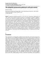
Fig. 1 Temporal separation of S and M phases in a typical cell cycle. DNA replication
(S-phase) and cell division (mitosis, M phase) are separated by distinct gap phases
Physical and temporal separation of DNA synthesis (S-phase) and mito-
sis (M phase) implies existence of gap phases separating these events (Fig. 1).
Gap phase 1 (G1) is defined as the period from end of mitosis to initiation
of DNA synthesis. Gap phase 2 (G2) separates end of DNA synthesis from
initiation of mitosis (Mitchison 1971). Time spent in G1 varies between cell
types and in different situations, but in mammalian cells it usually accounts
for a significant amount of total cycling time. A typical mammalian cell might
require 24 h to make a copy of itself and spend half this time in G1. How-
ever, in some specialized situations such as early development, G1 is absent
and cells go directly from M phase to synthesizing DNA (Murray and Hunt
1993). These extremes provide important clues about why separating the end
of mitosis from initiation of DNA synthesis is sometimes necessary and de-
sirable. In such cases it becomes important to understand how this period is
traversed, but before discussing this issue, the relationship between distinct
cell cycle phases must be further defined.
2.2
Maintaining Order
Continuity through multiple cell divisions requires that each new daughter
receive a complete and accurate copy of the genome. Chromosomes must
be duplicated once and only once before mitosis; conversely, mitosis must
be completed before DNA replication is re-initiated (Fig. 2) (DePamphilis
Fig. 2 Checkpoint control of S and M phase initiation. In pathway 1, ongoing DNA replica-
tion transmits a signal that blocks beginning of M phase (M
begin
). In pathway 2, ongoing
mitosis transmits a signal that blocks start of S-phase (S
begin
)
4 S.J. Moeller · R.J. Sheaff
2003). Cells also continually monitor for and repair the inevitable DNA dam-
age occurring throughout the division cycle (Kastan and Bartek 2004). In
all these situations order is maintained by checkpoints, wherein initiation of
later events is dependent on successful completion of earlier ones (Hartwell
1974; Hartwell and Weinert 1989). Temporally and spatially separate events
are linked via signaling components, which transmit information to elicit de-
sired responses (Nurse 2000b). By monitoring and linking events required for
cell division and repair, checkpoints help maintain genomic integrity essen-
tial for survival and continuation of the cell lineage.
Checkpoints represent an elegant solution to the problem of ordering DNA
synthesis and cell division, while at the same time raising additional ques-
tions. What drives progression through the cell cycle, and how is this process
regulated? These controls are distinct from machinery replicating DNA and
dividing the cell, which must receive instructions to initiate and complete
these tasks properly. Addressing such thorny issues required a paradigm shift
from observation of cell duplication to analysis of molecular events. Break-
throughs came from disparate but ultimately complementary approaches:
biochemical analysis of S to M phase cycling reproduced in a cell free system
derived from frog oocytes, generation and analysis of yeast mutants defec-
tive in cell division control, and analysis of protein expression patterns in
sea urchin extracts (Nurse 1990; Nasmyth 2001). These seminal investigations
(along with other important contributions) led inexorably to identification of
critical cell cycle machinery.
2.3
Cell Cycle Machinery
Recognition that specific protein catalysts are responsible for diverse cellu-
lar processes such as fermentation (late 1800s) suggested that cell growth and
proliferation would be similarly controlled (Nurse 2000a). Division of the cell
cycle into temporally ordered, discrete steps implied different proteins regu-
late specific cell cycle transitions (Hartwell 1974). If so, then factors advancing
cell cycle progression might be rate limiting (Nurse 1975). These concepts
gave birth to the idea of a cell cycle engine that both drives and controls
progression through the division cycle (Murray and Hunt 1993).
Biochemical and genetic approaches in different systems converged to
identify what we now know as the cell cycle machinery. A key discovery
was that nuclear division in frog oocytes is controlled by a “maturation pro-
moting factor”, or MPF (Masui and Markert 1971). Around the same time,
genetic screens identified yeast mutants defective in cell division or prema-
turely entering mitosis (Hartwell et al. 1973; Nurse et al. 1976). Rate limiting
components of S to M phase cycling were eventually isolated from frog egg
extracts, and the Deus ex machina turned out to be a kinase in association
with a regulatory subunit called cyclin (Evans et al. 1983; Lohka et al. 1988).
G1 Phase: Components, Conundrums, Context 5
These cyclin-dependent kinases (Cdks) transfer gamma-phosphate from ATP
to a specific protein substrate (Morgan 1995). However, the kinase subunit
alone is inactive because the bound ATP is not properly oriented, and ac-
cess of the protein substrate is blocked by a section of Cdk called the T-loop
(DeBondt et al. 1993). These impediments are removed by association with
cyclin and T-loop phosphorylation by the multicomponent Cdk-activating ki-
nase (CAK) (Russo et al. 1996).
The key to ordering and controlling cell cycle progression is thought to
lie in periodic expression of different cyclins, which associate with Cdks at
defined intervals and determine their specificity (Murray and Hunt 1993).
These unique cyclin–Cdk complexes must phosphorylate specific substrates
at the proper time to drive controlled progression through the cell cycle. After
completing their task, complexes are disassembled and cyclin degraded as
a prerequisite for subsequent steps (Murray et al. 1989). Temporal order is
achieved and maintained by linking cyclin expression to completion of pre-
vious events, then regulating activity of the resulting cyclin–Cdk complex.
Controlling complexes can be accomplished by removing activating modifica-
tions, inhibitory phosphorylation of the Cdk subunit, or tight binding of Cdk
inhibitory proteins (CKIs) (Morgan 1995). Cdk activity can also be modulated
by altering its location and/or accessibility to substrates, although these reg-
ulatory mechanisms are less well characterized (Murray 2004). Together, this
molecular circuitry provides a mechanistic explanation of cell cycle progres-
sion during G1.
Basic underlying principles derived from these investigations are: 1) cell
cycle machinery is evolutionarily conserved, 2) transitions between cell cycle
phases are catalyzed by Cdks, 3) cell cycle machinery is highly regulated, and
4) cell cycle components are an obvious target in proliferative diseases like
cancer (Murray and Hunt 1993).
3
G1 Progression in Cultured Cells
If John Donne were a developmental biologist, he might have penned: “In
multicellular organisms no cell is an island, entire of itself; each must be
responsive to the external environment”. Cells receive specific signals to sur-
vive, nutrients to grow, and additional signals to proliferate. After each di-
vision a G1 phase cell must re-evaluate its overall situation and determine
whether continued proliferation is desirable and feasible (Pardee 1974). Al-
though precisely how cell cycle machinery regulates G1 progression remains
poorly understood, a generally accepted working model has been constructed
from investigations in many different experimental systems. It posits that
unique G1 cyclin/Cdks are temporally expressed in response to extracellular
6 S.J. Moeller · R.J. Sheaff
signaling (Sherr and Roberts 1995). These complexes phosphorylate specific
substrates to promote required events and remove negative impediments to
G1 progression.
3.1
Coordinating Cell Growth and Division
A non-proliferating cell maintains a relatively constant size by establishing
homeostasis between cellular processes such as protein synthesis and degra-
dation (Neufeld and Edgar 1998). In contrast, conservation of mass requires
that a proliferating cell at some point duplicate its cellular contents (i.e. grow)
to maintain cell size; otherwise, it will become progressively smaller and
smaller until survival is untenable. This problem could be avoided by exactly
doubling cell components before each division, or by a stochastic process
averaging the required mass increase over several division cycles. Although
at some level proliferation must be coordinated with an increase in mass,
manipulating this relationship is crucial for development of multicellular or-
ganisms (Su and O’Farrell 1998a,b).
DNA replication and segregation can occur much faster than mass in-
creases, so a newly formed daughter cell must grow to become competent
for S-phase (Saucedo and Edgar 2002). Although growth is not rigorously
confined to a specific period like DNA synthesis and mitosis, much of the
necessary mass increase in mammalian cells occurs during its lengthy G1.
Consistent with these ideas, depriving cultured cells of growth factors or
amino acids causes a reduction in the rate of protein synthesis and cell cycle
arrest in G1. This result implies existence of a G1 checkpoint linking cell
growth with cell cycle progression, as in yeast (Campisi et al. 1982; Rupes
2002). A sizing mechanism, such as overall increase in mass (reflected in pro-
tein synthesis) or production of a specific molecule(s), could determine when
a critical size threshold is reached.
It is encouraging to see several recent reports re-invigorating the contro-
versy about whether mammalian cells contain an active sizing mechanism.
Rate of growth and division appears to be two separable and independently
controlled processes in rat Schwann cells, because reductions in cell volume
require several division cycles to re-establish homeostasis (Conlon and Raff
2003). In this case, size was determined by the net effect of how much growth
and division occurred. In contrast, a number of other cell types (e.g. human,
mouse, and chicken erythoblasts and fibroblasts) respond to size alterations
by compensatory shortening of the subsequent G1 phase (Dolznig et al. 2004).
These results provide evidence of a G1 size threshold that adjusts length of the
next cell cycle to maintain balance between growth and division.
Additional work is clearly required to explain the differing conclusions
reached in these two studies. One possibility is that generating cultured cell
lines compromises or alters the link between growth and proliferation; alter-
G1 Phase: Components, Conundrums, Context 7
natively, the extent or mechanics of coordination may vary depending on cell
type or situation. Regardless, identifying cells in which a sizing mechanism
is operational means that experiments can now be designed to identify its
molecular components.
3.2
Information Integration
G1 phase of the cell cycle is organized around the concept of a restriction
point (R point; called START in yeast) (Hartwell et al. 1973; Pardee 1974;
Blagosklonny and Pardee 2002). Before this G1 checkpoint, the cell receives
and interprets information from a variety of internal and external sources.
A decision is then made whether or not to continue with the cycle and initiate
another round of cell division. If conditions are not appropriate for prolifera-
tion, or the cell receives orders to adopt an alternative fate, it withdraws from
the cycle into a G0 resting state. It can remain in this position until prolifera-
tive conditions are re-established, or initiate an alternative program resulting
in differentiation, senescence, or apoptosis (Fig. 3).
The idea of a restriction point arose from analyzing how newly gener-
ated mammalian fibroblasts respond to nutrient and growth factor starvation
(Zetterberg et al. 1982). If serum is removed up to an experimentally de-
termined point, cells halt cell cycle progression in G1 phase. Upon serum
re-addition, completion of the cell division cycle is significantly extended
compared with continually fed cells. Thus, starvation not only blocks cell
cycle progression, but causes cells to exit the cycle and enter G0. However,
if serum is removed after this point, cells continue through the cycle unhin-
dered (Zetterberg and Larsson 1985). Subsequent analysis identified other
criteria that differ before and after this period in G1. Up until the R point,
cells stop cycling in response to low concentrations of cyclohexamide (a pro-
Fig. 3 Restriction point in G1 phase. The restriction point describes a position at which
the cell irreversibly commits to completing the division cycle. Up until the R point the
cell can withdraw to a quiescent state called G0. It can re-enter the cycle if conditions for
proliferation are favorable, or pursue an alternative fate
8 S.J. Moeller · R.J. Sheaff
tein synthesis inhibitor), while after the R point they are resistant (Pardee
1989). These observations suggested a molecular switch (such as an unstable
protein) might define R point control (Zetterberg and Larsson 1991).
3.3
The Cyclin-Cdk Engine
G1 progression is promoted and controlled by cyclin/Cdk complexes, so they
are often described as engines driving this process. Yeasts have only one
Cdk (originally Cdc28; now called Cdk1), while 11 distinct versions have
been identified in mammalian cells (van den Heuvel and Harlow 1994). Cdks
accomplish their overall mission by promoting positive events, overcoming
negative impediments, and policing themselves. In mammalian cells passage
through G1 is controlled by ordered expression of the D and E type cyclins,
which associate with Cdk4/6andCdk2/3, respectively (Fig. 4) (Sherr 1994).
There are three members of the cyclin D family and two of cyclin E, each of
which is expressed in a tissue-specific manner (Murray 2004). Current under-
standing of their regulation and function has emerged largely from the study
of how cultured mammalian cells respond to serum starvation/refeeding.
When an asynchronous population of proliferating mammalian cells is
deprived of serum, those located in G1 phase before the R point initiate a con-
certed shutdown of Cdk activity (Zetterberg and Larsson 1991; Sherr and
Roberts 1995). Cyclin expression is inhibited and its destruction promoted.
Any remaining cyclin/Cdk complexes are inhibited by phosphorylating Cdk
and/or association of tight binding inhibitors (Sherr and Roberts 1995). Cells
located after the R point when serum is removed complete the cycle and
then exit G1 by similar mechanisms. In order to re-enter the cell cycle Cdk
inhibition must be reversed. Refeeding G0 cells provides nutrients, growth
factors, and mitogens, resulting in rapid activation of cell surface receptors
and downstream signaling pathways like Ras/Map (also called Erk) kinase.
Fig. 4 Model of cyclin/Cdk activity during G1 phase. Ordered G1 progression in cultured
cells involves temporal and transient expression of different cyclins, which bind their
Cdk partners and determine specificity. The resulting complexes phosphorylate specific
substrates required for regulated movement through the cycle
G1 Phase: Components, Conundrums, Context 9
Activated Map kinase translocates to the nucleus, where it phosphorylates
specific targets to promote transcription of genes required for growth, cell
cycle progression, and upcoming S-phase (Alberts et al. 1994; Frost et al.
1994). Early mRNAs are induced within 30 min of refeeding cells and are in-
sensitive to protein synthesis inhibitors, indicating components required for
their production are already present. In contrast, late mRNAs are sensitive to
these inhibitors because they depend on unstable products of early response
genes. Identifying molecular events controlling this transcriptional program
was essential for further defining R point control.
In fibroblasts and many other cell types a key consequence of activating
Ras/Map kinase is rapid upregulation of cyclin D1 transcription; cyclin D1
proteinthenassociateswithCdk4/6 and initiates G1 progression (Winston
and Pledger 1993; Albanese et al. 1995). The Map kinase pathway also in-
fluences cyclin D1 localization, its association with Cdk4/6, and activation
of the complex by the Cdk-activating kinase (CAK). These multiple levels of
regulatory control help ensure cyclin D-Cdk4/6 is not inappropriately acti-
vated (Roussel et al. 1995). Mitogen dependence is maintained in part because
cyclin D1 is a very unstable protein degraded by the ubiquitin/proteasome
system (Matsushime et al. 1992; Diehl et al. 1997). Removing serum before
the R point inhibits cyclin D transcription, resulting in rapid disappearance
of cyclin D protein and subsequent exit from the cell cycle.
3.4
Removing Impediments: Inactivating Rb
A main target of activated cyclin D-Cdk4/6istheretinoblastomaprotein
(Rb), so called because it was first identified as a tumor suppressor whose
function is lost in a rare form of childhood retinal cancer (Friend et al. 1987).
Rb siblings include p130 and p107, and this family occupies a central pos-
ition in G1 control (Weinberg 1995). Rb acts in part as a repressor inhibiting
members of the E2F transcription factor family (Bartek et al. 1996). E2Fs
associate with Dp1 or Dp2 to form an active transcription factor complex
upregulating a wide array of gene products required for growth, cell cycle
progression, and upcoming S-phase (Stevaux and Dyson 2002). Rb can inhibit
E2F-Dp complexes in a number of ways, including sequestration away from
DNA and/or by forming active repressor complexes blocking DNA accessibil-
ity (Liu et al. 2004). This latter function is accomplished in part by recruiting
histone deacetylases that alter chromatin structure (Harbour and Dean 2000).
In addition to its well-characterized role inhibiting E2F, Rb interacts with
many different proteins and clearly regulates other processes in addition to
transcription. It helps block global protein synthesis in response to nutri-
tional deprivation by inhibiting expression of RNA polymerases I and III,
which are responsible for synthesizing ribosomal RNAs needed for protein
production (White 1994). Dual regulation of growth and cell cycle progres-
10 S.J. Moeller · R.J. Sheaff
Fig. 5 Control of G1 progression by cyclin/Cdk complexes. Mitogens generate cyclin D-
Cdk4 which phosphorylates Rb to release E2F. E2F transcriptionally upregulates cyclin E,
which in association with Cdk2 inactivates additional Rb to generate more cyclin E. This
positive feedback loop may represent the switch to mitogen independence (DK4: cyclin
D/Cdk4;EK2:cyclinE/Cdk2; AK2: cyclin A/Cdk2)
sion by Rb may help coordinate these two processes during division or
development.
As expected, Rb is highly regulated during the cell cycle. It is underphos-
phorylated (i.e. hypophosphorylated) in G0 cells and so binds E2F-Dp1 to
prevent transcription (Weinberg 1995). Re-feeding generates active cyclin D1-
Cdk4/6 that specifically phosphorylates Rb at a subset of available sites (Chen
et al. 1989). Activated E2F-Dp1 then upregulates cyclin E, which associates
with its partner Cdk2 and further phosphorylates Rb at distinct sites (Fig. 5)
(Dynlacht et al. 1994). The resulting spike in E2F-Dp1 activity causes a burst
of cyclin E synthesis and functional cyclin E/Cdk2 required for G1 pro-
gression (Ohtani et al. 1995). This positive feedback loop may represent the
transition to mitogen independence during G1 (Hatakeyama et al. 1994). The
burst is transient because cyclin E/Cdk2 marks its own cyclin subunit for
ubiquitination by SCF
Fbw7
and subsequent degradation by the proteasome
(Clurman et al. 1996). Continued Rb inactivation during this period likely
contributes to E2F-Dp1 dependent synthesis of cyclin A necessary for upcom-
ing S-phase (Stevaux and Dyson 2002). As cells proceed through the cycle, Rb
is dephosphorylated to reset the system (Buchkovich et al. 1989).
3.5
Removing Impediments: Inactivating p27
kip1
The Cdk inhibitor p27
kip1
(p27) is an anti-mitogenic gene activated in re-
sponse to serum starvation of proliferating cells (Sherr and Roberts 1995). It
participates in cell cycle exit and helps maintain the G0 state by ensuring that
cyclin/Cdk complexes remain inactive. High p27 levels in quiescent cells es-
tablish an inhibitory threshold that must be reduced for cell cycle re-entry
(Roberts et al. 1994; Sherr and Roberts 1995). Cyclin D1–Cdk4/6playsanim-
G1 Phase: Components, Conundrums, Context 11
portant role in this process by sequestering p27 away from cyclin E-Cdk2 in
early G1 (Reynisdottir et al. 1995).
In a satisfying twist, cyclin E/Cdk2 phosphorylates p27 later in G1 and
targets it for recognition by SCF (Skp2, Csk1, Cul1), a ubiquitin ligase com-
plex marking p27 for proteasomal degradation (Sheaff et al. 1997; Tsvetkov
et al. 1999). Thus, p27 elimination may contribute to the rapid burst of cy-
clin E/Cdk activity and transversal of the R point. Recent evidence in support
of this switch-like behavior comes from the discovery that Skp2 stability is
controlled by APC
Cdh1
(Bashir et al. 2004). This is a form of the APC/C
(anaphase promoting complex/cyclosome) that operates as a major ubiqui-
tin ligase in M phase. APC
Cdh1
remainsactiveintoG1andtargetsSkp2for
degradation, eventually running out of substrates and turning on itself. Skp2
protein can then accumulate and so p27 is degraded in a cyclin E/Cdk2 de-
pendent manner. Thus, APC
Cdh1
may contribute to maintenance and timing
of G1 progression.
3.6
Preparing for the Future
ThemainfunctionofcyclinD1-Cdk4/6 is inactivating Rb because these com-
plexes are dispensable in an Rb-/- background (Lukas et al. 1995). In contrast,
cyclin E/Cdk2 has other G1 targets since it is still required under these con-
ditions. In addition to helping remove negative impediments such as Rb and
p27, cyclin E/Cdk2 carries out various tasks required for upcoming S and
M phases. It helps license replication origins to ensure DNA is only dupli-
cated once per cell cycle. This process involves assembly of a pre-replicative
complex (PRC) after mitosis, which then recruits MCM (minichromosome
maintenance) helicases onto the DNA (Coverley et al. 2002; Diffley and Labib
2002). Cyclin E/Cdk2 may then participate in subsequent origin firing during
S-phase (Woo and Poon 2003).
Cyclin E/Cdk2 has also been implicated in modulating chromatin struc-
ture. Histone H1 continues to serve as a substrate for purified cyclin E/Cdk2,
but the physiological relevance of this reaction remained enigmatic. Recent
evidence suggests cyclin E/Cdk2 phosphorylates histone H1 in cells, desta-
bilizing its interaction with chromatin (Contreras et al. 2003). In addition,
cyclin E/Cdk2 influences chromatin structure by phosphorylating Rb and
altering its association with histone deacetylases (HDAC) at E2F promoters
(Takaki et al. 2004). Finally, cyclin E/Cdk2 helps mediate centrosome dupli-
cation in preparation for upcoming M phase (Hinchcliffe et al. 1999). These
microtubule organizing centers will relocate to opposite ends of the nucleus
during mitosis, after which microtubules will create the spindles separating
replicated chromosomes to daughter cells (Alberts et al. 1994).
12 S.J. Moeller · R.J. Sheaff
4
Ablating G1 Regulators in Mice
If cyclin/Cdk complexes are engines driving and controlling G1 progression,
then deleting their genes should result in very early lethality because cells
cannot proceed through the cycle. In the absence of cyclin D-Cdk4/6cells
should not respond to mitogens or inactivate Rb, and thus fail to initiate the
E2F transcriptional program required for G1 progression, S-phase, and divi-
sion. Cyclin E/Cdk2 should also be absolutely required, since it controls the
G1/S-phase transition by promoting key events required for cell cycle pro-
gression and DNA replication.
Similarly, deleting negative impediments such as Cdk inhibitors and Rb
was predicted to result in very early lethality due to disruption of G1 tim-
ing. In the absence of Rb cells would be expected to inappropriately activate
E2F/Dp1 and prematurely upregulate transcription of genes promoting G1
progression and S-phase entry. If p27 sets a threshold controlling S-phase
entry, its absence should compromise the critical G1 to S-phase transition.
4.1
Cyclin D-Cdk4/6
Cdk4 and Cdk6 are closely related kinases associating with D-type cyclins
to initiate G1 progression in response to proliferative signals (Sherr 1994).
This idea arose from extensive work in cultured cells showing: 1) overex-
pressed cyclin D1 shortens G1 phase, suggesting activity of cyclin D1/Cdk4 is
rate limiting for G1 progression, 2) cyclin D1 overexpression overcomes a G1
arrest caused by DNA damage or the unfolded protein response, 3) microin-
jected cyclin D1 antibodies, cyclin D1 antisense, inhibitory peptides derived
from p16
INK4a
, and small drug inhibitors all result in G1 arrest (Sherr and
Roberts 1999).
Mice lacking Cdk4 are viable and display proliferative defects in a limited
range of endocrine cell types, indicating Cdk4 is dispensable for prolifer-
ation in most situations (Tsutsui et al. 1999; Moons et al. 2002). Likewise,
Cdk6-/- mice are also viable and for the most part develop normally (Malum-
bres et al. 2004). Cdk6 is preferentially expressed in hematopoietic cells, and
its absence leads to delayed G1 progression in lymphocytes but not mouse
embryo fibroblasts (MEFs) (Meyerson et al. 1992). Viability of single knock-
outs and their normal cell proliferation was initially thought to reflect com-
pensation by the remaining family member.
Cdk4/6 double knockouts have now been generated and reveal that the
above interpretation is only partially correct (Malumbres et al. 2004). Em-
bryos lacking Cdk4 and Cdk6 die during late stages of embryogenesis due
to severe anemia. However, they display normal organogenesis and most cell