Cell Cycle Regulation in Mammalian Germ Cells
Bạn đang xem bản rút gọn của tài liệu. Xem và tải ngay bản đầy đủ của tài liệu tại đây (542.79 KB, 31 trang )
Results Probl Cell Differ (42)
P. Kaldis: Cell Cycle Regulation
DOI 10.1007/003/Published online: 20 December 2005
© Springer-Verlag Berlin Heidelberg 2005
Cell Cycle Regulation in Mammalian Germ Cells
Changanamkandath Rajesh · Douglas L. Pittman (✉)
Department of Physiology and Cardiovascular Genomics, Medical University of Ohio,
Toledo, Ohio 43614, USA
Abstract Meiosis is a unique form of cellular division by which a diploid cell produces
genetically distinct haploid gametes. Initiation and regulation of mammalian meiosis dif-
fers between the sexes. In females, meiosis is initiated during embryo development and
arrested shortly after birth during prophase I. In males, spermatogonial stem cells ini-
tiate meiosis at puberty and proceed through gametogenesis with no cell cycle arrest.
Mouse genes required for early meiotic cell cycle events are being identified by compar-
ative analysis with other eukaryotic systems, by virtue of gene knockout technology and
by mouse mutagenesis screens for reproductive defects. This review focuses on mouse re-
productive biology and describes the available mouse mutants with defects in the early
meiotic cell cycle and prophase I regulatory events. These research tools will permit rapid
advances in such medically relevant research areas as infertility, embryo lethality and
developmental abnormalities.
1
Introduction
Meiosisisadevelopmentalpathwayusedtoreducethechromosomenumberby
one-half, resulting in the production of haploid gametes and permitting sex-
ual reproduction (Fig. 1). Given that chromosomal aneuploidy is the leading
genetic cause of pregnancy loss and birth defects, it is critical to dissect out mei-
otic cell cycle checkpoints and proper chromosome segregation (Hassold and
Hunt 2001). However, understanding cell cycle regulation in mammalian germ
cells has considerably lagged behind that of the mitotic cell cycle. Challenges
to investigations of meiosis in mammals have included the long reproduc-
tive cycles, cell migration patterns, somatic/germ cell interaction and the
inability to propagate germ cells in culture for extended time. Understand-
ing mammalian meiosis has benefited considerably by general principles being
conserved across species. Much of our knowledge is built upon studies in model
organisms such as fungi, Arabidopsis and Drosophila (Engebrecht 2003; Page
and Orr-Weaver 1997; Wilson and Yang 2004). Tools are now available in mouse
genetics to make tremendous strides towards a thorough understanding of cell
cycle regulatory genes during meiosis and gametogenesis.
Mammalian meiosis and differentiation take place in multicellular and
hormonally regulated environments. During the gastrulation stage of mouse
344 C. Rajesh · D.L. Pittman
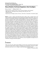
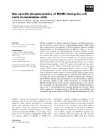
Fig. 1 Diagram illustrating the principle features of mammalian germ cell development.
Following proliferation and migration to the genital ridge, primordial germ cells differ-
entiate towards the spermatogonial or oogonial pathways, which differ in the number
and timing of meiotic arrests. In male mice, spermatogenesis starts shortly after birth
from the spermatogonia and once initiated, continues without interruption until haploid
spermatid formation. Meiotic divisions of each spermatogonia generate four spermatids
which mature by spermiogenesis to spermatozoa (not illustrated). Spermatogonia divide
continuously after birth to maintain a constant supply of spermatocytes. In the females,
oocyte development is initiated before birth and is arrested at the dictyate stage of mei-
otic prophase I. An oocyte resumes meiosis in response to hormonal stimuli only to be
arrested a second time at metaphase II. This arrest is overcome if fertilization ensues,
which triggers completion of the meiotic division. Cell division in females is unequal,
generating three non-functional polar bodies and an egg. The stages of meiosis are the
same in both sexes with the first division (meiosis I) resulting in reduction in the number
of chromosomes per cell. The prophase I is sub-divided into five sub-stages (leptotene,
zygotene, pachytene, diplotene and diakinesis) as illustrated. The second division (meio-
sis II) conserves the number of chromosomes in a mitosis-like process. Only a set of
homologous chromosomes is illustrated in the figure
Cell Cycle Regulation in Mammalian Germ Cells 345
embryonic development, primordial germ cells (PGCs) begin to form at
7.0 days postconception (dpc) and migrate from the base of the allantois
through the gut, up the dorsal mesentery and into the genital ridges (An-
derson et al. 2000; Nagy 2003). In males, germ cells undergo a G1 mitotic
arrest at 14.0 dpc (McLaren 2003). During the early stages of spermatogene-
sis, stem cells, termed type A spermatogonia, appear 3–5 days post-partum
and undergo self-renewal to produce type B spermatogonia. The type B sper-
matogonia divide and generate primary diploid (2n) spermatocytes capable
of initiating meiosis and forming four haploid (n) spermatids per spermato-
cyte, which eventually produce mature sperm (de Rooij and de Boer 2003).
These events are supported by Sertoli cells in the seminiferous tubules of the
testis, which are non-dividing diploid cells that form the blood-testis barrier.
Theentireprocessfromspermatogonialstemcellstomaturespermatozoa
takes approximately 5 weeks (Handel 1987; Russell 1990).
In females, meiosis is initiated following PGC migration and undergoes
a number of checkpoint regulated arrests (Mintz and Russell 1957). At
14.0 dpc in mouse, the peak number of oocytes are formed and meiosis be-
gins. After birth, oocytes arrest during the first meiotic prophase, followed
by a loss of more than one-third of the germ cell population by apoptosis
(atresia). Shortly before ovulation, the meiotic cell cycle is activated and an-
other, meiosis II, arrest ensues; activation and completion of oogenesis is then
dependent upon fertilization (Fig. 1). The first meiotic division is asymmet-
rical, resulting in the production of a large secondary oocyte and a small
non-functional polar body. The second meiotic division, if completed, re-
sults in four final haploid cells (three polar bodies and one egg). With recent
evidence that oogonial stem cells may proliferate and replenish the follicle
pool in adult mice, efforts to develop new technologies for identifying genes
involved during cell cycle regulation is even more alluring (Johnson et al.
2004). The possible replenishment from the oogonial stem cells will be critical
for addressing issues regarding infertility, preserving fertility and potentially
prolonging the reproductive years.
The genetic program that triggers meiosis and maintains the cycle through
checkpoints require some mechanisms not present during the mitotic cell
cycle. Until the past decade, the availability of mouse models for meiosis
studies were limited by the number of spontaneous meiosis mutants, most
having pleiotropic phenotypes (Handel 1987). Currently, there are four pri-
mary approaches for identifying meiosis regulatory genes in the mouse: cell
cycle genes having meiotic properties (Wolgemuth et al. 2004), homology
searches and compiling encyclopedic compendiums (Critchlow et al. 2004),
gene products associating with meiotic chromosomes (Heyting and Diet-
rich 1991) and phenotype-driven approaches (Reinholdt et al. 2004). This
chapterreviewsknownmousegenesinvolvedincellcycleregulationdur-
ing early meiosis, focusing on entry through meiosis I. Taking advantage of
database and literature searches, 53 genes for which mouse mutants are avail-
346 C. Rajesh · D.L. Pittman
Table 1 Gene knockouts affecting germ cell development in mouse
Gene Protein Name Effect on germ cell development Refs.
A. Cellular Proliferation and Differentiation
Ccna1 Cyclin A1 Arrest of germ cells prior to first meiotic division. Liu et al. 1998
Cdc25b Cell cycle protein Cell cycle protein involved in activation of MPF. Lincoln et al. 2002;
Deficiency leads to oocyte arrest at prophase Spruck et al. 2003
→ metaphase transition.
Cdk4 Cyclin dependant Decreased number of spermatocytes and spermatogonia Moons et al. 2002
kinase 4 and defective luteal function in females.
Cdk2 Cyclin dependant Sterile males arrest at G
2
/M transition stage of cell cycle, Ortega et al. 2003;
kinase 2 while females lack follicular development Berthet et al. 2003
due to breakdown at the pachytene stage.
CyclinD2 Cell cycle Absence of germ cells Sicinski et al. 1996
regulating D-cyclin due to lack of proliferation.
CyclinE2 Cyclin E2 Absence confers abnormal Geng et al. 2003;
synaptonemal complex formation. Parisi et al. 2003
Gcd/Pog Proliferation of Drastic depletion of germ cells in the developing ridges Agoulnik et al. 2002
germ cells (POG) due to proliferation defects
during PGC migration to genital ridges.
p18
INK4C
, Cyclin D-dependant Defects in transition from a pre-leptotene to the leptotene Zindy et al. 2001
p19
INK4D
kinase inhibitors stage in spermatogenesis, results in decreased sperm numbers.
p27
Kip1
p27
Kip1
Factor responsible for the G
1
/G
0
arrest Beumer et al. 1999;
a Cdk inhibitor in oocytes and absence results in defects in pre-leptotene Fero et al. 1996
to leptotene transition. Female sterility from aberration in
follicular maturation. Knockout male mice show
a large number of pre-leptotene stage spermatocytes.
Cell Cycle Regulation in Mammalian Germ Cells 347
Table 1 (continued)
Gene Protein Name Effect on germ cell development Refs.
B. Transcriptional and Translational Factors
Ahch Dax1 A transcriptional factor that has a role Yu et al. 1998
in gonadal differentiation and sex determination.
Lack of which leads to complete loss of germ cells
due to progressive degeneration of germinal epithelium.
Cpeb Cytoplasmic These RNA binding proteins regulate translation during Tay and Richter 2001
polyadenylation oocyte maturation. The absence of which results in vestigial
element binding protein ovaries with immature oocytes arrested at the pachytene stage.
Dazla Dazla protein (Deleted in RNA binding protein that affects translational control. Ruggiu et al. 1997;
azoospermia phenotype) Knockout results in lack of prenatal germ cells (females) Schrans-Stassen et al. 2001
or spermatogenic arrest by blockage of spermatogonial
differentiation (male).
Egr4 Early growth response A zinc finger transcriptional factor involved in cell Tourtellotte et al. 1999
protein (EGR) 4, differentiation and growth. In its absence incomplete block
NGFI-C, pAT133 of germ cell maturation at mid-pachytene stage occurs.
Also causes sperm with abnormal morphology.
Eif2s3y Eukaryotic translation Involved in the early steps of protein synthesis, Mazeyrat et al. 2001
initiation factor 2, causes spermatogonial proliferation impairment in its absence.
subunit 3,
structural gene Y-linked
Fox3a Foxo Factor involved in metabolism, cellular stress Castrillon et al. 2003
transcription factors response and aging. The absence leads to early
activation of follicles leading to low functional follicles.
Hspa2 Heat shock-related Chaperone protein involved in protein folding, Dix et al. 1996
70 kDa protein 2 (HSP70-2) leads to spermatogenic arrest at metaphase stage in knockouts.
348 C. Rajesh · D.L. Pittman
Table 1 (continued)
Gene Protein Name Effect on germ cell development Refs.
Taf 4b TAF4B RNA Transcription factor important in RNA Freiman et al. 2001
polymerase II, polymerase II machinery. Impaired folliculogenesis is seen
TAFII 105 in knockouts leading to lack of mature follicles.
Tiar TIAR An RNA-recognizing motif involved in splicing, transport, Beck et al. 1998
translation and stability of mRNA. Knockout results in decrease
in the survival of germ cells at the genital ridge.
Tls/Fus Translated in RNA-binding protein reported to contribute to N-terminal Kuroda et al. 2000
liposarcoma (TLS/FUS) half of fusion proteins in liposarcomas and leukemias.
Absence leads to increase in unpaired and mispaired
chromosomal axes in pre-meiotic spermatocytes leading
to apoptosis.
Trcp1 F-box protein Required for mitotic progression. Lack of gene leads Guardavaccaro et al. 2003
to accumulation β-Trcp1 of spermatocytes arrested at metaphase-I.
C. Cell Signalling
Bmp15 Bone morphogenetic Growth factor required for ovarian function, shows defects Yan et al. 2001
protein 15 in ovarian folliculogenesis and ovulation.
(growth differentiation
factor 9b)
Cit-k CIT-K A serine/threonine kinase interacting with Rho. Cunto et al. 2002
(citron kinase) Knockouts show embryonic and postnatal loss of germ cells leading
to complete lack of spermatocytes. Female effects not studied.
Cks2 CKS2 (mammalian homolog Essential for the biological function of Spruck et al. 2003
of the yeast cyclin dependent kinases by binding to its catalytic subunit.
Cdk1-binding protein) The lack of the gene results in germ cell arrest at metaphase-I.
Cell Cycle Regulation in Mammalian Germ Cells 349
Table 1 (continued)
Gene Protein Name Effect on germ cell development Refs.
Crem Cyclic AMP response Binds to the cAMP response element (CRE), loss of which leads Blendy et al. 1996
element modulator to interruption of spermatogenesis at early haploid stage
(CREM) (Round spermatids).
Gja1/Cx43 Connexin 43 These intracellular signaling molecules in their absence cause Juneja et al. 1999
impaired folliculogenesis resulting in a germ cell deficiency
within the gonads.
Gja4/Cx37 Connexin 37 Intracellular signaling molecules located at the gap junctions Simon et al. 1997
preventing the maturation of the follicles beyond meiotic
competence in their absence.
Madh1/5 MAD homolog Signal mediators for bone morphogenetic proteins. Loss leads Chang and Matzuk 2001;
Smad 1/5 to greatly reduced or absence of primordial germ cells in embryos. Tremblay et al. 2001
Steel
panda
Steel factor Serves as a ligand for tyrosine kinase in c-kit protooncogene, Huang et al. 1993
knockout shows folliculogenesis defects and
reduced germ cell number.
D. Cytoplasmic and Apoptotic Factors
Apaf1 APAF1 (apoptotic The protein has a role in cytochrome c-mediated apoptosis, Honarpour et al. 2000
protease activating absence of which leads to spermatogonial degeneration.
factor 1)
AR Androgen receptor Sertoli cell specific knockout of this receptor resulted Chang et al. 2004
(AR) in spermatogenic arrest predominantly at the diplotene stage.
Also results in low serum testosterone levels leading
to azoospermia and infertility.
350 C. Rajesh · D.L. Pittman
Table 1 (continued)
Gene Protein Name Effect on germ cell development Refs.
Bcl2/Bclx B-cell leukemia/ Anti-apoptotic protein controlling cell survival. The absence Ratts et al. 1995;
lymphoma 2/X of which leads to severe loss of the primordial germ cells Rucker et al. 2000
and hence absence of spermatogonia in testis
and depletion of follicles in the post-natal ovary.
Bcl2l2 (Bclw) Bcl2 like 2 A cytoplasmic protein that promotes cell survival and has a role Ross et al. 1998
in maintenance of reproductive germ cells. The knockout leads to
progressiveloss of germ cells, Sertoli cells and Leydig cells and cause
extensive testicular degradation.
Bsg Basigin A protein belonging to the immunoglobulin superfamily important Toyama et al. 1999;
for pre-implantation development and spermatogenesis. Igakura et al. 1998
The knockout mice show azoospermia due to metaphase-I arrest.
Casp2 Caspase 2 Intracellular death effectors, the absence of which significantly increase Bergeron et al. 1998
the primordial follicles in the ovary due to the absence of cell death.
c-mos c-mos, An essential part of cytostatic factor (CSF), Colledge et al. 1994
proto-oncogene lack of which results in oocyte maturation from second meiotic
product MOS metaphase arrest, without any activation, resulting in ovarian teratomas.
Miwi Miwi (Murine homolog Cytoplasmic protein expressed in spermatocytes and spermatids, Deng and Lin 2002
of piwi- P-element absence of which can lead to spermatogenic arrest at beginning
induced wimpy testis) of spermatid stage.
Tlf/Trf2 TATA box Physiological function not known but absence results in arrest Martianov et al. 2001
binding protein-like during spermiogenesis at transition from round spermatids,
factor (TLF/TRF2) leading to apoptosis.
Cell Cycle Regulation in Mammalian Germ Cells 351
Table 1 (continued)
Gene Protein Name Effect on germ cell development Refs.
E. Prophase-I Regulation
Atm Ataxia A nuclear protein with a role in cell cycle and DNA repair. Disrupted Barlow et al. 1996
telangiectasia cell division leading to lack of ovarian follicles and spermatids.
Brca1 BRCA1 Absence leads to prophase I arrest of spermatocytes accompanied Xu et al. 2003
by p53 dependent and independent apoptosis.
Brca2 BRCA2 Plays role in meiotic recombination, chromosome pairing Sharan et al. 2004
and synapsis during spermatogenesis. Absence of
Brca 2 leads to prophase-I arrest of spermatocytes.
Dmc1 Disrupted meiotic Gene involved in homologous recombination, lack of which leads Pittman et al. 1998;
cDNA (DMC) 1 to defects in chromosomal synapsis. Yoshida et al. 1998
Ercc1/Xpf DNA repair Involved in recombination, double strand break repair Hsia et al. 2003
proteins and repair of interstrand cross-links. The lack of Ercc1
leads to oocyte degeneration and low number of oocytes.
Fkbp6 FK506 binding Synaptonemal complex component essential for sex-specific fertility Crackower et al. 2003
protein 6 and chromosome pairing. Male knockouts have complete spermatogenesis
block and death of meiotic spermatocytes.
H2afx Histone H2A.X Facilitates specific DNA-specific DNA-repair complex assembly Celeste et al. 2002
on damaged DNA. Absence leads to pachytene stage arrest of spermatocytes.
Mei1 Meiosis Homozygote mutants of both sexes are sterile due to meiotic Libby et al. 2002;
defective 1 arrest arising from defects in recombinational repair Libby et al. 2003
and chromosomal synapsis.
Mlh1 Mismatch Non-viable oocyte or spermatocytes. Edelmann et al. 1996
repair enzyme
352 C. Rajesh · D.L. Pittman
Table 1 (continued)
Gene Protein Name Effect on germ cell development Refs.
Msh4/5 Mut S Post replicative DNA mismatch repair gene whose disruption leads Kneitz et al. 2000
homologue 4/5 to loss of oocytes and spermatocytes. Abnormal chromosomal pairing
occurs at zygotene phase of prophase-I in spermatocytes.
Nbs1 Nijmegen breakage Component of Mre11 complex involved in DNA strand break repair. Kang et al. 2002
syndrome Female knockout mice are sterile due to oogenesis failure.
(NBS 1) protein
Siah1a Ubiquitin ligase The protein is required for completion of meiosis I, Dickins et al. 2002
component defects are observed in synaptonemal complex formation
and progression from metaphase I in knockouts.
Sycp1 Synaptonemal complex Component of the transverse filament of the synaptonemal complex. de Vries et al. 2005
protein 1 (SCP1) Absence leads to male and female sterility with disruption
of spermatogenesis primarily at pachytene.
Scp3 Synaptonemal complex Component of axial/lateral element of the Yuan et al. 2000;
protein 3 (SCP3) synaptonemal complex and is associated with the centromeres, Yuan et al. 2002
lack of which leads to disruption of spermatogenesis at zygotene stage,
massive cell death and female germ cell aneuploidy.
Spo11 Spo11 protein Protein involved in initiation of genetic recombination. Romanienko and
Arrest prior to pachytene stage of meiosis I in mutants. Camerini-Otero 2000;
Baudat et al. 2000
Cell Cycle Regulation in Mammalian Germ Cells 353
able are listed in Table 1. Though a strict classification is not possible due to
the lack of in-depth knowledge concerning complete mechanisms, the genes
are arranged into 5 categories based upon the cell cycle process affected:
initiation and maintenance of meiosis, transcriptional and translational fac-
tors, cellular signaling, cytoplasmic and apoptotic factors and prophase I
regulation.
2
Cell Cycle Regulatory Genes Required
for Initiation and Maintenance of Meiosis
During germ cell development, spermatogonia or oogonia stem cells undergo
a series of mitotic divisions leading to the formation of gametocytes that
go through a final interphase and enter meiotic prophase I. The triggers for
meiosis entry are not known, but like the mitotic cell cycle (G
1
to S, S progres-
sion and G
2
to M), transition is controlled, at least in part, by phosphorylation
events catalyzed by cyclin dependant kinase (Cdk) complexes. In this section,
Cdks, cyclins and Cdk inhibitors and activators are discussed.
The G
2
/M transition in meiosis is controlled by the Cdc2 kinase, Cdk1 and
the B-type cyclin complex, called the M-phase-promoting factor (MPF) (Choi
et al. 1991; Masui and Markert 1971). The build up of cyclin B and its degra-
dation at the end of the cell cycle is a hallmark of MPF activity guiding the
initiation and termination of the cycle (Pines and Hunter 1989). MPF activ-
ity in oocytes peaks once at the time of meiotic initiation and again at the
meiotic arrest in metaphase. The MPF is involved in several features of cell
division such as disassembly of the nucleus, chromosome condensation, cy-
toskeletal rearrangements and transcriptional activity (Moreno and Nurse
1990).
At least 11 distinct Cdks (Cdk1-11) and 11 cyclins (cyclins A–J, T) exist in
higher eukaryotes. Cyclin A1, Ccna, was one of the first cyclins demonstrated
by targeted disruption in mouse as having a definite role in meiosis (Liu
et al. 1998). Otherwise healthy males were sterile due to a block in sper-
matogonia before the first meiotic division, while females were fully fertile.
The disruption did not have significant effect on either Cdk1 or Cdk2 ki-
nase activity, suggesting that cyclin A1 affects downstream targets such as the
MPF complex and other protein kinases or phosphatases for G
2
/M transition.
Additionally, cyclin B/Cdk1 activity was insufficient to bypass cyclin A1 for
entry into meiosis (Liu et al. 2000).
In addition to mitotic cell cycle control, Cdk2 was implicated as having
a role during meiosis by localization at telomeres during spermatogenesis
when axial elements form and at synaptic sites corresponding to recombi-
nation events (Ashley et al. 2001). Surprisingly, Cdk2 knockout mice were
viable, apparently being compensated by another kinase, but male and female
354 C. Rajesh · D.L. Pittman
mice were sterile (Berthet et al. 2003; Ortega et al. 2003). Females lack follic-
ular development, suggesting that meiotic arrest occurred at the early stages
of meiosis. Cdk2-deficient oocytes developed up to the pachytene stage but
break down at the dictyate stage, suggesting a failure to maintain chromo-
some synapsis. In spermatocytes, chromosomes also fail to synapse.
Cdk4 localizes along synaptonemal complexes of newly synapsed bivalents
and disappear by mid-pachytene (Ashley et al. 2001). Cdk4 and its cata-
lytic partner, cyclin D, are responsible for the initial phosphorylation of the
Rb protein by regulating cyclin E/Cdk2 and lifting the S-phase inhibition by
preventing binding of its inhibitor, p27 (Tsutsui et al. 1999). Cdk4/cyclin D2
have an important role in follicle stimulating hormone-induced proliferation
of granulosa cells in females (Moons et al. 2002) and in decreased numbers of
spermatogonia leading to sterility in males (Wolgemuth 2003).
Cdk1 function is regulated by phosphorylation at several sites. For ex-
ample, human Cdk1 phosphorylation at Thr-14 and Tyr-15 are inhibitory
while Thr-160 is activating (Pines 1999). The activating threonine phospho-
rylation, opens up a catalytic cleft, which is catalyzed by a Cdk activating
kinase comprising Cdk7 and cyclin H. The phosphate is removed after cy-
clin degradation by a phosphatase KAP. The Cdc25 family of phosphatases
are activators of Cdk complexes by removal of the phosphates from the
ATP-binding site of Cdk1, but their function may differ between sexes. Fe-
males deficient for Cdc25b are sterile and oocytes arrest at prophase, but
males are fertile (Lincoln et al. 2002).
Two classes of Cdk inhibitors modulate Cdk activity during the mitotic cell
cycle. Members of the INK4 family (p15
INK4b
,p16
INK4a
,p18
INK4c
,p19
INK4d
)
specifically bind and regulate cyclin D dependant kinases. The Kip/Cip fam-
ily (p21
Cip1/Wa f 1
,p27
Kip1
and p57
Kip2
) binds and regulates cyclin A, D and E
dependant kinases (Sherr and Roberts 1999). p27
Kip1
is an important factor
guiding the G
1
/G
0
arrest in germ cells as suggested by their high expression
levels at 16 dpc. In adult males, p27
Kip1
expression is observed in the Ser-
toli cells and Leydig cells, probably to mediate spermatocyte development.
p27
Kip1
knockout male mice show a large number of pre-leptotene stage sper-
matocytes indicating a role in regulation of spermatogonial proliferation and
onset of meiotic prophase (Beumer et al. 1999). The pre-leptotene spermato-
cytes enter mitosis instead of meiosis, leading to speculations about their role
in male germ cell tumorigenesis. In females, sterility results from aberration
in follicular maturation and corpora lutea formation (Fero et al. 1996).
The p19
INK4d
gene is expressed during meiotic prophase and in post-
meiotic spermatids (Zindy et al. 2001). The p19
INK4d
knockouts are fertile but
display markedly lower sperm counts owing to testicular atrophy associated
with apoptosis in seminiferous tubules. p18
INK4c
null mice are also fertile
but develop Leydig cell hyperplasia and pituitary tumors. The p19
INK4d
and
p18
INK4c
double mutant knockout mice have lower fertility due to a decrease
of spermatocytes, while females were fertile. A delay in the mitosis to meio-