human endometrial programming and lessons in health and disease
Bạn đang xem bản rút gọn của tài liệu. Xem và tải ngay bản đầy đủ của tài liệu tại đây (788.52 KB, 14 trang )
REVIEWS
Fertile ground: human endometrial
programming and lessons in health
and disease
Jemma Evans1–3, Lois A. Salamonsen1,2,4, Amy Winship1,2, Ellen Menkhorst1,2,
Guiying Nie1,2,5, Caroline E. Gargett4,6 and Eva Dimitriadis1,2,7
Abstract | The human endometrium is a highly dynamic tissue that is cyclically shed, repaired,
regenerated and remodelled, primarily under the orchestration of oestrogen and progesterone,
in preparation for embryo implantation. Humans are among the very few species that menstruate
and that, consequently, are equipped with unique cellular and molecular mechanisms controlling
these cyclic processes. Many reproductive pathologies are specific to menstruating species, and
studies in animal models rarely translate to humans. Abnormal remodelling and regeneration of
the human endometrium leads to a range of reproductive complications. Furthermore, the
processes regulating endometrial remodelling and implantation, including those controlling
hormonal impact, breakdown and repair, stem/progenitor cell activation, inflammation and
cell invasion have broad applications to other fields. This Review presents current knowledge
regarding the normal and abnormal function of the human endometrium. The development of
biomarkers for prediction of uterine diseases and pregnancy disorders and future avenues
of investigation to improve fertility and enhance endometrial function are also discussed.
Centre for Reproductive
Health, Hudson Institute of
Medical Research, Clayton,
3168, Australia.
2
Department of Molecular
and Translational Medicine,
Monash University, Clayton,
3800, Australia.
3
Department of Physiology,
Monash University, Clayton,
3800, Australia.
4
Department of Obstetrics
and Gynaecology, Monash
University, Clayton, 3800,
Australia.
5
Department of Biochemistry
and Molecular Biology,
Monash University, Clayton,
3800, Australia.
6
The Ritchie Centre, Hudson
Institute of Medical Research,
Clayton, 3168, Australia.
7
Department of Anatomy and
Developmental Biology,
Monash University, Clayton,
3800, Australia.
1
Correspondence to E.D.
evdokia.dimitriadis@hudson.
org.au
doi:10.1038/nrendo.2016.116
Published online 22 Jul 2016
The transformative processes that prepare the endometrium for embryo implantation are unique to menstruating
species, and are thought to underlie the evolution of
menstruation. Although rodent species, which are easy
to manipulate, are common experimental models for
studies of endometrial receptivity and embryo implantation, findings obtained with these animals often cannot
be directly translated to humans.
Biological processes that have developed in the
human endometrium during the evolution of menstruation are specialized versions of processes that are found
in other tissues, altered to regulate endometrial biology.
Understanding how the human endometrium undergoes controlled and spatially limited tissue destruction,
resolution of inflammation, scar-free repair and re‑
epithelialization followed by regeneration and transformation can inform our understanding of processes that
occur in other tissues.
In this Review, we describe the remodelling of the
endometrium before it becomes receptive for embryo
implantation, the dynamic fetal–maternal communication that contributes to successful implantation, the
endometrial defects that result in infertility and miscarriage and the detection and treatment of these disorders.
We also identify missing links, both experimental and
clinical, which should be investigated to enable progress
in the field, and areas where understanding of endometrial biology might influence other fields and the
development of therapeutics.
Evolution of human menstruation
Unlike other organs, the human endometrium does not
have a single, constant function from birth to death.
The endometrium exists to provide a ‘fertile ground’
for implantation of an embryo and development of a
highly invasive placenta, which is achieved by an orderly
sequence of development and transformation within each
menstrual cycle, under the influence of the ovarian steroid
hormones1. The endometrial cells become terminally differentiated during each menstrual cycle; in the absence of
conception, tissue shedding and regeneration for subsequent fertile cycles occurs. In menstruating species, decidualization is spontaneous, rather than embryo-mediated.
Decidualization is the process of the transformation or
differentiation of human endometrial stromal fibroblasts
to secretory ‘epithelioid’ cells, which occurs under the
influence of the hormones oestrogen and progesterone,
along with cAMP and local paracrine factors.
The evolution of spontaneous decidualization is
thought to have occurred when genes that were ancestrally expressed in other organs and tissue systems were
expressed in the endometrium. Transposable elements,
NATURE REVIEWS | ENDOCRINOLOGY
ADVANCE ONLINE PUBLICATION | 1
.
d
e
v
r
e
s
e
r
s
t
h
g
i
r
l
l
A
.
d
e
t
i
m
i
L
s
r
e
h
s
i
l
b
u
P
n
a
l
l
i
m
c
a
M
6
1
0
2
©
REVIEWS
Key points
•The human endometrium is a unique, dynamic tissue that is cyclically shed, repaired,
regenerated and remodelled, in preparation for embryo implantation
•Decidualization in women occurs spontaneously (regardless of the presence of an
embryo) during the mid‑to‑late luteal phase, necessitating endometrial shedding and
subsequent regeneration in the absence of conception
•Endometrial remodelling occurs primarily under the orchestration of oestrogen and
progesterone, but is influenced by many factors, including epigenetic signals and
stem/progenitor cells
•Abnormalities in endometrial remodelling lead to pathologies including infertility,
endometriosis and pregnancy disorders
•Understanding the processes that operate in the endometrium could provide
information that is applicable to nonreproductive pathologies such as cancer and
wound healing
for instance, contributed to the origin of decidualization
by conferring progesterone responsiveness to numerous
genes across the genome2. The evolutionary transformation of the endometrial regulatory landscape has been
mapped and found to explain the developments within
the human uterus that support its unique pregnancy
phenotype, of which decidualization and menstruation
are central2.
Decidualization probably evolved because it provided protection to uterine tissues from the hyper
inflammation and oxidative stress associated with deep
haemochorial placentation3,4. However, menstruation as
a consequence of decidualization is equally important
in the human adaptation to haemochorial placentation.
Repeated cycles of decidualization and shedding prepare
human uterine tissues by physiological preconditioning
for the stress of haemochorial placentation4. In an
adolescent who is pregnant (and has experienced few
menstrual cycles), extensive preconditioning has not
occurred, which results in a higher risk of major obstetric
complications associated with defective placentation
than is seen in older pregnant women 3. Menstrual
cycles are hypothesized to undergo their own ‘evolution’ throughout the reproductive lifespan, with the
endometrium transitioning from a fairly progesteroneresistant, immature tissue at menarche to become more
responsive because of the cumulative effects of cyclic
menstruation and inflammatory signalling 3,5. The lack
of preconditioning and, thus, the absence of these cyclederived changes is proposed to contribute to the aetiology of pregnancy complications in adolescents who have
not yet developed progesterone responsiveness.
This evolution of spontaneous decidualization and
menstruation, and the dysfunction associated with these
processes, has given rise to human-specific reproductive
complications, including recurrent early pregnancy loss
and placental pathologies such as pre-eclampsia, in addition to menstrual problems such as heavy or abnormal
bleeding.
Mechanisms of endometrial remodelling
Endometrial luminal epithelium. The endometrium
undergoes substantial remodelling under the influence
of ovarian steroid hormones, and becomes receptive
for only a few days in the mid-secretory phase of the
menstrual cycle (FIG. 1). The luminal epithelium is the
first uterine point of contact for blastocysts, and differentiates considerably during the receptive phase to
facilitate embryo attachment and subsequent implantation. The transformation of the plasma membrane in
cells of the luminal epithelium from a nonadhesive to an
adhesive surface encompasses remodelling of elements
that contribute to the endometrial barrier function,
including the glycocalyx, epithelial polarity, epithelial–mesenchymal transition and the lateral junctional
complexes (FIG. 1)6. Importantly, in humans the placental
trophoblasts invade between epithelial cells, without the
epithelial destruction that is observed in other species
with haemochorial placentation7. Defects in interactions between embryos and the endometrial epithelium
contribute substantially to infertility and implantation
failure8.
The known molecular changes that occur in human
endometrial luminal epithelium in relation to receptivity affect the integrins, osteopontin, Notch signalling,
heparin-binding EGF-like growth factor, cell-surfaceassociated mucins, glycodelin and ion channels, which
have been reviewed elsewhere9. Some cytokines probably also have important roles in endometrial epithelial
receptivity. For example, levels of IL‑11 are lower in
endometrial luminal epithelium in infertile women
than in fertile women10. IL‑11 regulates the adhesiveness of epithelial cells in vitro, probably by upregulating
expression of the plasma membrane proteins annexin A2
and flotillin‑111, which are proposed to be essential for
receptivity and embryo attachment 11.
The results of transcriptomic profiling studies have
identified large numbers of genes that are upregulated
or downregulated in the induction of receptivity, but
data sets vary considerably between studies (as has been
reviewed elsewhere12), which suggests post-translational
regulation of proteins at the endometrial epithelial surface is important (FIG. 1). Studies on the serine protease
proprotein convertase subtilisin/kexin type 5 (PC6)
have revealed that, in endometrial epithelium, PC6 is
maximally expressed during the receptive phase, but its
expression is lower in women with implantation failure
than in reproductively healthy women13. PC6, via its
proteolytic activity, post-translationally regulates antiadhesion molecules and the organization of the plasma
membrane in human endometrial epithelium, altering
the apical architecture to provide a receptive surface13,14.
Decidualization. In human endometrial stromal cells
(ESCs), decidualization is the process of spontaneous,
terminal differentiation that occurs in the mid‑to‑late
secretory phase of each menstrual cycle, whereas in
nonmenstruating species, this process is initiated during pregnancy (FIG. 1). In a menstrual cycle that does not
result in conception, the terminally differentiated cells
are shed during menses. However, if pregnancy occurs,
decidual cells promote the invasion of fetal extravillous
trophoblasts that (along with uterine natural killer
(uNK) cells) facilitate spiral-artery remodelling and
protect the conceptus by conferring maternal immunotolerance of the fetal allograft 15. The decidual cells also
2 | ADVANCE ONLINE PUBLICATION
www.nature.com/nrendo
.
d
e
v
r
e
s
e
r
s
t
h
g
i
r
l
l
A
.
d
e
t
i
m
i
L
s
r
e
h
s
i
l
b
u
P
n
a
l
l
i
m
c
a
M
6
1
0
2
©
REVIEWS
Blastocyst
Trophectoderm
Glycocalyx
Post-translational
regulation of the
surface molecules
1
4
Inner
cell mass
Pinopode
5
Adherens
Tight
junction
junction
Cell surface
adhesion factors
EMT
Luminal
epithelium
6
Mesenchymal
stem cell
Differentiation
Decidual cell
Macrophage
7
Progesterone
2
Glandular
epithelium
MET
Perivascular cell
Blood
vessel
Stromal
fibroblast
uNK
Ovulation
15
8
3
16
17
18
19
20
21
22
23
24
25
Day
Pre-receptive
Receptive
Post-receptive
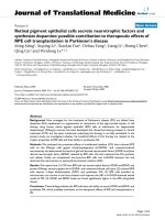
Figure 1 | The pre-receptive, receptive and post-receptive endometrium. The pre-receptive
epithelium
(1) is
Natureluminal
Reviews
| Endocrinology
nonadhesive, owing to the presence of antiadhesive factors, including the glycocalyx, a polarized epithelium and lateral
junctions that anchor cells tightly together. During the pre-receptive phase, the glandular epithelium becomes highly
secretory (2), uterine natural killer (uNK) cells proliferate and macrophages influx into the endometrium (3). To become
receptive, the luminal epithelium undergoes considerable changes (4): epithelial and blastocyst-secreted enzymes
post-translationally modify the glycocalyx; the epithelium undergoes epithelial–mesenchymal transition (EMT), becoming
less polarized with fewer lateral junctions, and the adhesion-factor repertoire on the luminal epithelial surface changes.
Pinopodes (5) appear on the surface of the luminal epithelium at the initiation of receptivity, but their role in blastocyst–
epithelium adhesion is currently unclear. Communication between blastocysts and uterine luminal epithelium further
enhances receptivity (6). Decidualization (7) is initiated by progesterone in stromal cells adjacent to blood vessels, and in
vascular mesenchymal stem cells. These cells undergo mesenchymal–epithelial transition (MET) to become rounded,
secretory cells expressing the decidual markers prolactin and insulin-like growth factor-binding protein 1. Decidual cells
secrete factors (such as hormones, cytokines, chemokines, lipids and noncoding RNAs) that act synergistically or
additively to create a wave of decidualization (8) throughout the endometrium.
shield the conceptus from environmental stress signals16,
and ‘sense’ embryo quality to facilitate maternal rejection
of developmentally incompetent embryos17.
Progesterone induces decidualization in stromal cells
adjacent to spiral arterioles (FIG. 1). In vitro, decidualized
stem-cell-like perivascular stromal cells produce higher
levels of cytokines and chemokines that are involved
in promoting decidualization and the recruitment
of trophoblasts than nonperivascular stromal cells16.
Decidualization also requires cAMP18, and involves
reprogramming of ESCs, which ensures that different
genes are expressed at specific stages of differentiation19.
After the initiation of decidualization, local paracrine factors create a ‘wave’ of decidualization that
spreads from spiral arterioles throughout the endometrium (FIG. 1). Decidual regulation has been investigated
predominantly in studies of individual molecules; more
comprehensive studies of the proteome and secretome20
NATURE REVIEWS | ENDOCRINOLOGY
ADVANCE ONLINE PUBLICATION | 3
.
d
e
v
r
e
s
e
r
s
t
h
g
i
r
l
l
A
.
d
e
t
i
m
i
L
s
r
e
h
s
i
l
b
u
P
n
a
l
l
i
m
c
a
M
6
1
0
2
©
REVIEWS
and microRNA (miRNA) signature21 of decidualization
have not added substantially to the repertoire of processes that are known to be involved in decidualization.
This repertoire has been reviewed elsewhere22. Although
progesterone drives decidualization, other steroid receptors (specifically, oestrogen receptor (ER), glucocorticoid receptor, mineralocorticoid receptor and androgen
receptor) also have distinct roles22,23, and might confer
specificity of hormone action.
Few in vitro studies have investigated the role of
other cell types in the progress of decidualization. The
results of studies in mice that lack uterine glands show
that these glands are essential for decidualization24, but
whether they are similarly important in women is not
known. Human uterine glands secrete many factors that
are known to drive decidualization in vitro, but in vivo
secretion of these factors into the stroma has not been
confirmed25. Leukocytes, including uNK cells, mast
cells, T cells and dendritic cells, are essential for decidual
angiogenesis during the initiation of pregnancy (FIG. 1).
However, their function in decidualization is less clear.
Murine models of dendritic-cell depletion indicate that
these cells are required for decidual proliferation and differentiation26. Similarly, uNK cells in mice seem to maintain decidual integrity 27. However, the results of both
mouse and human in vitro co‑culture experiments with
epithelial and stromal cells have not provided evidence
that uNK cells initiate or promote decidualization28.
Decidual leukocytes have specific phenotypes, and
express distinct markers of differentiation and function
compared with peripheral leukocytes. Decidualized stromal cells secrete mediators that can act on, and influence
the function and differentiation of, resident leukocytes29.
Menstrual breakdown and repair. Menstruation is initiated by the withdrawal of oestrogen and progesterone
support in the absence of implantation and pregnancy,
and is governed by a complex cascade of endocrine and
paracrine signalling within the endometrium (FIG. 2).
In macaques, which are menstruating nonhuman primates, the onset of menstruation can be blocked by
progesterone replacement within 36 h of hormone
withdrawal, but replacement after 36 h has no effect 30.
This result suggests a biphasic activation of menstruation, in which endocrine signalling to cells expressing
progesterone receptor initiates paracrine signalling to
cells without progesterone receptor, which facilitates
progesterone-independent effects that lead to menses.
Intriguingly, endometrial tissue destruction and re‑
epithelialization occur simultaneously; re‑epithelialization is generally considered to start ~36 h after the onset
of menses, and is complete within a further 48 h. The
results of hysteroscopic analysis of the menstrual endometrium emphasize that menstrual shedding is a zonal
event; areas undergoing breakdown can be observed
adjacent to intact tissues from the previous cycle and
areas that have already undergone re-epithelialization31.
Decidualized stromal cells (FIG. 1) are essential for
responding to endocrine cues and transmitting paracrine
signals during menstruation, as they express the progesterone receptor premenstrually 32 and detect progesterone
withdrawal33. Hormone withdrawal from decidualized
stromal cells in vitro enhances inflammatory reactive
oxygen species via inhibition of superoxide dismutase
activity, which upregulates nuclear factor κB (NF‑κB)
and prostaglandin G/H synthase 2 (PTGS2, also known
as COX‑2) signalling relative to levels in the presence of
progesterone and results in production of inflammatory
factors, including prostaglandin F2α (REFS 33,34) (FIG. 2).
Hormone withdrawal triggers the recruitment of
inflammatory cells into the perimenstrual endometrium
via alterations in chemokines derived from decidualized
stromal cells33,35 (FIG. 2). Secretion of proteolytic enzymes
by leukocytes results in tissue breakdown at menses,
as reviewed elsewhere35, and local tissue lysis simultaneously results in the production of cues for repair 36.
Expression of proteases and gene products involved
in extracellular matrix synthesis and repair is elevated
specifically in stromal cells derived from areas of the
endometrium that have undergone lysis36,37.
Oestrogen is not required for endometrial repair, as
demonstrated by evidence from the study of ovariectomized women and women in natural menopause38.
In vitro human studies have defined ‘wound-healing’
factors, including activin, vascular endothelial growth
factor (VEGF), cysteine-rich secretory protein 3 and
galectin‑7, along with development-related pathways, such as Wnt signalling pathways and mesenchymal–epithelial transition (FIG. 2), which contribute
to re‑epithelialization and endometrial wound repair
independently of oestrogen 37,39–42. However, once
the endometrial surface is re‑epithelialized, oestrogen is required to stimulate glandular and stromal
regeneration (FIG. 2).
Menstrual endometrium demonstrates the opposing
processes of tissue destruction and repair simultaneously
in an inflammatory environment; both processes are initiated by similar physiological cues. Understanding how
the menstrual endometrium limits inflammation, modulates immune-cell activity, rapidly repairs and remains
scar-free has implications for the development of
treatments for a number of pathologies (BOX 1).
Stem/progenitor cells in regeneration. Small populations of adult stem/progenitor cells with classic
stem-cell properties of clonogenicity, self-renewal and
differentiation have been identified in human endometrium43 (TABLE 1); these cells contribute to the ability of
the endometrium to regenerate during each menstrual
cycle (FIG. 2). Specific stem/progenitor cell types, including epithelial progenitors, mesenchymal stem cells and
side-population cells (which are characterized by the
efflux of DNA-binding dyes, a universal property of
adult stem cells) might be involved in the regeneration
of different endometrial cellular compartments43.
Epithelial progenitor cells have been identified in
human endometrium as clonogenic cells that differentiate into large, gland-like structures44, and in mice as
label-retaining cells that proliferate in response to oestrogen, despite lacking ERs (TABLE 1). ERα-expressing
niche cells that are closely associated with epithelial
progenitor cells probably transmit the oestrogen signal
4 | ADVANCE ONLINE PUBLICATION
www.nature.com/nrendo
.
d
e
v
r
e
s
e
r
s
t
h
g
i
r
l
l
A
.
d
e
t
i
m
i
L
s
r
e
h
s
i
l
b
u
P
n
a
l
l
i
m
c
a
M
6
1
0
2
©
REVIEWS
Uterine bleeding
Late secretory (days 24–28)
• No conception
• Corpus luteum demise
• Hormone withdrawal
Luminal epithelium
Repair (days 2–5)
• Chemokines
• Growth factors
• Wnt signalling
• MET
Regeneration (days 5–14)
• Epithelial progenitor cells
• Mesenchymal stem cells
• Wnt signalling
• Notch signalling
11
1
Blood
vessel
8
Tissue
destruction
NF-κB
4
COX-2
Growth
factor
activation
• Growth factors
• Chemokines
Endometrial growth
9
5
Vasoconstriction
• PGE2
• PGF2α
10
3
Stromal fibroblast
2
Decidualized
stromal cell
Menstruation (days 1–5)
Proteolytic
enzymes 7
6
Functionalis
Basalis
Eosinophil
Neutrophil
Macrophage
Perivascular
cell
Epithelial
progenitor
cell
Mesenchymal
stem cell
Figure 2 | Endometrial decidualization, menstruation, repair and regeneration. Endometrial
(1) and
Naturestromal
Reviewscells
| Endocrinology
mesenchymal stem cells (2) undergo decidualization under the influence of oestrogen and progesterone. In the absence
of conception and implantation (3), endometrial stromal cells ‘sense’ hormone withdrawal upon the demise of the corpus
luteum, and upregulate intracellular inflammatory signalling (4) and the release of inflammatory factors that contribute to
vasoconstriction of uterine blood vessels (5), recruitment of leukocytes (6) and propagation of the menstrual cascade.
However, these inflammatory and growth factors (4), proteolytic enzymes (7) and recruited immune cells (8) also
contribute to repair after menstruation, in concert with processes such as mesenchymal–epithelial transition (MET) (9)
and Wnt signalling, to restore endometrial homeostasis (10). Activation of endometrial epithelial progenitor cells and
perivascular mesenchymal stem cells (11), possibly involving Wnt signalling or Notch signalling, drives cellular
replacement in the glands and stroma respectively, to mediate regeneration of the endometrium. COX‑2, prostaglandin
G/H synthase 2 (PTGS2); NF‑κB, nuclear factor κB.
to these ERα-negative cells. Epithelial progenitors are
thought to be located in the basalis region of the uterine
glands (FIG. 1), where a high level of telomerase activity (a feature of adult stem cells) has been detected43.
Specific markers identifying epithelial progenitor cells
are required to facilitate delineation of their role in
endometrial proliferative disorders (BOX 2).
Human endometrium also contains a small population
of mesenchymal stem cells (eMSCs)43 (TABLE 1). Specific
surface markers of clonogenic eMSCs demonstrate their
perivascular localization in the endometrial functionalis and basalis45,46 (FIG. 1), as well as their presence within
shed fragments in menstrual fluid43. eMSCs have been
identified by the co‑expression of CD146 and plateletderived growth factor receptor β (PDGFRβ) markers as
pericytes45. A single marker, sushi domain-containing
protein 2 (SUSD2, also known as W5C5)) identified
4% of endometrial stromal cells in 34 samples of stromal cells as eMSCs46 (TABLE 1). Gene profiling of fresh
CD146+PDGFRβ+ cells47 and cultured SUSD2+ cells16
confirmed that eMSCs have a pericytic, perivascular
signature, which suggests that eMSCs have an additional
role in angiogenesis during stromal regeneration and
placentation43. These endometrial perivascular cells are
distinct from the stromal fibroblast (CD146−PDGFRβ+)
and endothelial (CD146+PDGFRβ−) populations47.
Side-population cells48 are also present within human
endometrium; these populations are a mix of ERβexpressing endothelial cells with some epithelial and
stromal cells that do not express ERα or progesterone
receptor 49,50 (TABLE 1). In xenografts, the side-population
cells regenerate human ‘endometrium’ consisting mainly
NATURE REVIEWS | ENDOCRINOLOGY
ADVANCE ONLINE PUBLICATION | 5
.
d
e
v
r
e
s
e
r
s
t
h
g
i
r
l
l
A
.
d
e
t
i
m
i
L
s
r
e
h
s
i
l
b
u
P
n
a
l
l
i
m
c
a
M
6
1
0
2
©
REVIEWS
Box 1 | Translating endometrial biology to other pathologies
Skin wounds
Chronic skin wounds commonly exhibit deficient re‑epithelialization. Understanding
how the endometrium undergoes rapid repair after menstruation could lead to novel
insights into the development of treatments to promote repair of chronic wounds.
Chronic inflammatory diseases
The endometrium limits inflammation during menstruation to prevent excessive tissue
destruction. Translating the mechanism by which inflammation is restricted could aid
the resolution of chronic inflammation.
Stem-cell dysfunction
Cyclic activation of stem cells is required for endometrial regeneration after
menstruation. This process occurs monthly for an average of 450 menstrual cycles,
but stem-cell senescence occurs in women with recurrent pregnancy loss.
Delineation of the factors and mechanisms involved in cyclic activation could aid the
treatment of recurrent pregnancy loss and other diseases associated with stem-cell
dysfunction.
Fibrotic diseases
Repair of the endometrium following menstrual shedding is scar-free. Understanding
how the endometrium remains scar-free despite inflammation and tissue destruction
each month could lead to novel therapies for fibrosis.
of stromal and vascular tissue, with occasional epithelial
gland-like structures49–51. Similarly, SUSD2+ eMSCs generate stromal tissue in xenografts46. However, in human
endometrium in vivo, whether one or more stem/
progenitor cell type regenerates endometrial tissue, or
a stem/progenitor cell hierarchy exists, is not known.
In an experimental model of wound repair, eMSCs
modulated chronic inflammation outside the uterus,
which suggests these cells have a role in communication and regulation of macrophages52. Determination
of the function of these cells in endometrial physiology
has the potential to identify their roles in endometrial
disorders (BOX 2).
Endometrium–embryo crosstalk
The pre-implantation microenvironment. Uterine fluid
provides the natural environment for sperm transport
and blastocyst hatching together with pre-implantation
development, as well as peri-implantation embryonic–
maternal interactions. The fluid contains not only the
nutrients necessary for blastocyst growth, but also
important regulatory molecules and microvesicles53.
Specific proteins secreted from the endometrium interact with the blastocyst to facilitate implantation25,54,55
(FIG. 1). miRNAs in uterine fluid are taken up by preimplantation mouse embryos and alter embryonic
mRNA expression in vitro 56. Uterine fluid must also
contain factors to protect the mother and embryo from
bacteria and other pathogens57.
Many classes of molecules, from simple salts and
amino acids through to proteins, steroids and lipids are
contained in uterine fluid. These molecules are derived
from multiple sources, including endometrial epithelial
secretions, selective transudation from blood, leukocyte
activation and possibly Fallopian tubal secretions and
peritoneal fluid.
Glucose, lactate and pyruvate are required for human
blastocyst development58. Alterations in the levels of these
factors might also alter the pH of the local environment.
Proteins in uterine fluid include leukaemia inhibitory
factor (LIF), VEGF, IL‑11 and other chemokines and
cytokines that are probably synthesized in the endometrium and secreted into the uterine cavity 54, establishing a
complex milieu to facilitate implantation. The amino acid
profile of uterine fluid has been determined, but the full
molecular composition is not yet known59. The mechanisms involved in the regulation of levels of nutrients
and ions, and the relationships between these components and their relative importance in the establishment
of pregnancy, are still to be determined.
Blastocysts and endometrial epithelium. Successful
implantation and pregnancy outcome require both a
receptive endometrium and an appropriately developed
blastocyst. Blastocysts enter the uterine cavity during the
receptive phase and remain for up to 72 h before implantation (FIG. 1). After blastocyst hatching from the zona
pellucida, the trophectoderm comes into close contact
with, and firmly adheres to, the receptive endometrial
luminal epithelium, which initiates implantation (FIG. 1).
The influence of blastocysts on receptivity and implantation is poorly defined in humans, although hormonal,
epigenetic and metabolomic cues have been identified.
Blastocysts communicate with the endometrium
via cell-surface proteins and secreted factors60 (FIG. 1).
Human chorionic gonadotropin (hCG) is secreted
by hatched human blastocysts in close apposition to
the endometrial epithelium61. Treatment of primary
human endometrial epithelial cells (EECs) with hCG, as
well as infusion of hCG into the uterine cavity of humans
and baboons, mediates the production of factors that are
associated with endometrial receptivity, including LIF,
VEGF, IL‑11 and prokineticin‑1 (REFS 62–65).
Human blastocysts require glucose metabolism, but
exhibit an idiosyncratic metabolic mechanism that produces high levels of lactate in close proximity to the uterine epithelium, creating a low pH environment 66. This
process is thought to promote local endometrial tissue
disaggregation, facilitating trophectoderm cell invasion
into the endometrium via modulation of epithelial
VEGF production54.
Human blastocysts regulate EEC adhesion and gene
expression via secreted regulators67. Culture media
derived from blastocysts generated by in vitro fertilization (IVF) that subsequently implant (resulting in a live
birth) enhance primary human EEC adhesion, unlike
IVF blastocysts that do not successfully implant 68.
Human blastocysts that are determined by morphology
to be of high quality during IVF culture, but that do not
subsequently implant after transfer, secrete miRNAs that
are not secreted by blastocysts that implant 68. miRNAs
secreted by IVF blastocysts during culture might reflect
their quality and implantation potential.miR‑661, bound
to the RNA-binding protein argonaute‑1, is secreted specifically by human IVF blastocysts that do not implant68.
miR‑661 is also taken up by primary human EECs in
culture and blocks their adhesive capacity 68,69. This antiadhesion effect of miR‑661 is mediated, at least in part,
by downregulation of the production of nectin‑1 in
primary human EECs68.
6 | ADVANCE ONLINE PUBLICATION
www.nature.com/nrendo
.
d
e
v
r
e
s
e
r
s
t
h
g
i
r
l
l
A
.
d
e
t
i
m
i
L
s
r
e
h
s
i
l
b
u
P
n
a
l
l
i
m
c
a
M
6
1
0
2
©
REVIEWS
Table 1 | Properties of endometrial stem/progenitor cells
Stem/progenitor Stem-cell property
cell
Cell types and markers
Frequency among
endometrial cells
Clonogenic cells
(human)
Ability of a single cell to
form a colony when seeded
at low density in culture
Epithelial progenitor cells
<1%
44
Mesenchymal stem cells
1–5%
44
Mesenchymal
stem cells
(human)
Differentiate into multiple
mesodermal cell types
(fibroblasts, adipocytes,
chondrocytes, osteocytes
and smooth-muscle cells)
CD146 PDGFRβ
1.5%
45
SUSD2 (W5C5)
4%
46
Clonogenic
1–5%
44
Side-population cells
0.4%
49
Efflux DNA-binding
dyes (Hoechst) because
of high expression of
plasma-membrane
transporter molecules
Mixed cell population
<5%
48
Endothelial cells (ERβ )
51%*
50
Epithelial cells (ERα PR )
27%
50
CD146 PDGFRβ
10–14%
Side-population
cells (human)
Label-retaining
cells (mouse)
+
+
+
+
–
+
–
+
Quiescent, proliferate rarely Epithelial (ERα–); proliferate in
and retain DNA-synthesis
response to oestradiol
label for a long time
Stromal (84% ERα–, 16% ERα+);
12% proliferate in response to
oestradiol
Refs
50
3%
172,173
2–6%
172,173
ER, oestrogen receptor; PDGFRβ, platelet-derived growth factor receptor β; PR, progesterone receptor; SUSD2, sushi
domain-containing protein 2. *Frequency of cell types in the heterogeneous side-population cell population.
Human blastocysts, via their secreted mediators, alter
endometrial receptivity. Secretion of specific signalling
molecules that influence receptivity is probably affected
by the quality of blastocysts. As lactate and noncoding
RNAs have roles in cancer development and invasion,
determining how they function and are regulated in
the highly controlled process of embryo invasion has
implications for our understanding of cancer biology.
Endometrial pathologies and treatments
Endometriosis. Endometriosis is characterized by the
growth of ectopic endometrial tissue outside the uterus;
this tissue cycles similarly to eutopic endometrium,
undergoing inflammation, shedding and regeneration in
response to hormonal changes across the menstrual cycle.
Endometriosis affects ~10% of women of reproductive
age; affected women commonly present with pelvic pain
and dysfunctional menstrual bleeding.
Sampson’s theory of retrograde menstruation70 is the
most widely accepted cause of endometriosis. This theory
posits that fragments of shed endometrium reflux
through the Fallopian tubes into the peritoneal cavity.
As most women experience retrograde menstruation,
alterations in the function of tissue fragments or an inability of the immune system to clear the fragments could
exist in women who develop endometriosis. Altered ER
function has been proposed to modulate apoptosis and
inflammasome activation, possibly enabling fragments
of refluxed endometrial tissue to survive71 and establish
endometriosis. Alternatively, women with endometriosis
might have variants of susceptibility genes that have been
identified in genome-wide association studies (GWAS),
or they could have epigenetic alterations72.
In support of Sampson’s theory, endometrial stem/
progenitor cells together with niche cells in shed tissue
fragments can establish endometriotic lesions (BOX 2).
Clonogenic, self-renewing endometrial cells are present in endometriotic lesions in adult women73, and in a
subset who had a neonatal ‘menstrual bleed’, these cells
might have been present from birth74, remaining viable
and dormant until rising oestrogen levels at menarche
initiated the growth of ectopic endometrium.
Establishment of ectopic endometriotic lesions probably alters gene expression and cellular function within
the eutopic endometrium. A proliferative transcriptomic
fingerprint is maintained within the early-secretory eutopic
endometriotic endometrium75; eutopic ESCs from women
with endometriosis do not undergo decidualization, and
they have alterations in gene expression suggestive of
resistance to progesterone-mediated differentiation76.
However, even in the absence of progesterone stimulation
in vitro, endometriotic eutopic stromal cell gene expression
is altered compared with endometrial stromal cells isolated
from women without endometriosis, and this underlying
difference could contribute to the altered responses to
progesterone stimulation in vitro76. The phenomenon of
progesterone resistance might not be restricted to endometriosis5, as similar genetic alterations have been observed in
ESCs isolated from women with recurrent pregnancy loss
and polycystic ovary syndrome (PCOS)77,78. Evidence from
a transcriptomic analysis suggests that eutopic endometrial
gene expression is more dysregulated in severe endometriosis than in mild endometriosis79. By contrast, pilot data
with the endometrial receptivity array suggest that eutopic
endometrial gene expression is not altered in different
stages of endometriosis80. However, levels of noncoding
RNA and protein production, as well as post-translational
modifications within the eutopic endometrium, might be
altered, mediating functional cellular changes81.
Despite the association of endometriosis with infertility, embryo implantation proceeds normally in women
with endometriosis who receive supplementary steroid
NATURE REVIEWS | ENDOCRINOLOGY
ADVANCE ONLINE PUBLICATION | 7
.
d
e
v
r
e
s
e
r
s
t
h
g
i
r
l
l
A
.
d
e
t
i
m
i
L
s
r
e
h
s
i
l
b
u
P
n
a
l
l
i
m
c
a
M
6
1
0
2
©
REVIEWS
Box 2 | Potential roles of endometrial stem/progenitor cells in endometrial disorders43
Endometriosis
Normal stem/progenitor cells are shed into the peritoneal cavity by retrograde menstruation and probably establish
ectopic clonal endometriotic growths170. In early-onset endometriosis, retrograde neonatal uterine bleeding resulting
from withdrawal of maternal hormones might seed the pelvic cavity with endometrial stem/progenitor cells that survive
and become activated as oestrogen levels begin to rise at puberty74.
Adenomyosis
The presence of normal endometrial stem/progenitor cells in an abnormal niche might enable ectopic endometrium to
grow in the myometrium. Inappropriate differentiation of endometrial mesenchymal stem cells into smooth-muscle cells
could account for the associated smooth-muscle hyperplasia.
Endometrial cancer
Mutations in the genome or epigenome of endometrial epithelial stem/progenitor cells might generate cancer stem cells
that are responsible for tumour initiation, progression, metastasis and recurrence171.
Thin, dysfunctional endometrium
Diminished activity of normal endometrial stem/progenitor cells, with an inability to respond to oestrogen stimulation,
results in an atrophic endometrium (<7 mm thick) that is insufficient for embryo implantation and subsequent
establishment of pregnancy43.
Asherman syndrome
Damage or loss of normal endometrial stem/progenitor cells from injury to the endometrial basalis layer or
postpartum infection in a setting of low oestrogen levels results in complete obliteration of the endometrium
by fibrous tissue43.
Endometrial ablation
Heat-induced damage to normal endometrial stem/progenitor cells prevents future growth of endometrial tissue.
hormones82. Oocyte-donation studies have not demonstrated any association between endometriosis, embryo
implantation and pregnancy outcome82,83. Endometriosis
does not seem to negatively affect pregnancy outcome
when standard endometrial-priming protocols are followed84,85, although it should be noted that these studies
did not examine different stages of endometriosis. In
two meta-analyses86,87, severe endometriosis was found
to negatively affect the probability of pregnancy success
in women undergoing IVF.
Diagnosis of endometriosis currently requires the
direct surgical visualization of lesions at laparoscopy, so
the identification of appropriate biomarkers is the subject of considerable interest. Noninvasive or minimally
invasive biomarkers are urgently required, particularly
for adolescent endometriosis. The existence of a heritable component to endometriosis is well supported, as
the risk of disease is elevated in first-degree relatives
of women with severe endometriosis, and concordance
is high for disease incidence and stage in monozygotic
twins88. However, despite extensive GWAS, no diagnostic genetic signature is available yet, which suggests
the involvement of epigenetic, rather than genetic,
regulation89.
Single and combined biomarkers, along with global
techniques such as miRNA analysis, transcriptomics and
proteomics, have been investigated in relation to endometriosis, as reviewed elsewhere90. However, no single
biomarker or biomarker panel has been independently
confirmed and tested for sensitivity and specificity. A lack
of concordance between the results of different studies has
prompted the establishment of guidelines for standardization of surgical and clinical data collection and biological
sample collection and storage in endometriosis research91.
Assisted reproductive technology. Ovarian-stimulation
protocols and embryo-culture techniques have improved
dramatically since the birth of the first ‘test-tube’ baby
in 1978, but pregnancy success rates with assisted
reproductive technology (ART) have not significantly
altered. ART success in stimulated cycles remains
around 25–30%92.
Endometrial receptivity is abnormal in women
undergoing ART93. The altered hormonal milieu that
results from controlled ovarian hyperstimulation (COH)
affects the development and timing of endometrial
receptivity 94. Women treated by ART with COH who do
not become pregnant after fresh-embryo transfers tend
to have prematurely advanced endometrial histology
on day 2 after hCG treatment for oocyte maturation95.
This abnormal histology correlates with the presence
of markers of decidualization, and elevated leukocyte
numbers and activation; these alterations are not normally seen until later in the menstrual cycle95. Similarly,
implantation that occurs late in natural cycles, when the
endometrium is highly differentiated, is more likely to
result in miscarriage than earlier implantation96. A study
of endometrial gene expression during IVF demonstrated that ovarian stimulation with follicle-stimulating
hormone (FSH) and gonadotropin-releasing hormone
(GnRH) antagonist leads to the occurrence of gene
expression characteristic of a decidualized, late-secretory
endometrium earlier in the cycle than in the absence of
ovarian stimulation97.
A promising approach to maximize reproductive
success in ovulatory infertile couples is embryo freezing, with transfer of a thawed embryo into a natural
cycle98. This strategy bypasses the detrimental effects of
COH and supraphysiological hormone levels, enabling
8 | ADVANCE ONLINE PUBLICATION
www.nature.com/nrendo
.
d
e
v
r
e
s
e
r
s
t
h
g
i
r
l
l
A
.
d
e
t
i
m
i
L
s
r
e
h
s
i
l
b
u
P
n
a
l
l
i
m
c
a
M
6
1
0
2
©
REVIEWS
natural endometrial development. The combination
of this approach with endometrial-receptivity testing
could optimize reproductive success. Biomarkers for
receptivity have been reviewed elsewhere99,100. Both
endometrial tissue and uterine fluid have been investigated by genomic, proteomic and metabolomic
methods in attempts to find a marker, or a cohort of
markers, to use as a fingerprint to define a receptive
endometrium. Current effort is being directed to the
validation of an RNA-based endometrial receptivity
array 101, which detected a nonreceptive endometrium
in ~25% of women with implantation failure during the
expected time of endometrial receptivity, indicating a
displacement of the implantation window. The remaining women with implantation failure were classified as
having a receptive endometrium during the expected
window. However, although such a test is useful, it cannot be performed in the implantation cycle because it
requires an endometrial biopsy specimen, as well as
time to complete the analysis. Development of a rapid,
noninvasive test that can be performed before embryo
transfer, to facilitate clinical decision-making, is required
to overcome these limitations.
PCOS. Up to 20% of women of reproductive age have
PCOS102. The syndrome presents as hyperandrogenism, either clinical (hirsutism) or biochemical, with
oligo-ovulation or anovulation, as well as polycystic ovaries, and is often associated with hyperinsulinaemia and
obesity. Infertility affects 40% of women with PCOS103,
mainly as a result of ovulation failure, with progesterone
deficiency resulting from the lack of corpus luteum formation, which subsequently affects endometrial development. However, even when ovulation is restored,
women with PCOS have low pregnancy rates and
high miscarriage rates (~73%)104, which are attributed
to the elevation of levels of oestrogen and androgens,
and the presence of the metabolic syndrome (including
hyperinsulinaemia and obesity), all of which affects the
endometrium.
The endometrium in women with PCOS is progesterone-resistant, exhibiting defective decidualization78,105,106
and altered levels of inflammatory mediators, which
are likely to contribute to pregnancy complications and
reductions in fertility 107,108. In a gene-array analysis,
466 genes were differentially regulated in the midsecretory endometrium in women with PCOS compared with unaffected women, and the expression pattern was indicative of progesterone resistance in PCOS78.
Endometrial receptivity factors, such as glycodelin
and LIF, are also dysregulated in the endometrium in
women with PCOS78,104. Pregnancy success in IVF cycles
is affected, because of endometrial insulin resistance109.
Specifically, the insulin-regulated facilitated glucose
transporters GLUT1 and GLUT4 and the insulin receptor
substrate (IRS) 1 are downregulated in the endometrium
in women with PCOS110.
Interventions, including lifestyle changes and treatment with the insulin-sensitizing drug metformin,
have been tested to determine their potential to restore
menstrual cyclicity and fertility 111. Results indicate that
restoration of ovulation and improvement in menstrual function, along with normalization of hormonal parameters (free testosterone, FSH and luteinizing
hormone) can be achieved in women with PCOS by
treatment with metformin112–114 or by intervention with
hypocaloric diets and physical activity 110,115,116. These
interventions, which ranged from 6 weeks to 6 months
in duration, also improved endometrial function and
expression of receptivity markers. Endometrial blood
flow 112,113, GLUT4, GLUT1 and IRS1 (REFS 110,117) and
the ERα:ERβ ratio116 were all improved from the levels
in untreated women with PCOS. Evidence also indicates that metformin treatment throughout pregnancy
continues to affect endometrial function, which results
in a decreased risk of miscarriage and an increased rate
of pregnancy and live birth compared with untreated
women118–120.
Receptivity is characterized by low androgen levels,
and the elevation of androgens in women with PCOS
is likely to alter receptivity 121. The endometrium in
women with PCOS does not exhibit the expected
downregulation of the androgen receptor during the
receptive phase121. However, compared with untreated
PCOS, metformin treatment and lifestyle interventions
can reduce levels of free testosterone and endometrial
expression of the androgen receptor, normalizing the
endometrial androgen environment 114,116,117.
Thin endometrium and Asherman syndrome. Repair
mechanisms and endometrial stem/progenitor cell
function are potentially compromised in women with
thin (<7–8 mm), dysfunctional endometrium that fails
to regenerate sufficiently for embryo implantation, and
in Asherman syndrome (intrauterine scarring), even
with long-term use of oestrogen to stimulate endometrial growth43 (BOX 2). Endometrial stem/progenitor
cells might be lacking, or unable to respond to oestrogen via niche cells expressing ER122. In two case studies
of Asherman syndrome, autologous bone-marrow cells
were administered into the sub-endometrial zone via a
needle123, or infused into the uterine arterioles under
ultrasonographic guidance. These cell-based treatments
were followed by oestrogen replacement for several
months, but the results were modest in terms of endometrial receptivity, and the lack of controls necessitates
caution in interpretation124.
Endometrial ‘scratch’. Local endometrial injury (for
example, by biopsy or a scratch) is receiving attention
for its potential effect on pregnancy success in IVF
cycles. This procedure is now recommended by up to
83% of IVF clinicians125, and the administration of an
injury during the cycle preceding embryo transfer is
proposed to double live-birth rates126.
An important issue in the application of this technique
is to define the clinical population for whom endometrial
injury could prove beneficial. The initial results127, published in 2003, demonstrated a beneficial effect of endometrial injury on pregnancy outcome in IVF cycles in
women with recurrent implantation failure who were classified as good responders to hormonal stimulation. Since
NATURE REVIEWS | ENDOCRINOLOGY
ADVANCE ONLINE PUBLICATION | 9
.
d
e
v
r
e
s
e
r
s
t
h
g
i
r
l
l
A
.
d
e
t
i
m
i
L
s
r
e
h
s
i
l
b
u
P
n
a
l
l
i
m
c
a
M
6
1
0
2
©
REVIEWS
Box 3 | Future directions in reproductive research
•Understanding epigenetic mechanisms that alter intergenerational inheritance and
contribute to endometrial pathologies.
•Developing personalized medicine.
•Developing platforms for timely, noninvasive assessment of endometrial receptivity,
endometriosis and early pre-eclampsia.
•Developing models to study environmental toxins and endocrine disrupters,
implantation and early post-implantation pregnancy disorders.
•Refining the use of endometrial stem/progenitor cells to enhance receptivity and
treat endometrial disorders.
•Defining the dialogue between the blastocyst and the endometrium, and using this
information to enhance receptivity and treat infertility.
•Developing models to recapitulate human endometrial disorders.
•Establishing a biobank of material from patient cohorts with standardized collection
procedures.
•Altering the microbiome to improve reproductive health.
•Developing targeted treatment-delivery strategies.
then, a large number of randomized, controlled trials
(RCTs) and observational studies have been conducted,
with results that either dispute128–130 or corroborate131–133
those of the original study. A Cochrane meta-analysis126 of
14 RCTs provided evidence that endometrial injury doubles live-birth rates in IVF cycles in women with recurrent
implantation failure. However, this analysis could have
underestimated the overall effect of endometrial injury,
because women in the control groups could also have
undergone some degree of endometrial manipulation126.
The number and quality of the studies that were included
in the meta-analysis has been criticized, and the number
of participants in individual studies was generally low 134.
Overall, results suggest that endometrial damage
might only be effective in women undergoing a freshembryo transfer cycle who have experienced recurrent
implantation failure (two or more failures), which suggests
that these women have abnormal receptivity 135. The positive effect of endometrial injury on embryo implantation
was not replicated in oocyte recipients129, or in the results
of a well-designed RCT of women without recurrent
implantation failure130. However, subgroup analysis in
oocyte recipients suggested that endometrial injury
is beneficial as the number of previous failed embryo
transfers increases129. In a study of women with recurrent
implantation failure who underwent endometrial
injury 132, increased maternal age, elevated FSH during
the proliferative phase of previous cycles and diminished ovarian reserve were negatively associated with
pregnancy outcomes.
The mechanisms underlying the effects of local endometrial injury are unknown. One possibility is that injury
induces an inflammatory reaction within the uterus,
improving synchronicity between the endometrium and
the embryo126. However, as the endometrial damage is
caused in the cycle preceding ovarian stimulation and
embryo transfer, how it affects the subsequent cycle is
unclear.
Well-controlled studies are now required, focusing on
women with recurrent implantation failure, determining
the optimal timing of injuries and number of injuries per
menstrual cycle. These studies present ethical challenges,
but are necessary to prevent the withholding of a beneficial procedure, or the provision of an unproven one.
Future avenues of endometrial research
The results of research in several areas relating to endometrial biology have suggested the potential benefits of
future investigations (BOX 3).
The endometrial microbiome. The concept of the sterile
uterus is no longer considered valid. Indeed, the uterus
and other tissues (including lung and bladder) widely
cited as being free of bacteria are now known to harbour
unique microbiota. In the endometrium, deep sequencing of a hypervariable region of the 16S ribosomal RNA
gene identified 15 phylotypes that were present in each
of 19 samples from nonpregnant women136. In 90% of
these samples, Bacteroides spp. were dominant, and
Proteobacteria spp. and Firmicutes spp. were also common, presenting a unique uterine core microbiome136
that is quite different from that of the vagina. Notably,
Bacteroides spp. regulate certain mechanisms in the gut
that are relevant to the endometrium, including epithelial-cell maturation and maintenance, mucosal-barrier
reinforcement and interactions with the host immune
system to control other bacteria. However, the low level
microbial presence in the uterus is not associated with
inflammation137.
The endometrial epithelial surface and uterine fluid
contain hormonally-regulated immunomodulatory
molecules that are important to control infection57 and
to maintain the uterine microenvironment in a noninflammatory state to enable its functions, including
sperm chemotaxis, embryo development and implantation. Among the immunomodulatory molecules within
uterine fluid, the oestrogen-regulated antileukoproteinase, a whey-acidic-protein-motif protein138 and human
β‑defensin‑2 (REF. 139) (one of four β‑defensins with different cyclical expression profiles in the endometrium140)
have antibacterial activity against both Gram-positive
and Gram-negative bacteria141. Interferon‑ε, the only
oestrogen-regulated interferon in the endometrium,
is secreted from human uterine epithelial cells142, and
might provide essential antiviral activity. Neutrophils are
also a source of antimicrobials, and are abundant during
menstruation, when the epithelial layer is not intact 140.
Epigenetics and noncoding RNA. Evidence suggests
that human endometrial remodelling, receptivity and
the development of epigenetic pathologies are epigenetically regulated. Epigenetics describes heritable changes
that do not alter the genomic DNA sequence, but
involve stable modifications of chromatin, DNA, protein or noncoding RNA143. Emerging evidence indicates
that hormonal and local paracrine responses within
the endometrium are, in part, epigenetically regulated.
Endometrial global histone acetylation varies across the
menstrual cycle, which suggests epigenetic regulation
of gene expression. Abnormal epigenetic modifications might be associated with impaired receptivity and
implantation failure144.
10 | ADVANCE ONLINE PUBLICATION
www.nature.com/nrendo
.
d
e
v
r
e
s
e
r
s
t
h
g
i
r
l
l
A
.
d
e
t
i
m
i
L
s
r
e
h
s
i
l
b
u
P
n
a
l
l
i
m
c
a
M
6
1
0
2
©
REVIEWS
Noncoding RNA could also contribute to endometrial remodelling and endometrial disorders. Noncoding
RNAs are classified according to their size, structure
and regulatory properties, from long noncoding RNA
(lncRNA) >200 nucleotides to small or medium noncoding RNA, including miRNA of 18–20 nucleotides145.
The miRNAs are the most well studied noncoding RNAs
in the endometrium, and their expression changes
throughout the menstrual cycle146, which indicates that
they are subject to hormonal regulation. Some miRNAs
are released into the uterine cavity 56, and are potential markers of receptivity and the phase of the cycle.
Endometrial miRNA expression profiles are altered in
women with infertility, endometriosis, recurrent miscarriage146–148 and implantation failure149. In addition,
miRNAs are involved in regulating decidualization,
and could affect embryo attachment to the endometrial
surface68,150. The imprinted lncRNA H19 is expressed
at lower levels in eutopic endometrium of women with
endometriosis than in normal controls, contributing
to reduced proliferation of human ESCs151. Expression
patterns of lncRNAs have also been associated with
spontaneous pregnancy loss152. Whether the targeting
of epigenetic regulation is useful in the treatment of
endometrial pathologies remains to be determined.
Extracellular vesicles. Endocrine and paracrine signalling are well-known mechanisms of intercellular communication. A fairly new paradigm is that of cell‑to‑cell
signalling via extracellular vesicles that transmit functional cargo between cells even at a distance. Exosomes
(30–150 μm in diameter) and the slightly larger micro
vesicles (150 μm to ~300 μm) are particles of endocytic
origin that are released by most cells into the extracellular space. They comprise a lipid bilayer membrane that
encases an organelle-free cytosol, and they contain a
diverse array of nucleic acids, proteins, lipids and metabolites that are specific to the cell of origin153. Extracellular
vesicles are taken up by cells, whereupon they release
their contents, influencing the function of the recipient
cell154. Extracellular vesicles have essential roles in many
processes including cell‑to‑cell communication155 and
immune regulation156.
Extracellular vesicles are present in human uterine
lavage and aspirate56,157, and are released from both primary EECs56 and EEC lines157–159. Analysis of a murine
endometrial miRNA (hsa-miR‑30d) showed that it was
maximally expressed in the mid-secretory phase, and
was taken up by mouse-embryo trophectoderm and by
JEG3 choriocarcinoma cells, resulting in indirect overexpression of adhesion molecules and an increase in
adhesion to endometrial epithelial monolayers56.
Proteomic analysis of exosomes from ECC1 human
EECs treated with oestrogen or oestrogen plus progesterone159 identified hormonally regulated proteins,
which suggests that endometrial exosome contents
vary across the cycle. The exosomal cargo derived from
stimulation by oestrogen plus progesterone was internalized by human trophoblast-derived HTR8 cells, and
altered their adhesion properties. Whether the com
position of endometrial exosomal cargo is unique to the
endometrium, and whether endometrial exosomes can
influence cell function in other tissues, is not known.
Blastocysts are also likely to produce exosomes, which
could contribute to the communication between the
embryo and endometrium.
Toxicology. Evidence indicates that endocrinedisrupting chemicals (EDCs), including environmental
xenoestrogens, have detrimental effects on the female
reproductive tract. EDCs include hundreds of exogenous
chemicals that interfere with many aspects of hormone
action, such as bisphenol A, phthalates, herbicides,
pesticides and industrial chemicals such as polychlorinated biphenyls and polybrominated diethyl ethers160.
Although observations and correlations relating to the
effects of EDCs can be made in women, mechanistic
understanding requires data from animal studies.
Experimental and epidemiological evidence suggests
that EDCs are associated with reduced fertility, infertility,
endometriosis and fibroids.
Diethylstilbestrol was administered widely from
the late 1940s to the 1970s to prevent miscarriage.
Disastrously, it provided no such benefit, and in addition,
both men and women who are exposed to diethylstilbestrol in utero have an increased incidence of reproductive
tract aberrations, infertility and reproductive cancers as
adults, compared with the general population161. Women
exposed to diethylstilbestrol commonly have vaginal
adenosis, with the presence of vaginal glandular mucosa
that is typical of endometrium162. In studies in mice,
diethylstilbestrol, pesticides and xenoestrogens alter the
expression of important reproductive tract patterning
genes (such as Hoxa10), but no studies have yet been
performed in women162.
Targeting the uterus. Cancer research has led the way
in developing targeted pharmaceutical delivery strategies, and this approach could be exploited to advance
reproductive therapies by specifically targeting the endometrium. Conditionally replicative adenoviruses can be
targeted to malignant cells, enabling viral replication
and selective induction of tumour-cell apoptosis, and
avoiding cell death in healthy host tissues, as reviewed
elsewhere163. This strategy has been applied to induce cell
death in VEGF-expressing human ectopic endometriotic
explants, by inducing targeted adenovirus-mediated
apoptosis directed by the VEGF promoter 164. However,
the success of this approach was limited by toxicity to
nontarget cells and tropism of adenoviral vectors to the
liver in a mouse model164.
Developments in research involving polymer-based
biodegradable nanoparticles165 have demonstrated that
this approach is safe, efficacious and capable of delivering cargo to implantation sites in mice166. Homing
peptides, which exploit the existence of a tissue-specific
‘vascular identification’ system, have also expanded
targeted delivery strategies. In vivo phage display has
enabled the profiling of vascular heterogeneity, identifying peptides that are targeted to specific organs.
Although homing peptides have been identified that
are specific to the mouse uterus167, none have yet been
NATURE REVIEWS | ENDOCRINOLOGY
ADVANCE ONLINE PUBLICATION | 11
.
d
e
v
r
e
s
e
r
s
t
h
g
i
r
l
l
A
.
d
e
t
i
m
i
L
s
r
e
h
s
i
l
b
u
P
n
a
l
l
i
m
c
a
M
6
1
0
2
©
REVIEWS
validated in women, and the use of homing peptides
and nanoparticles for delivery of compounds to the
endometrium has not been investigated.
The benefits of developing new, targeted delivery
strategies include the ability to selectively transport
contraceptives to act directly on the endometrium. This
approach could be useful for the delivery of nonhormonal factors that have off-target adverse effects, or a
short half-life in serum. For example, a LIF antagonist
can block embryo implantation in mice, but reduces
bone density when administered systemically 168,169.
Delivery methods could include local application
via the vagina using slow-release delivery platforms.
Similar approaches could potentially be used to treat
endometriotic lesions, uterine fibroids, abnormal uterine bleeding and abnormal receptivity. In relation to
receptivity, the potential for off-target uptake of nano
particles by the blastocyst, or cargo-induced paracrine
effects from the endometrium to the fetus, must be
characterized. Targeted delivery might be restricted
to the endometrial epithelium, limiting the potential to
mediate changes in the endometrial stroma or decidua.
For these techniques to be efficacious and to successfully translate to women, they must first be tested using
Finn, C. A. Menstruation: a nonadaptive consequence of
uterine evolution. Q. Rev. Biol. 73, 163–173 (1998).
2.Lynch, V. J. et al. Ancient transposable elements
transformed the uterine regulatory landscape and
transcriptome during the evolution of mammalian
pregnancy. Cell Rep. 10, 551–561 (2015).
3.Brosens, I. et al. The perinatal origins of major
reproductive disorders in the adolescent: research
avenues. Placenta 36, 341–344 (2015).
4. Brosens, J. J., Parker, M. G., McIndoe, A.,
Pijnenborg, R. & Brosens, I. A. A role for menstruation
in preconditioning the uterus for successful pregnancy.
Am. J. Obstet. Gynecol. 200, 615.e1–e6 (2009).
5. Al‑Sabbagh, M., Lam, E. W. & Brosens, J. J.
Mechanisms of endometrial progesterone resistance.
Mol. Cell. Endocrinol. 358, 208–215 (2012).
6. Murphy, C. R. Uterine receptivity and the plasma
membrane transformation. Cell Res. 14, 259–267
(2004).
7. Li, Y., Sun, X. & Dey, S. K. Entosis allows timely
elimination of the luminal epithelial barrier for embryo
implantation. Cell Rep. 11, 358–365 (2015).
8. Revel, A. Defective endometrial receptivity. Fertil.
Steril. 97, 1028–1032 (2012).
9. Davidson, L. M. & Coward, K. Molecular mechanisms
of membrane interaction at implantation. Birth
Defects Res. C Embryo Today 108, 19–32 (2016).
10.Dimitriadis, E. et al. Interleukin‑11, IL‑11 receptorα
and leukemia inhibitory factor are dysregulated in
endometrium of infertile women with endometriosis
during the implantation window. J. Reprod. Immunol.
69, 53–64 (2006).
11. Yap, J., Foo, C. F., Lee, M. Y., Stanton, P. G. &
Dimitriadis, E. Proteomic analysis identifies interleukin
11 regulated plasma membrane proteins in human
endometrial epithelial cells in vitro. Reprod. Biol.
Endocrinol. 9, 73–87 (2011).
12. Haouzi, D., Dechaud, H., Assou, S., De Vos, J. &
Hamamah, S. Insights into human endometrial
receptivity from transcriptomic and proteomic data.
Reprod. Biomed. Online 24, 23–34 (2012).
13. Heng, S., Hannan, N. J., Rombauts, L. J.,
Salamonsen, L. A. & Nie, G. PC6 levels in uterine
lavage are closely associated with uterine receptivity
and significantly lower in a subgroup of women with
unexplained infertility. Hum. Reprod. 26, 840–846
(2011).
14.Heng, S. et al. Posttranslational removal of
α‑dystroglycan N terminus by PC5/6 cleavage is
important for uterine preparation for embryo
implantation in women. FASEB J. 29, 4011–4022
(2015).
1.
in vitro culture models and in vivo animal models,
and assessments should be made of biodistribution,
pharmacokinetics and toxicity, both local and systemic.
Conclusions
Human endometrium is a unique tissue that sheds,
regenerates and differentiates on a monthly basis
throughout a woman’s reproductive life, providing the
‘fertile ground’ for embryo implantation. Much can be
learned from the study of events that are specific to the
endometrium, and this knowledge can be translated to
an understanding of disease states in other organs. The
abilities of the endometrium to sense and respond to the
reproductive quality of embryos, and to restrain invasion
of the trophoblast, have implications for cancer biology,
whereas repetitive, scar-free endometrial repair is relevant to wound healing, particularly in other mucosal
tissues. Abnormalities in the unique remodelling of the
human endometrium lead to reproductive disorders
that affect considerable numbers of women. Research
into diseases related to endometrial dysfunction has the
potential for direct benefits to women’s health and quality of life, as well as indirect benefits from insight into
mechanisms of human disease.
15.Croxatto, D. et al. Stromal cells from human decidua
exert a strong inhibitory effect on NK cell function and
dendritic cell differentiation. PLoS ONE 9, e89006
(2014).
16.Murakami, K. et al. Decidualization induces a
secretome switch in perivascular niche cells of the
human endometrium. Endocrinology 155,
4542–4553 (2014).
17.Teklenburg, G. et al. Natural selection of human
embryos: decidualizing endometrial stromal cells
serve as sensors of embryo quality upon implantation.
PLoS ONE 5, e10258 (2010).
18. Brosens, J. J., Hayashi, N. & White, J. O. Progesterone
receptor regulates decidual prolactin expression in
differentiating human endometrial stromal cells.
Endocrinology 140, 4809–4820 (1999).
19.Popovici, R. M. et al. Gene expression profiling of human
endometrial-trophoblast interaction in a coculture
model. Endocrinology 147, 5662–5675 (2006).
20.Garrido-Gomez, T. et al. Modeling human endometrial
decidualization from the interaction between
proteome and secretome. J. Clin. Endocrinol. Metab.
96, 706–716 (2011).
21.Estella, C. et al. miRNA signature and Dicer
requirement during human endometrial stromal
decidualization in vitro. PLoS ONE 7, e41080 (2012).
22. Gellersen, B. & Brosens, J. J. Cyclic decidualization of
the human endometrium in reproductive health and
failure. Endocr. Rev. 35, 851–905 (2014).
23.Kuroda, K. et al. Induction of 11β‑HSD 1 and
activation of distinct mineralocorticoid receptor- and
glucocorticoid receptor-dependent gene networks in
decidualizing human endometrial stromal cells. Mol.
Endocrinol. 27, 192–202 (2013).
24. Filant, J. & Spencer, T. E. Endometrial glands are
essential for blastocyst implantation and decidualization
in the mouse uterus. Biol. Reprod. 88, 93 (2013).
25. Filant, J. & Spencer, T. E. Uterine glands: biological
roles in conceptus implantation, uterine receptivity
and decidualization. Int. J. Dev. Biol. 58, 107–116
(2014).
26.Plaks, V. et al. Uterine DCs are crucial for decidua
formation during embryo implantation in mice. J. Clin.
Invest. 118, 3954–3965 (2008).
27. Ashkar, A. A., Di Santo, J. P. & Croy, B. A. Interferon γ
contributes to initiation of uterine vascular
modification, decidual integrity, and uterine natural
killer cell maturation during normal murine pregnancy.
J. Exp. Med. 192, 259–270 (2000).
28.Gong, X. et al. Insights into the paracrine effects of
uterine natural killer cells. Mol. Med. Rep. 10,
2851–2860 (2014).
12 | ADVANCE ONLINE PUBLICATION
29.Nancy, P. et al. Chemokine gene silencing in decidual
stromal cells limits T cell access to the maternal-fetal
interface. Science 336, 1317–1321 (2012).
30. Slayden, O. D. & Brenner, R. M. A critical period of
progesterone withdrawal precedes menstruation in
macaques. Reprod. Biol. Endocrinol. 4 (Suppl. 1), S6
(2006).
31. Garry, R., Hart, R., Karthigasu, K. A. & Burke, C.
A re‑appraisal of the morphological changes within the
endometrium during menstruation: a hysteroscopic,
histological and scanning electron microscopic study.
Hum. Reprod. 24, 1393–1401 (2009).
32. Mote, P. A., Balleine, R. L., McGowan, E. M. &
Clarke, C. L. Heterogeneity of progesterone receptors
A and B expression in human endometrial glands and
stroma. Hum. Reprod. 15 (Suppl. 3), 48–56 (2000).
33. Evans, J. & Salamonsen, L. A. Decidualized human
endometrial stromal cells are sensors of hormone
withdrawal in the menstrual inflammatory cascade.
Biol. Reprod. 90, 14 (2014).
34. Sugino, N., Karube-Harada, A., Taketani, T., Sakata, A.
& Nakamura, Y. Withdrawal of ovarian steroids
stimulates prostaglandin F2α production through
nuclear factor‑κB activation via oxygen radicals in
human endometrial stromal cells: potential relevance to
menstruation. J. Reprod. Dev. 50, 215–225 (2004).
35. Evans, J. & Salamonsen, L. A. Inflammation,
leukocytes and menstruation. Rev. Endocr. Metab.
Disord. 13, 277–288 (2012).
36. Gaide Chevronnay, H. P. et al. Spatiotemporal
coupling of focal extracellular matrix degradation
and reconstruction in the menstrual human
endometrium. Endocrinology 150, 5094–5105
(2009).
37. Evans, J., Kaitu‘u‑Lino, T. & Salamonsen, L. A.
Extracellular matrix dynamics in scar-free
endometrial repair: perspectives from mouse in vivo
and human in vitro studies. Biol. Reprod. 85,
511–523 (2011).
38. Maybin, J. A. & Critchley, H. O. Steroid regulation of
menstrual bleeding and endometrial repair. Rev.
Endocr. Metab. Disord. 13, 253–263 (2012).
39.Cousins, F. L. et al. Evidence from a mouse model that
epithelial cell migration and mesenchymal-epithelial
transition contribute to rapid restoration of uterine
tissue integrity during menstruation. PLoS ONE 9,
e86378 (2014).
40.Evans, J. et al. Endometrial CRISP3 is regulated
throughout the mouse estrous and human menstrual
cycle and facilitates adhesion and proliferation of
endometrial epithelial cells. Biol. Reprod. 92, 99
(2015).
www.nature.com/nrendo
.
d
e
v
r
e
s
e
r
s
t
h
g
i
r
l
l
A
.
d
e
t
i
m
i
L
s
r
e
h
s
i
l
b
u
P
n
a
l
l
i
m
c
a
M
6
1
0
2
©
REVIEWS
41.Evans, J. et al. Galectin‑7 is important for normal
uterine repair following menstruation. Mol. Hum.
Reprod. 20, 787–798 (2014).
42.Fan, X. et al. Dynamic regulation of Wnt7a expression
in the primate endometrium: implications for
postmenstrual regeneration and secretory
transformation. Endocrinology 153, 1063–1069
(2012).
43. Gargett, C. E., Schwab, K. E. & Deane, J. A.
Endometrial stem/progenitor cells: the first 10 years.
Hum. Reprod. Update 22, 137–163 (2016).
44. Gargett, C. E., Schwab, K. E., Zillwood, R. M.,
Nguyen, H. P. & Wu, D. Isolation and culture of
epithelial progenitors and mesenchymal stem cells
from human endometrium. Biol. Reprod. 80,
1136–1145 (2009).
45. Schwab, K. E. & Gargett, C. E. Co‑expression of two
perivascular cell markers isolates mesenchymal stemlike cells from human endometrium. Hum. Reprod.
22, 2903–2911 (2007).
46. Masuda, H., Anwar, S. S., Bühring, H. J., Rao, J. R. &
Gargett, C. E. A novel marker of human endometrial
mesenchymal stem-like cells. Cell Transplant. 21,
2201–2214 (2012).
47.Spitzer, T. L. et al. Perivascular human endometrial
mesenchymal stem cells express pathways relevant to
self-renewal, lineage specification, and functional
phenotype. Biol. Reprod. 86, 58 (2012).
48.Masuda, H. et al. Endometrial side population cells:
potential adult stem/progenitor cells in endometrium.
Biol. Reprod. 93, 84 (2015).
49.Cervelló, I. et al. Reconstruction of endometrium from
human endometrial side population cell lines. PLoS
ONE 6, e21221 (2011).
50.Miyazaki, K. et al. Stem cell-like differentiation
potentials of endometrial side population cells as
revealed by a newly developed in vivo endometrial
stem cell assay. PLoS ONE 7, e50749 (2012).
51.Masuda, H. et al. Stem cell-like properties of the
endometrial side population: implication in endometrial
regeneration. PLoS ONE 5, e10387 (2010).
52.Ulrich, D. et al. Human endometrial mesenchymal
stem cells modulate the tissue response and
mechanical behavior of polyamide mesh implants for
pelvic organ prolapse repair. Tissue Eng. Part A 20,
785–798 (2014).
53. Salamonsen, L. A., Evans, J., Nguyen, H. P. &
Edgell, T. A. The microenvironment of human
implantation: determinant of reproductive success.
Am. J. Reprod. Immunol. 75, 218–225 (2016).
54.Hannan, N. J. et al. Analysis of fertility-related soluble
mediators in human uterine fluid identifies VEGF as a
key regulator of embryo implantation. Endocrinology
152, 4948–4956 (2011).
55. Robertson, S. A., Chin, P. Y., Glynn, D. J. &
Thompson, J. G. Peri-conceptual cytokines—setting
the trajectory for embryo implantation, pregnancy and
beyond. Am. J. Reprod. Immunol. 66 (Suppl. 1), 2–10
(2011).
56.Vilella, F. et al. Hsa-miR‑30d, secreted by the human
endometrium, is taken up by the pre-implantation
embryo and might modify its transcriptome.
Development 142, 3210–3221 (2015).
57. Wira, C. R., Rodriguez-Garcia, M. & Patel, M. V. The
role of sex hormones in immune protection of the
female reproductive tract. Nat. Rev. Immunol. 15,
217–230 (2015).
58. Gardner, D. K., Wale, P. L., Collins, R. & Lane, M.
Glucose consumption of single post-compaction
human embryos is predictive of embryo sex and live
birth outcome. Hum. Reprod. 26, 1981–1986 (2011).
59.Kermack, A. J. et al. Amino acid composition of
human uterine fluid: association with age, lifestyle and
gynaecological pathology. Hum. Reprod. 30,
917–924 (2015).
60. Van Sinderen, M., Menkhorst, E., Winship, A.,
Cuman, C. & Dimitriadis, E. Preimplantation human
blastocyst-endometrial interactions: the role of
inflammatory mediators. Am. J. Reprod. Immunol. 69,
427–440 (2013).
61.Butler, S. A. et al. Human chorionic gonadotropin
(hCG) in the secretome of cultured embryos:
hyperglycosylated hCG and hCG-free beta subunit are
potential markers for infertility management and
treatment. Reprod. Sci. 20, 1038–1045 (2013).
62.Evans, J. et al. Prokineticin 1 mediates fetal-maternal
dialogue regulating endometrial leukemia inhibitory
factor. FASEB J. 23, 2165–2175 (2009).
63. Licht, P., Fluhr, H., Neuwinger, J., Wallwiener, D. &
Wildt, L. Is human chorionic gonadotropin directly
involved in the regulation of human implantation?
Mol. Cell. Endocrinol. 269, 85–92 (2007).
64.Paiva, P. et al. Human chorionic gonadotrophin
regulates FGF2 and other cytokines produced by
human endometrial epithelial cells, providing a
mechanism for enhancing endometrial receptivity.
Hum. Reprod. 26, 1153–1162 (2011).
65.Sherwin, J. R. et al. Identification of novel genes
regulated by chorionic gonadotropin in baboon
endometrium during the window of implantation.
Endocrinology 148, 618–626 (2007).
66. Gardner, D. K. Lactate production by the mammalian
blastocyst: manipulating the microenvironment for
uterine implantation and invasion? Bioessays 37,
364–371 (2015).
67.Cuman, C. et al. Preimplantation human blastocysts
release factors that differentially alter human
endometrial epithelial cell adhesion and gene
expression relative to IVF success. Hum. Reprod. 28,
1161–1171 (2013).
68.Cuman, C. et al. Human blastocyst secreted microRNA
regulate endometrial epithelial cell adhesion.
EBioMedicine 2, 1528–1535 (2015).
69. Cano, A. & Gomez, R. Mir‑661: a key factor in
embryo-maternal dialog with potential clinical
application to predict implantation outcome?
EBioMedicine 2, 1312–1313 (2015).
70. Sampson, J. A. Metastatic or embolic endometriosis,
due to the menstrual dissemination of endometrial
tissue into the venous circulation. Am. J. Pathol. 3,
93–110 (1927).
71.Han, S. J. et al. Estrogen receptor β modulates
apoptosis complexes and the inflammasome to drive
the pathogenesis of endometriosis. Cell 163,
960–974 (2015).
72.Rahmioglu, N. et al. Genetic variants underlying risk
of endometriosis: insights from meta-analysis of eight
genome-wide association and replication datasets.
Hum. Reprod. Update 20, 702–716 (2014).
73. Chan, R. W., Ng, E. H. & Yeung, W. S. Identification of
cells with colony-forming activity, self-renewal capacity,
and multipotency in ovarian endometriosis. Am.
J. Pathol. 178, 2832–2844 (2011).
74.Gargett, C. E. et al. Potential role of endometrial stem/
progenitor cells in the pathogenesis of early-onset
endometriosis. Mol. Hum. Reprod. 20, 591–598
(2014).
75.Burney, R. O. et al. Gene expression analysis of
endometrium reveals progesterone resistance and
candidate susceptibility genes in women with
endometriosis. Endocrinology 148, 3814–3826
(2007).
76.Aghajanova, L. et al. Unique transcriptome,
pathways, and networks in the human endometrial
fibroblast response to progesterone in
endometriosis. Biol. Reprod. 84, 801–815 (2011).
77.Salker, M. et al. Natural selection of human embryos:
impaired decidualization of endometrium disables
embryo-maternal interactions and causes recurrent
pregnancy loss. PLoS ONE 5, e10287 (2010).
78.Savaris, R. F. et al. Progesterone resistance in PCOS
endometrium: a microarray analysis in clomiphene
citrate-treated and artificial menstrual cycles. J. Clin.
Endocrinol. Metab. 96, 1737–1746 (2011).
79. Aghajanova, L. & Giudice, L. C. Molecular evidence
for differences in endometrium in severe versus
mild endometriosis. Reprod. Sci. 18, 229–251
(2011).
80.Garcia-Velasco, J. A. et al. Is endometrial receptivity
transcriptomics affected in women with
endometriosis? A pilot study. Reprod. Biomed. Online
31, 647–654 (2015).
81. Rai, P., Kota, V., Deendayal, M. & Shivaji, S.
Differential proteome profiling of eutopic
endometrium from women with endometriosis to
understand etiology of endometriosis. J. Proteome
Res. 9, 4407–4419 (2010).
82.Díaz, I. et al. Impact of stage III‑IV endometriosis on
recipients of sibling oocytes: matched case-control
study. Fertil. Steril. 74, 31–34 (2000).
83.Budak, E. et al. Improvements achieved in an oocyte
donation program over a 10‑year period: sequential
increase in implantation and pregnancy rates and
decrease in high-order multiple pregnancies. Fertil.
Steril. 88, 342–349 (2007).
84. Polat, M., Boynukalin, F. K., Yarali, I., Esinler, I. &
Yarali, H. Endometriosis is not associated with inferior
pregnancy rates in in vitro fertilization: an analysis of
616 patients. Gynecol. Obstet. Invest. 78, 59–64
(2014).
85.Vernaeve, V. et al. Endometrial receptivity after oocyte
donation in recipients with a history of chemotherapy
and/or radiotherapy. Hum. Reprod. 22, 2863–2867
(2007).
NATURE REVIEWS | ENDOCRINOLOGY
86. Barnhart, K., Dunsmoor‑Su, R. & Coutifaris, C. Effect
of endometriosis on in vitro fertilization. Fertil. Steril.
77, 1148–1155 (2002).
87. Harb, H. M., Gallos, I. D., Chu, J., Harb, M. &
Coomarasamy, A. The effect of endometriosis on
in vitro fertilisation outcome: a systematic review and
meta-analysis. BJOG 120, 1308–1320 (2013).
88. Hadfield, R. M., Mardon, H. J., Barlow, D. H. &
Kennedy, S. H. Endometriosis in monozygotic twins.
Fertil. Steril. 68, 941–942 (1997).
89.Dyson, M. T. et al. Genome-wide DNA methylation
analysis predicts an epigenetic switch for GATA factor
expression in endometriosis. PLoS Genet. 10,
e1004158 (2014).
90.Fassbender, A. et al. Update on biomarkers for the
detection of endometriosis. Biomed. Res. Int. 2015,
130854 (2015).
91.Fassbender, A. et al. World endometriosis research
foundation endometriosis phenome and biobanking
harmonisation project: IV. Tissue collection,
processing, and storage in endometriosis research.
Fertil. Steril. 102, 1244–1253 (2014).
92.Kupka, M. S. et al. Assisted reproductive technology
in Europe, 2010: results generated from European
registers by ESHRE†. Hum. Reprod. 29, 2099–2113
(2014).
93.Evans, J. et al. Fresh versus frozen embryo transfer:
backing clinical decisions with scientific and clinical
evidence. Hum. Reprod. Update 20, 808–821 (2014).
94. Fauser, B. C. & Devroey, P. Reproductive biology and
IVF: ovarian stimulation and luteal phase
consequences. Trends Endocrinol. Metab. 14,
236–242 (2003).
95. Evans, J., Hannan, N. J., Hincks, C., Rombauts, L. J. &
Salamonsen, L. A. Defective soil for a fertile seed?
Altered endometrial development is detrimental to
pregnancy success. PLoS ONE 7, e53098 (2012).
96. Wilcox, A. J., Baird, D. D. & Weinberg, C. R. Time of
implantation of the conceptus and loss of pregnancy.
N. Engl. J. Med. 340, 1796–1799 (1999).
97. Macklon, N. S., van der Gaast, M. H., Hamilton, A.,
Fauser, B. C. & Giudice, L. C. The impact of ovarian
stimulation with recombinant FSH in combination with
GnRH antagonist on the endometrial transcriptome in
the window of implantation. Reprod. Sci. 15,
357–365 (2008).
98.Shapiro, B. S. et al. Matched-cohort comparison of
single-embryo transfers in fresh and frozen-thawed
embryo transfer cycles. Fertil. Steril. 99, 389–392
(2013).
99. Edgell, T. A., Rombauts, L. J. & Salamonsen, L. A.
Assessing receptivity in the endometrium: the need
for a rapid, non-invasive test. Reprod. Biomed. Online
27, 486–496 (2013).
100.Vilella, F. et al. PGE2 and PGF2α concentrations in
human endometrial fluid as biomarkers for embryonic
implantation. J. Clin. Endocrinol. Metab. 98,
4123–4132 (2013).
101.Ruiz-Alonso, M. et al. The endometrial receptivity
array for diagnosis and personalized embryo transfer
as a treatment for patients with repeated implantation
failure. Fertil. Steril. 100, 818–824 (2013).
102.Sirmans, S. M. & Pate, K. A. Epidemiology, diagnosis,
and management of polycystic ovary syndrome. Clin.
Epidemiol. 6, 1–13 (2013).
103. Teede, H., Deeks, A. & Moran, L. Polycystic ovary
syndrome: a complex condition with psychological,
reproductive and metabolic manifestations that impacts
on health across the lifespan. BMC Med. 8, 41 (2010).
104.Bellver, J. et al. Endometrial gene expression in the
window of implantation is altered in obese women
especially in association with polycystic ovary
syndrome. Fertil. Steril. 95, 2335–2341 (2011).
105.Li, X., Feng, Y., Lin, J. F., Billig, H. & Shao, R.
Endometrial progesterone resistance and PCOS.
J. Biomed. Sci. 21, 2 (2014).
106.Piltonen, T. T. et al. Endometrial stromal fibroblasts
from women with polycystic ovary syndrome have
impaired progesterone-mediated decidualization,
aberrant cytokine profiles and promote enhanced
immune cell migration in vitro. Hum. Reprod. 30,
1203–1215 (2015).
107.Matteo, M. et al. Reduced percentage of natural killer
cells associated with impaired cytokine network in the
secretory endometrium of infertile women with
polycystic ovary syndrome. Fertil. Steril. 94,
2222–2227 (2010).
108.Piltonen, T. T. et al. Mesenchymal stem/progenitors
and other endometrial cell types from women with
polycystic ovary syndrome (PCOS) display
inflammatory and oncogenic potential. J. Clin.
Endocrinol. Metab. 98, 3765–3775 (2013).
ADVANCE ONLINE PUBLICATION | 13
.
d
e
v
r
e
s
e
r
s
t
h
g
i
r
l
l
A
.
d
e
t
i
m
i
L
s
r
e
h
s
i
l
b
u
P
n
a
l
l
i
m
c
a
M
6
1
0
2
©
REVIEWS
109.Chang, E. M. et al. Insulin resistance does not affect
early embryo development but lowers implantation
rate in in vitro maturation‑in vitro fertilization-embryo
transfer cycle. Clin. Endocrinol. (Oxf.) 79, 93–99
(2013).
110.Ujvari, D. et al. Lifestyle intervention up‑regulates
gene and protein levels of molecules involved in
insulin signaling in the endometrium of overweight/
obese women with polycystic ovary syndrome. Hum.
Reprod. 29, 1526–1535 (2014).
111. Jakubowicz, D. J., Iuorno, M. J., Jakubowicz, S.,
Roberts, K. A. & Nestler, J. E. Effects of metformin on
early pregnancy loss in the polycystic ovary syndrome.
J. Clin. Endocrinol. Metab. 87, 524–529 (2002).
112.Palomba, S. et al. Uterine effects of metformin
administration in anovulatory women with polycystic
ovary syndrome. Hum. Reprod. 21, 457–465 (2006).
113. Mohsen, I. A., Elkattan, E., Nabil, H. & Khattab, S.
Effect of metformin treatment on endometrial
vascular indices in anovulatory obese/overweight
women with polycystic ovarian syndrome using threedimensional power doppler ultrasonography. J. Clin.
Ultrasound 41, 275–282 (2013).
114.Ito-Yamaguchi, A. et al. Effects of metformin on
endocrine, metabolic milieus and endometrial
expression of androgen receptor in patients with
polycystic ovary syndrome. Gynecol. Endocrinol. 31,
44–47 (2015).
115.Palomba, S. et al. Six weeks of structured exercise
training and hypocaloric diet increases the probability
of ovulation after clomiphene citrate in overweight
and obese patients with polycystic ovary syndrome: a
randomized controlled trial. Hum. Reprod. 25,
2783–2791 (2010).
116. Hulchiy, M., Nybacka, A., Sahlin, L. &
Hirschberg, A. L. Endometrial expression of estrogen
receptors and the androgen receptor in women with
polycystic ovary syndrome: a lifestyle intervention
study. J. Clin. Endocrinol. Metab. 101, 561–571
(2016).
117.Li, X. et al. Reversing the reduced level of endometrial
GLUT4 expression in polycystic ovary syndrome: a
mechanistic study of metformin action. Am. J. Transl.
Res. 7, 574–586 (2015).
118.Kollmann, M. et al. Strategies to improve the
outcomes of assisted reproduction in women with
polycystic ovarian syndrome: a systematic review and
meta-analysis. Ultrasound Obstet. Gynecol.
(2016).
119. Feng, L., Lin, X. F., Wan, Z. H., Hu, D. & Du, Y. K.
Efficacy of metformin on pregnancy complications in
women with polycystic ovary syndrome: a metaanalysis. Gynecol. Endocrinol. 31, 833–839 (2015).
120.Al‑Biate, M. A. Effect of metformin on early pregnancy
loss in women with polycystic ovary syndrome. Taiwan
J. Obstet. Gynecol. 54, 266–269 (2015).
121.Gonzalez, D. et al. Loss of WT1 expression in the
endometrium of infertile PCOS patients: a
hyperandrogenic effect? J. Clin. Endocrinol. Metab.
97, 957–966 (2012).
122.Gargett, C. E. & Ye, L. Endometrial reconstruction
from stem cells. Fertil. Steril. 98, 11–20 (2012).
123.Singh, N. et al. Autologous stem cell transplantation
in refractory Asherman‘s syndrome: a novel cell
based therapy. J. Hum. Reprod. Sci. 7, 93–98
(2014).
124.Gargett, C. E. & Healy, D. L. Generating receptive
endometrium in Asherman‘s syndrome. J. Hum.
Reprod. Sci. 4, 49–52 (2011).
125.Lensen, S., Sadler, L. & Farquhar, C. Endometrial
scratching for subfertility: everyone‘s doing it. Hum.
Reprod. 31, 1241–1244 (2016).
126.Nastri, C. O. et al. Endometrial injury in women
undergoing assisted reproductive techniques.
Cochrane Database Syst. Rev. .
org/10.1002/14651858.CD009517.pub2
(2012).
127.Barash, A. et al. Local injury to the endometrium
doubles the incidence of successful pregnancies in
patients undergoing in vitro fertilization. Fertil. Steril.
79, 1317–1322 (2003).
128.Werner, M. D. et al. Endometrial disruption does not
improve implantation in patients who have failed the
transfer of euploid blastocysts. J. Assist. Reprod.
Genet. 32, 557–562 (2015).
129.Dain, L. et al. Effect of local endometrial injury on
pregnancy outcomes in ovum donation cycles. Fertil.
Steril. 102, 048–1054 (2014).
130.Yeung, T. W. et al. The effect of endometrial injury on
ongoing pregnancy rate in unselected subfertile women
undergoing in vitro fertilization: a randomized
controlled trial. Hum. Reprod. 29, 2474–2481 (2014).
131.Hayashi, T. et al. Single curettage endometrial biopsy
injury in the proliferative phase improves reproductive
outcome of subsequent in vitro fertilization-embryo
transfer cycle in infertile patients with repeated
embryo implantation failure. Clin. Exp. Obstet.
Gynecol. 40, 323–326 (2013).
132.Kitaya, K. et al. Clinical background affecting
pregnancy outcome following local endometrial injury
in infertile patients with repeated implantation failure.
Gynecol. Endocrinol. />3590.2016.1144742 (2016).
133.Tada, Y. et al. A pilot survey on obstetric
complications in pregnant women with a history of
repeated embryo implantation failure and those
undergoing single local endometrial injury. Clin. Exp.
Obstet. Gynecol. 42, 176–178 (2015).
134.Simón, C. & Bellver, J. Scratching beneath ‘The
Scratching Case‘: systematic reviews and metaanalyses, the back door for evidence-based medicine.
Hum. Reprod. 29, 1618–1621 (2014).
135.Gibreel, A. et al. Endometrial scratching for
women with previous IVF failure undergoing IVF
treatment. Gynecol. Endocrinol. 31, 313–316
(2015).
136.Verstraelen, H. et al. Characterisation of the human
uterine microbiome in non-pregnant women through
deep sequencing of the V1–2 region of the 16S rRNA
gene. PeerJ 4, e1602 (2016).
137.Mitchell, C. M. et al. Colonization of the upper genital
tract by vaginal bacterial species in nonpregnant
women. Am. J. Obstet. Gynecol. 212, 611.e1–611.e9
(2015).
138.King, A. E., Critchley, H. O. & Kelly, R. W. Presence of
secretory leukocyte protease inhibitor in human
endometrium and first trimester decidua suggests an
antibacterial protective role. Mol. Hum. Reprod. 6,
191–196 (2000).
139.Fahey, J. V. et al. Estradiol selectively regulates innate
immune function by polarized human uterine
epithelial cells in culture. Mucosal Immunol. 1,
317–325 (2008).
140.King, A. E., Critchley, H. O. & Kelly, R. W. Innate
immune defences in the human endometrium.
Reprod. Biol. Endocrinol. 1, 116 (2003).
141.Fahey, J. V. & Wira, C. R. Effect of menstrual status on
antibacterial activity and secretory leukocyte
protease inhibitor production by human uterine
epithelial cells in culture. J. Infect. Dis. 185,
1606–1613 (2002).
142.Fung, K. Y. et al. Interferon‑ε protects the female
reproductive tract from viral and bacterial infection.
Science 339, 1088–1092 (2013).
143.Jirtle, R. L. & Skinner, M. K. Environmental
epigenomics and disease susceptibility. Nat. Rev.
Genet. 8, 253–262 (2007).
144.Munro, S. K., Farquhar, C. M., Mitchell, M. D. &
Ponnampalam, A. P. Epigenetic regulation of
endometrium during the menstrual cycle. Mol. Hum.
Reprod. 16, 297–310 (2010).
145.Taft, R. J., Pang, K. C., Mercer, T. R., Dinger, M. &
Mattick, J. S. Non-coding RNAs: regulators of
disease. J. Pathol. 220, 126–139 (2010).
146.Galliano, D. & Pellicer, A. MicroRNA and
implantation. Fertil. Steril. 101, 1531–1544
(2014).
147.Hull, M. L. & Nisenblat, V. Tissue and circulating
microRNA influence reproductive function in
endometrial disease. Reprod. Biomed. Online 27,
515–529 (2013).
148.Siristatidis, C. et al. Review: microRNAs in assisted
reproduction and their potential role in IVF failure. In
Vivo 29, 169–175 (2015).
149.Kang, Y. J. et al. MiR‑145 suppresses embryoepithelial juxtacrine communication at implantation
by modulating maternal IGF1R. J. Cell Sci. 128,
804–814 (2015).
150.Zhang, Q. et al. MicroRNA‑181a is involved in the
regulation of human endometrial stromal cell
decidualization by inhibiting Kruppel-like factor 12.
Reprod. Biol. Endocrinol. 13, 23 (2015).
151.Ghazal, S. et al. H19 lncRNA alters stromal cell
growth via IGF signaling in the endometrium of
women with endometriosis. EMBO Mol. Med. 7,
996–1003 (2015).
152.Wang, H. et al. LncRNA-regulated infection and
inflammation pathways associated with pregnancy
loss: genome wide differential expression of lncRNAs
in early spontaneous abortion. Am. J. Reprod.
Immunol. 72, 359–375 (2014).
153.Raposo, G. & Stoorvogel, W. Extracellular vesicles:
exosomes, microvesicles, and friends. J. Cell Biol.
200, 373–383 (2013).
14 | ADVANCE ONLINE PUBLICATION
154.Mathivanan, S., Ji, H. & Simpson, R. J. Exosomes:
extracellular organelles important in intercellular
communication. J. Proteom. 73, 1907–1920 (2010).
155.Simons, M. & Raposo, G. Exosomes—vesicular
carriers for intercellular communication. Curr. Opin.
Cell Biol. 21, 575–581 (2009).
156.Greening, D. W., Gopal, S. K., Xu, R., Simpson, R. J. &
Chen, W. Exosomes and their roles in immune
regulation and cancer. Semin. Cell Dev. Biol. 40,
72–81 (2015).
157.Ng, Y. H. et al. Endometrial exosomes/microvesicles in
the uterine microenvironment: a new paradigm for
embryo-endometrial cross talk at implantation. PLoS
ONE 8, e58502 (2013).
158.Braundmeier, A. G., Dayger, C. A., Mehrotra, P.,
Belton, R. J. Jr & Nowak, R. A. EMMPRIN is secreted
by human uterine epithelial cells in microvesicles and
stimulates metalloproteinase production by human
uterine fibroblast cells. Reprod. Sci. 19, 1292–1301
(2012).
159.Greening, D. W., Nguyen, H. P., Elgass, K.,
Simpson, R. J. & Salamonsen, L. A. Human
endometrial exosomes contain hormone-specific cargo
modulating trophoblast adhesive capacity: insights
into endometrial-embryo interactions. Biol. Reprod.
94, 38 (2016).
160.Gore, A. C. et al. Executive summary to EDC‑2: The
Endocrine Society‘s second scientific statement on
endocrine-disrupting chemicals. Endocr. Rev. 36,
593–602 (2015).
161.Newbold, R. R. Prenatal exposure to diethylstilbestrol
(DES). Fertil. Steril. 89 (Suppl. 2), e55–e56 (2008).
162.Taylor, H. S. Endocrine disruptors affect developmental
programming of HOX gene expression. Fertil. Steril.
89 (Suppl. 2), e57–e58 (2008).
163.Rein, D. T., Breidenbach, M. & Curiel, D. T. Current
developments in adenovirus-based cancer gene
therapy. Future Oncol. 2, 137–143 (2006).
164.Rein, D. T. et al. Treatment of endometriosis with a
VEGF-targeted conditionally replicative adenovirus.
Fertil. Steril. 93, 2687–2694 (2010).
165.Lu, X. Y., Wu, D. C., Li, Z. J. & Chen, G. Q. Polymer
nanoparticles. Prog. Mol. Biol. Transl. Sci. 104,
299–323 (2011).
166.Abd Ellah, N. et al. Development of non-viral,
trophoblast-specific gene delivery for placental
therapy. PLoS ONE 10, e0140879 (2015).
167.Kolonin, M. G. et al. Synchronous selection of homing
peptides for multiple tissues by in vivo phage display.
FASEB J. 20, 979–981 (2006).
168.Menkhorst, E. et al. Vaginally administered
PEGylated LIF antagonist blocked embryo
implantation and eliminated non-target effects on
bone in mice. PLoS ONE 6, e19665 (2011).
169.White, C. et al. Blocking LIF action in the uterus by
using a PEGylated antagonist prevents implantation:
A nonhormonal contraceptive strategy. Proc. Natl
Acad. Sci. USA 104, 19357–19362 (2007).
170.Hufnagel, D., Li, F., Cosar, E., Krikun, G. &
Taylor, H. S. The role of stem cells in the etiology and
pathophysiology of endometriosis. Semin. Reprod.
Med. 33, 333–340 (2015).
171.Hubbard, S. A. & Gargett, C. E. A cancer stem cell
origin for human endometrial carcinoma?
Reproduction 140, 23–32 (2010).
172.Chan, R. W., Kaitu‘u‑Lino, T. & Gargett, C. E. Role of
label retaining cells in estrogen-induced endometrial
regeneration. Reprod. Sci. 19, 102–114 (2012).
173.Chan, R. W. & Gargett, C. E. Identification of labelretaining cells in mouse endometrium. Stem Cells 24,
1529–1538 (2006).
Acknowledgements
The authors are grateful for funding from National Health
and Medical Research Council (NHMRC) of Australia Project
Grants to J.E., L.A.S., C.E.G., E.D. and E.M. (grants 1081944,
1085435, 1098321 and 1098332), a Senior Principal
Research Fellowship to L.A.S. (grant 1002028), Senior
Research Fellowships to E.D. (grant 1019826), G.N. (grant
494808) and C.E.G. (grant 1042298), a Cancer Council of
Victoria Fellowship to A.W. and the Victorian Infrastructure
Support Program and Australian Government NHMRC
Independent Research Institute Infrastructure Support Scheme.
Author contributions
All authors researched data for the article. J.E., L.A.S., G.N.,
C.E.G. and E.D. provided substantial contributions to discus‑
sions of the content. All authors contributed to writing the
article and to review and/or editing of the manuscript before
submission.
Competing interests statement
The authors declare no competing interests.
www.nature.com/nrendo
.
d
e
v
r
e
s
e
r
s
t
h
g
i
r
l
l
A
.
d
e
t
i
m
i
L
s
r
e
h
s
i
l
b
u
P
n
a
l
l
i
m
c
a
M
6
1
0
2
©