Ebook Lange pathology flash cards (2nd edition) Part 1
Bạn đang xem bản rút gọn của tài liệu. Xem và tải ngay bản đầy đủ của tài liệu tại đây (1.21 MB, 365 trang )
Lange Pathology Flash Cards
Second Edition
Suzanne J. Baron, MD
Christoph I. Lee, MD
Cardiology Fellow
Department of Medicine
Cardiology Division
Massachusetts General Hospital
Boston, Massachusetts
Resident Physician
Department of Radiology
Stanford University Hospital
Palo Alto, California
New York / Chicago / San Francisco / Lisbon / London / Madrid / Mexico City
Milan / New Delhi / San Juan / Seoul / Singapore / Sydney / Toronto
Notice
Medicine is an ever-changing science. As new research and clinical experience broaden our knowledge,
changes in treatment and drug therapy are required. The authors and the publisher of this work have
checked with sources believed to be reliable in their efforts to provide information that is complete and
generally in accord with the standards accepted at the time of publication. However, in view of the
possibility of human error or changes in medical sciences, neither the authors nor the publisher nor any
other party who has been involved in the preparation or publication of this work warrants that the
information contained herein is in every respect accurate or complete, and they disclaim all responsibility
for any errors or omissions or for the results obtained from use of the information contained in this
work. Readers are encouraged to confirm the information contained herein with other sources. For
example and in particular, readers are advised to check the product information sheet included in the
package of each drug they plan to administer to be certain that the information contained in this work
is accurate and that changes have not been made in the recommended dose or in the contraindications for
administration. This recommendation is of particular importance in connection with new or infrequently
used drugs.
Copyright © 2009 by The McGraw-Hill Companies, Inc. All rights reserved. Except as permitted under the United States Copyright Act of 1976, no part
of this publication may be reproduced or distributed in any form or by any means, or stored in a database or retrieval system, without the prior written
permission of the publisher.
ISBN: 978-0-07-179472-5
MHID: 0-07-179472-7
The material in this eBook also appears in the print version of this title: ISBN: 978-0-07-161305-7,
MHID: 0-07-161305-6.
All trademarks are trademarks of their respective owners. Rather than put a trademark symbol after every occurrence of a trademarked name, we use names
in an editorial fashion only, and to the benefit of the trademark owner, with no intention of infringement of the trademark. Where such designations appear in
this book, they have been printed with initial caps.
McGraw-Hill eBooks are available at special quantity discounts to use as premiums and sales promotions, or for use in corporate training programs. To contact
a representative please e-mail us at
International Edition ISBN 978-0-07-162241-7; MHID 0-07-162241-1
Copyright © 2009. Exclusive right by The McGraw-Hill Companies, Inc. for manufacture and export. This book cannot be reexported from the country to
which it is consigned by McGraw-Hill. The International Edition is not available in North America.
TERMS OF USE
This is a copyrighted work and The McGraw-Hill Companies, Inc. (“McGraw-Hill”) and its licensors reserve all rights in and to the work. Use of this work is
subject to these terms. Except as permitted under the Copyright Act of 1976 and the right to store and retrieve one copy of the work, you may not decompile,
disassemble, reverse engineer, reproduce, modify, create derivative works based upon, transmit, distribute, disseminate, sell, publish or sublicense the work or
any part of it without McGraw-Hill’s prior consent. You may use the work for your own noncommercial and personal use; any other use of the work is strictly
prohibited. Your right to use the work may be terminated if you fail to comply with these terms.
THE WORK IS PROVIDED “AS IS.” McGRAW-HILL AND ITS LICENSORS MAKE NO GUARANTEES OR WARRANTIES AS TO THE
ACCURACY, ADEQUACY OR COMPLETENESS OF OR RESULTS TO BE OBTAINED FROM USING THE WORK, INCLUDING ANY
INFORMATION THAT CAN BE ACCESSED THROUGH THE WORK VIA HYPERLINK OR OTHERWISE, AND EXPRESSLY DISCLAIM ANY
WARRANTY, EXPRESS OR IMPLIED, INCLUDING BUT NOT LIMITED TO IMPLIED WARRANTIES OF MERCHANTABILITY OR FITNESS
FOR A PARTICULAR PURPOSE. McGraw-Hill and its licensors do not warrant or guarantee that the functions contained in the work will meet your
requirements or that its operation will be uninterrupted or error free. Neither McGraw-Hill nor its licensors shall be liable to you or anyone else for any
inaccuracy, error or omission, regardless of cause, in the work or for any damages resulting therefrom. McGraw-Hill has no responsibility for the content of any
information accessed through the work. Under no circumstances shall McGraw-Hill and/or its licensors be liable for any indirect, incidental, special, punitive,
consequential or similar damages that result from the use of or inability to use the work, even if any of them has been advised of the possibility of such
damages. This limitation of liability shall apply to any claim or cause whatsoever whether such claim or cause arises in contract, tort or otherwise.
This page intentionally left blank
Contents
Preface
Acknowledgments
About the Authors
Abbreviations
1
2
3
4
5
6
7
8
9
10
11
11
13
iv
v
v
vi
Hematologic and Lymphoreticular System
Vascular System
The Heart
Respiratory System
Gastrointestinal System
Renal System
Endocrine System
Reproductive System
Nervous System
Musculoskeletal System
The Skin
Immune System
Chromosomal Disorders
Bibliography
Index
1–29
30–45
46–64
65–84
85–123
124–146
147–175
176–202
203–229
230–251
252–259
260–270
271–277
278
279–285
iii
This page intentionally left blank
Preface
When we began to prepare for the USMLE Step 1 early in our second year at Yale Medical School, we discovered
that a myriad of sources was available for pathology review but none that were both comprehensive and organized
in a fashion conducive to repetitive review. We found that the material in many of the resources was presented
in outline form, which failed to organize the etiology, pathology, clinical findings, and treatment options of each
disease in a consistent manner. Although we used the most highly rated pathology reviews, we still found ourselves
combining information from multiple sources to create a more comprehensive and convenient review tool.
Lange Pathology Flash Cards are the result of our trials and tribulations while trying to learn pathology
and review the subject for the boards. These cards offer the most complete, concise, and relevant high-yield
information for the major diseases tested on the USMLE Step 1 and in the second-year pathology course. The
content covers the most current and board-relevant information that can otherwise only be found by combining the content of several review sources. In this second edition, we have added more, up-to-date high-yield
disease processes and key information that is currently being tested on the Step 1.
We are excited to present this high-yield pathology content in a format designed for active review. Each
card provides a structured presentation of a specific disease and allows students to easily compare and contrast
diseases. The introductory cards for each system describe the basic physiologic and/or pathophysiologic principles
of the relevant structures and organs. Each disease-specific card contains both a clinical vignette and the
characteristics of the disorder. The vignette appears on the front side of the card and frames the pertinent
information in the context of a clinical application. The reverse side of the card includes information related
to the etiology and epidemiology of the disease; classical or defining pathologic, pathophysiologic, or histologic
iv
findings; clinical manifestations, classical presentations, and current medical treatments; and pearls. In
addition, the most salient features of each disease are highlighted in bold throughout each card for rapid review
purposes.
We would suggest using these cards as an adjunct to your pathology course materials early in your second
year. Familiarizing yourself with these cards as you tackle your course will be immensely helpful to you during
your Step 1 review. We would like to encourage you to jot down your own notes in the margins and make
these cards your personal pathology review. We also encourage you to make your own cards for low-yield conditions that you identify.
We hope that this second edition of the Lange Pathology Flash Cards will help prepare you for the boards
and serve as a resource that will bridge your basic pathology knowledge with the clinical aspects of disease
that you will soon be encountering firsthand on the wards.
Best of luck with Step 1, and feel free to contact us with any suggestions to improve this study tool in the
next edition.
Suzanne J. Baron, MD
Christoph I. Lee, MD
April 2009
iv
Acknowledgments
We would like to thank the many editors at McGraw-Hill without whom this publication would not have
become a reality.
We acknowledge the many physicians and basic science teaching faculty of the Yale University School of
Medicine, Harvard Medical School, and the Stanford University School of Medicine who provided the foundation of
knowledge and expertise for the relevant content in these review cards. We would like to thank our mentors,
Drs. Howard Forman, Terry Desser, and Lawrence Young for their constant encouragement and guidance of
our professional development.
Finally, we thank all of our family and friends who have provided constant reassurance and moral support
throughout this enriching experience. Special thanks to Fran and Joe Baron, John and Jay Lee, Monique
Mogensen, Bart Kenney, Bettina Lee, and Elena Paul.
v
About the Authors
Suzanne J. Baron, MD, earned an AB magna cum laude in psychology and biology from Harvard University
and her MD from Yale University School of Medicine. She completed her internal medicine residency at
Massachusetts General Hospital and is currently a cardiology fellow at Massachusetts General Hospital.
Suzanne is an accomplished pianist and has published her research in peer-reviewed journals including JAMA
and Circulation Research.
Christoph I. Lee, MD, earned an AB cum laude in English from Princeton University and his MD from Yale
University School of Medicine. He is currently completing a residency in diagnostic radiology at Stanford
University Hospital. In the past, Dr. Lee managed a global tuberculosis initiative for Ralph Nader and has
published several original research articles regarding health policy and diagnostic radiology in peer-reviewed
journals.
v
Abbreviations
5-HIAA: 5-hydroxyindoleacetic acid
ACA: anterior cerebral artery
ACE: angiotensin-converting enzyme
ACTH: adrenocorticotropic hormone
ADH: antidiuretic hormone
AFP: alpha-fetoprotein
AIHA: autoimmune hemolytic anemia
ALL: acute lymphoblastic leukemia
ALP: alkaline phosphatase
ALS: amyotrophic lateral sclerosis
ALT: alanine transaminase
AML: acute myelogenous leukemia
ANA: antinuclear antibody
ANCA: antineutrophil cytoplasmic antibody
Anti-TTG: Anti-transglutaminase
APC: antigen presenting cells
APKD: adult polycystic kidney disease
ARDS: acute respiratory distress syndrome
ARF: acute renal failure
ASD: atrial septal defect
ASO: antistreptolysin O
AST: aspartate transaminase
AZT: Zidovudine
BP: blood pressure
BPH: benign prostatic hypertrophy
BUN: blood urea nitrogen
CAD: coronary artery disease
CALLA: common acute lymphoblastic leukemia antigen
c-ANCA: cytoplasmic antineutrophilic cytoplasmic
antibodies
CCK: cholecystokinin
CD4: clusters of differentiation
CEA: carcinoembryonic antigen
CF: cystic fibrosis
CFTR: cystic fibrosis transmembrane conductance
regulator
CHF: congestive heart failure
chr: chromosome
CK: creatinine kinase
CK-MB: creatine kinase-myocardial band
Cl−: chloride ion
CLL: chronic lymphocytic leukemia
CML: chronic myelogenous leukemia
CMV: cytomegalovirus
vi
CNS: central nervous system
CO: cardiac output
COPD: chronic obstructive pulmonary disease
CRF: corticotropin releasing factor
CSF: cerebrospinal fluid
CVA: costovertebral angle
CXR: chest x-ray
DES: diethylstilbestrol
DEXA: dual energy X-ray absorptiomety
DHT: dihydrotestosterone
DIC: disseminated intravascular coagulation
DIP: distal interphalangeal
DM: diabetes mellitus
DNA: deoxyribonucleic acid
dsDNA: double-stranded DNA
DVT: Deep venous thrombosis
EBV: Epstein-Barr virus
ECG: electrocardiogram
EDV: end-diastolic volume
EEG: Electroencephalogram
EPO: erythropoietin
ESR: erythrocyte sedimentation rate
ESRD: end-stage renal disease
FEV1: forced expiratory volume in 1 second
FNA: Fine needle aspiration
FSH: follicule-stimulating hormone
FVC: forced vital capacity
GABA: Gamma-aminobutyric acid
GBM: glomerular basement membrane
G-CSF: granulocyte colony stimulating factor
GERD: gastroesophageal reflux disease
GFR: glomerular filtration rate
GGT: gamma glutamyl transpeptidase
GH: growth hormone
GHRH: growth hormone releasing hormone
GI: gastrointestinal
GnRH: gonadotropin releasing hormone
GU: genitourinary
HAV: hepatitis A virus
HBcAg: hepatitis B core antigen
HBeAg: hepatitis B e antigen
HBsAg: hepatitis B surface antigen
HBV: hepatitis B virus
hCG: human chorionic gonadotropin
HCO3−: bicarbonate ion
Hct: hematocrit
HCV: hepatitis C virus
HDV: hepatitis D virus
HEV: hepatitis E virus
HGPRT: hypoxanthine-guanine phosphoribosyltransferase
vi
HLA: human leukocyte antigen
HMG-CoA: 3-hydroxy-3-methylglutaryl-coenzyme A
HPV: human papilloma virus
HSV: Herpes simplex virus
HTN: hypertension
HUS: hemolytic uremic syndrome
IBD: inflammatory bowel disease
IgA: immunoglobulin A
IgE: immunoglobulin E
IgG: immunoglobulin G
IgM: immunoglobulin M
INH: isonicotinic acid hydrazide
IPKD: infantile polycystic kidney disease
IV: intravenous
IVC: inferior vena cava
JVD: jugular venous distention
JVP: jugular venous pressure
KSHV: Kaposi’s sarcoma-associated herpesvirus
LA: left atrium
LAD: left anterior descending coronary artery
LAP: leukocyte alkaline phosphatase
LDH: lactate dehydrogenase
LDL: low-density lipoprotein
LES: lower esophageal sphincter
LFTs: liver function tests
LH: luteinizing hormone
LLQ: left lower quadrant
LV: left ventricle
MAO: monoamine oxidase
MCA: middle cerebral artery
MCHC: mean corpuscular
hemoglobin concentration
MCP: metacarpophalangeal
MCV: mean corpuscular volume
MDS: myelodysplastic syndrome
MEN: multiple endocrine neoplasias
MHC: major histocompatibility complex
MI: myocardial infarction
MLF: medial longitudinal fasciculus
MPTP: 1-methyl-4-phenyl-1,2,3,6-tetrahydropyridine
MRI: magnetic resonance imaging
MS: multiple sclerosis
MSH: melanocyte-stimulating hormone
MTP: metatarsopharyngeal
NADPH: nicotinamide adenine dinucleotide phosphate
(reduced form)
NHL: non-Hodgkin lymphoma
NSAIDs: nonsteroidal anti-inflammatory drugs
OCP: oral contraceptive pills
OSA: obstructive sleep apnea
vii
P-ANCA: perinuclear pattern of antineutrophil
cytoplasmic antibodies
PAS: periodic acid-Schiff
PCA: posterior cerebral artery
PCP: Pneumocystis carinii pneumonia
PDA: patent ductus arteriosus
PGE: prostaglandin E
PIP: proximal interphalangeal
PNS: peripheral nervous system
PPD: purified protein derivative
PSA: prostate-specific antigen
PT: prothrombin time
PTH: parathyroid hormone
PTHrP: Parathyroid hormone related peptide
PTT: partial prothromboplastin time
PV: pemphigus vulgaris
RA: rheumatoid arthritis
RBC: red blood cell
RHD: rheumatic heart disease
RLQ: right lower quadrant
RNP: ribonucleoprotein
RPR: Rapid plasma reagin
RSV: respiratory syncytial virus
RUQ: right upper quadrant
RVH: right ventricular hypertrophy
RV: right ventricle
SIADH: syndrome of inappropriate antidiuretic
hormone secretion
SLE: systemic lupus erythematosus
STDs: sexually transmitted diseases
STEMI: ST-elevation myocardial infarction
SVC: superior vena cava
t(#;#): chromosomal translocation
TB: tuberculosis
TH: thyroid hormone
TIBC: total iron-binding capacity
TLC: total lung capacity
TMP-SMX: trimethoprim-sulfamethoxazole
TRAP: tartrate-resistant acid phosphatase
TRH: thyrotropin-releasing hormone
TSH: thyroid-stimulating hormone
UDP: uridine diphosphate
URI: upper respiratory infection
UTI: urinary tract infection
VDRL: venereal disease research laboratory
VHL: von Hippel-Lindau
VMA: vanillylmandelic acid
VSD: ventricular septal defect
VZV: varicella zoster virus
WBC: white blood cell
vii
1
Anemias
Hypochromic
and Microcytic
Iron Deficiency
Anemia
Thalassemias
Normocytic and
Normochromic
Hemolytic
Anemias
Megaloblastic
Other
Glucose-6-PhosphateDehydrogenase Deficiency
Anemia
Warm Antibody Autoimmune
Hemolytic Anemia and Cold
Agglutinin Disease
Hereditary
Spherocytosis
Erythroblastosis Fetalis
Sickle Cell Anemia
1
Aplastic Anemia
Anemia of Chronic
Inflammation
Vitamin B12
Deficiency and
Folate Deficiency
Anemia
Erythrocytes
Development
• Hematopoietic stem cell → proerythroblast → → reticulocyte → erythrocyte.
• Mature erythrocytes contain no nucleus or cytoplasmic organelles; thus, energy is derived from anaerobic
degradation of glucose.
• Lifespan is 120 days; old erythrocytes are removed from circulation by spleen and liver.
Structure
• Biconcave disk shape is maintained by submembrane cytoskeleton composed of spectrin, ankyrin, protein 4.1,
and other proteins.
• Biconcave shape results in increased surface area to volume ratio, thereby enhancing gas exchange.
Function
• Involved in oxygen and carbon dioxide transport via hemoglobin.
1
1
Neoplasms of the Hematologic
and Lymphoreticular System
Lymphomas
Myeloproliferative
Disorders
Leukemias
Plasma Cell
Dyscrasias
Hodgkin
Lymphoma
Chronic
Lymphocytic
Leukemia
Polycythemia Vera
Multiple Myeloma
Non-Hodgkin
Lymphoma
Acute
Myeloid
Leukemia
Myelofibrosis with
Myeloid Metaplasia
Waldenstrom
Macroglobulinemia
Hairy Cell
Leukemia
Acute
Lymphoblastic
Leukemia
Chronic
Myelogenous
Leukemia
2
Leukocytes
GRANULOCYTES
Neutrophil
• Structure: multilobed nucleus; granules containing lysozyme, myeloperoxidase, and hydrolytic enzymes
• Function: involved in acute inflammatory response
Basophil
• Structure: bilobed nucleus; dark blue granules containing heparin, histamine, leukotrienes
• Function: involved in mediating allergic reactions
•
•
Eosinophil
Structure: bilobed nucleus; pinkish granules containing major basic protein, histaminase, and arylsulfatase
Function: defends against parasitic infections; levels increased in asthma, allergic processes, neoplasm, collagen vascular disease
LYMPHOCYTES
B Cell
• Development: develops from lymphoblast in bone marrow; matures in bone marrow
• Structure: small cell with large, darkly staining nucleus
• Function: mediates humoral immune response; differentiates into antibody-producing plasma cells; can function
as antigen-presenting cell via MHC II
T Cell
• Development: develops from lymphoblast in bone marrow; matures in thymus
• Structure: small cell with large, darkly staining nucleus
• Function: mediates cellular immune response; differentiates into cytotoxic T cells (MHC I, CD8), helper
T cells (MHC II, CD4), and suppressor T cells
2
1
Disorders of Blood
Coagulation
Clotting Factor
Deficiencies
Hemophilia A and B
Platelet
Disorders
Idiopathic and
Thrombotic
Thrombocytopenic
Purpura
von Willebrand Disease
3
Other
Disorders
Disseminated
Intravascular
Coagulation
Hypercoagulable
States
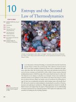
Platelets and the Coagulation Cascade
PLATELETS
COAGULATION CASCADE
Intrinsic Pathway
Extrinsic Pathway
Kallikrein
XII
XIIa
XI
XIa
IX
Tissue Factor
IXa
VIII
VIIa
VIIIa
X
VII
Xa
V
Va
Prothrombin
Thrombin
Fibrinogen
Fibrin
XIIIa
Stable Fibrin
3
• Development: derived from megakaryocytes
in the bone marrow
• Function: involved in thrombogenesis;
forms platelet plugs via adhesion and aggregation reactions; activates the coagulation cascade
1
A 26-year-old pregnant woman presents to your office for a checkup. She states that her pregnancy
has been proceeding smoothly, although she has been feeling more tired than she expected. Her
physical examination is largely unremarkable except for marked pallor. You order serum studies
and find that she has decreased hematocrit, decreased ferritin, and increased total iron-binding
capacity. Her peripheral blood smear shows red blood cells that are both microcytic and
hypochromic. You reassure her that these findings are most likely associated with her pregnancy
status and recommend iron supplements.
4
Iron Deficiency Anemia
Etiology
Chronic blood loss, most often caused by gastrointestinal bleeding or menorrhagia; dietary
deficiency (rare); malabsorption; pregnancy
Pathology
Peripheral blood smear: Hypochromic microcytic erythrocytes
Clinical
Manifestations
Fatigue, pallor, and dyspnea during exercise
Lab findings: Decreased hematocrit, decreased serum iron, decreased serum ferritin, increased
TIBC, decreased Fe/TIBC ratio (< 15%)
Treatment
Iron supplementation; identification of source of occult blood loss
Notes
Plummer-Vinson syndrome is a disease in which patients present with iron deficiency anemia,
esophageal webs, and glossitis. It is associated with an increased risk for developing esophageal
cancer.
Sideroblastic anemia is the result of defective heme biosynthesis within erythrocyte precursor
cells. It can be caused by hereditary enzymatic defects or acquired defects (ie, alcohol or lead,
MDS). Laboratory studies reveal increased iron and ferritin levels, but a normal TIBC. Ringed
sideroblasts are present in the bone marrow. Treatment is directed at underlying cause as well as
supportive with blood transfusions.
4
1
A 46-year-old man presents to your office complaining of weakness and a “pins and needles”
feeling in his extremities. You note that he is ataxic and has decreased vibration and position
sense in both his arms and legs. Upon further examination, you also observe that his tongue is
red and enlarged. When laboratory tests reveal a positive Schilling test and a macrocytic anemia,
you question the patient’s diet habits, drinking habits, and history of abdominal surgery.
5
Megaloblastic Anemias
Etiology
Vitamin B12 deficiency anemia (pernicious anemia): Autoimmune gastritis (results in the failure
to produce intrinsic factor); malabsorption or vegetarian diet; gastric resection or resection of the
ileum
Folate deficiency anemia: Malabsorption or dietary deficiency (often seen in alcoholics);
pregnancy; pharmacologic agents (methotrexate, sulfa drugs, phenytoin, AZT)
Pathology
Vitamin B12 deficiency: Demyelination of the posterior and lateral columns of the spinal cord.
Peripheral blood smear: pancytopenia; hypersegmented neutrophils; macrocytic erythrocytes
Folate deficiency: Peripheral blood smear: pancytopenia; hypersegmented neutrophils; macrocytic
erythrocytes
Clinical
Manifestations
Vitamin B12 deficiency: Neurologic abnormalities (ataxia, impaired proprioception, and vibratory
sensation); glossitis; symptoms of autoimmune gastritis. Lab findings: Decreased Hct, decreased
serum vitamin B12, anti-intrinsic factor antibodies, abnormal Schilling test (tests for decreased
absorption of oral vitamin B12).
Folate deficiency: Glossitis and diarrhea. Lab findings: Decreased Hct, decreased red blood cell
folate levels.
Treatment
Vitamin B12 deficiency: Vitamin B12 supplementation; intrinsic factor supplementation if anemia
caused by autoimmune gastritis
Folate deficiency: Folic acid supplementation
Notes
5