Ebook Essentials of critical care nursing A holistic approach Part 2
Bạn đang xem bản rút gọn của tài liệu. Xem và tải ngay bản đầy đủ của tài liệu tại đây (14.81 MB, 303 trang )
Respiratory System
FOUR
CHAPTER
15
Patient Assessment:
Respiratory System
OBJECTIVES
Based on the content in this chapter, the reader should be able to:
1 Describe the components of the history for respiratory assessment.
2 Explain the use of inspection, palpation, percussion, and auscultation for
respiratory assessment.
3 Explain the components of an arterial blood gas and the normal values for
each component.
4 Compare and contrast the arterial oxygen saturation and the partial pressure
of oxygen dissolved in arterial blood.
5 Compare and contrast the causes, signs, and symptoms of respiratory
acidosis, respiratory alkalosis, metabolic acidosis, and metabolic alkalosis.
6 Analyze examples of an arterial blood gas result.
7 Discuss the purpose of pulse oximetry, end-tidal carbon dioxide monitoring,
and mixed venous oxygen saturation monitoring.
8 Discuss the purpose of respiratory diagnostic studies and associated nursing
implications.
207
Morton_Chap15.indd 207
2/4/2012 3:12:16 PM
208
P A R T F O U R Respiratory System
TA B L E 1 5- 1 Sputum Assessment
History
Sputum Appearance
Significance
Yellow, green, brown
Clear, white
Yellow
Rust colored (yellow mixed with
blood)
Mucoid, viscid, blood streaked
Persistent, slightly blood
streaked
Clotted blood present
Bacterial infection
Absence of infection
Possible allergies
Possible tuberculosis
Principal symptoms to investigate in more detail
commonly include dyspnea, chest pain, sputum
production (Table 15-1), and cough. Because smoking has a significant impact on the patient’s respiratory health, the patient’s use of tobacco should
be quantified by amount and how long the patient
has smoked. Elements of the respiratory history are
summarized in Box 15-1. A pulmonary illness often
results in the production (or a change in the production) of sputum.
Viral infection
Carcinoma
Pulmonary infarct
Physical Examination
A
comprehensive pulmonary assessment allows
the nurse to establish the patient’s baseline status
and provides a framework for rapidly detecting
changes in the patient’s condition.
High-quality physical assessments often provide
information that can lead to the detection of complications or changes in the patient’s condition before
information from laboratory and diagnostic studies
is available.
B O X 1 5 - 1 Respiratory Health History
History of the Present Illness
Complete analysis of the following signs and symptoms
(using the NOPQRST format; see Chapter 12, Box 12-1):
• Dyspnea, dyspnea on exertion
• Shortness of breath
• Chest pain
• Cough
• Sputum production and appearance
• Hemoptysis
• Wheezing
• Orthopnea
• Clubbing
• Cyanosis
Past Health History
• Relevant childhood illnesses and immunizations:
whooping cough (pertussis), mumps, cystic fibrosis
• Past acute and chronic medical problems, including treatments and hospitalizations: streptococcal
infection of the throat, upper respiratory infections,
tonsillitis, bronchitis, sinus infection, emphysema,
asthma, bronchiectasis, tuberculosis, cancer, pulmonary hypertension, heart failure, musculoskeletal and
neurological diseases affecting the respiratory system
• Risk factors: age, obesity, smoking, allergens
• Past surgeries: tonsillectomy, thoracic surgery, coronary artery bypass grafting (CABG), cardiac valve
surgery, aortic aneurysm surgery, trauma surgery,
tracheostomy
• Past diagnostic tests and interventions: tuberculin
skin test, allergy tests, pulmonary function tests, chest
radiograph, computed tomography (CT) scan, magnetic resonance imaging (MRI), bronchoscopy, cardiac
Morton_Chap15.indd 208
stress test, ventilation–perfusion scanning, pulmonary
angiography, thoracentesis, sputum culture
• Medications, including prescription drugs,
over-the-counter drugs, vitamins, herbs, and
supplements: oxygen, bronchodilators, antitussives,
expectorants, mucolytics, anti-infectives, antihistamines, methylxanthine agents, anti-inflammatory
agents
• Allergies and reactions to medications, foods, contrast dye, latex, or other materials
• Transfusions, including type and date
Family History
• Health status or cause of death of parents and
siblings: tuberculosis, cystic fibrosis, emphysema,
asthma, malignancy
Personal and Social History
• Tobacco, alcohol, and substance use
• Environment: exposure to asbestos, chemicals, coal
dust, allergens; type of heating and ventilation system
• Diet
• Sleep patterns: use of pillows
• Exercise
Review of Other Systems
• HEENT: strep throat, sinus infections, ear infection,
deviated nasal septum, tonsillitis
• Cardiac: heart failure, dysrhythmias, coronary artery
disease (CAD), valvular disease, hypertension
• Gastrointestinal: weight loss, nausea, vomiting
• Neuromuscular: Guillain–Barré syndrome, myasthenia gravis, amyotrophic lateral sclerosis, weakness
• Musculoskeletal: scoliosis, kyphosis
2/4/2012 3:12:19 PM
Patient Assessment: Respiratory System C H A P T E R 1 5
Inspection
Inspection of the patient involves checking for the
presence or absence of several factors (Box 15-2).
• Central cyanosis (blueness of the tongue or lips)
usually means the patient has low oxygen tension. The presence of cyanosis is a late and often
ominous sign. Cyanosis is difficult to detect in a
patient with anemia. A patient with polycythemia may have cyanosis even if oxygen tension is
normal.
• Labored breathing is an important marker of
respiratory distress. As part of the inspection, the
nurse determines whether the patient is using the
accessory muscles of respiration (the scalene and
sternocleidomastoid muscles). Intercostal retractions (inward movement of the muscles between
the ribs) suggest that the patient is making a larger
effort at inspiration than normal. The nurse also
observes the patient for use of the abdominal muscles during the usually passive expiratory phase.
Sometimes, the number of words a patient can say
before having to gasp for another breath is a good
measure of the degree of labored breathing.
• Respiratory rate, depth, and pattern. These are
important parameters to follow and may be indicators of the underlying disease process (Table 15-2).
• Anterior–posterior diameter of the chest.
The size of the chest from front to back may be
increased in patients with obstructive pulmonary
disease (due to overexpansion of the lungs) and in
patients with kyphosis.
• Chest deformities and scars (eg, kyphoscoliosis
or flail chest from trauma) are important in helping to determine the reason for respiratory distress.
• Chest expansion is important to note. Causes of
abnormal chest expansion are listed in Box 15-3.
Asynchronous respiratory effort often precedes
the need for ventilatory support.
BOX 15-2
• Clubbing of the fingers (see Chapter 30, Fig. 30-2)
is seen in many patients with respiratory and cardiovascular diseases, especially chronic hypoxia.
Palpation
In addition to observing expansion of the chest
wall, the nurse palpates chest expansion by positioning the thumbs on the patient’s back, at the
level of the 10th rib, and observing the divergence
of the thumbs caused by the patient’s breathing.
Expansion of the chest wall should be symmetrical
(see Box 15-3).
To assess tactile fremitus (the ability to feel sound
on the chest wall), the nurse asks the patient to say
“ninety-nine” while palpating the posterior surfaces of the chest wall. Tactile fremitus is slightly
increased by the presence of solid substances, such
as the consolidation of a lung due to pneumonia,
pulmonary edema, or pulmonary hemorrhage.
Conditions that result in greater air volume in the
lung (eg, emphysema) are associated with decreased
or absent tactile fremitus, because air does not conduct sound well.
The nurse palpates for subcutaneous emphysema by moving the fingers in a gentle rolling
motion across the chest and neck to feel pockets of
air underneath the skin. Subcutaneous emphysema
may result from a pneumothorax or small pockets
of alveoli that have burst with increased pulmonary pressure, (eg, PEEP). In severe cases, the subcutaneous emphysema may spread throughout the
body.
Finally, the nurse palpates the position of the trachea. Pleural effusion, hemothorax, pneumothorax,
or a tension pneumothorax can cause the trachea to
move away from the affected side. Atelectasis, fibrosis, tumors, and phrenic nerve paralysis often pull
the trachea toward the affected side.
Components of the Inspection Process in the Physical Assessment of the Respiratory
System
General
• Mentation
• Anxiety level
• Speech
• Skin color (pallor, cyanosis)
• Weight (obese, malnourished)
• Body position (leaning forward, arms elevated)
Thorax
• Symmetry of thorax
• Anterior–posterior diameter (should be less than
transverse by at least half)
• Rate, pattern, rhythm, and duration of breathing
• Use of accessory muscles
Morton_Chap15.indd 209
209
• Synchrony of chest and abdomen movement
• Alignment of spine
Head and Neck
• Nasal flaring
• Pursed-lip breathing
• Mouth breathing versus nasal breathing
• Use of neck and shoulders
• Tracheal position
• Central cyanosis
Extremities
• Clubbing
• Edema
• Peripheral cyanosis
2/4/2012 3:12:19 PM
210
P A R T F O U R Respiratory System
TA B L E 1 5- 2 Respiration Patterns
Type
Description
Normal
12–20 breaths/min and
regular
Normal breathing pattern
Tachypnea
Greater than 24 breaths/
min and shallow
Bradypnea
Less than 10 breaths/min
and regular
Hyperventilation
Increased rate and
increased depth
May be a normal response to fever,
anxiety, or exercise
Can occur with respiratory insufficiency,
alkalosis, pneumonia, or pleurisy
May be normal in well-conditioned
athletes
Can occur with medication-induced
depression of the respiratory center,
diabetic coma, neurologic damage
Extreme exercise, fear, or anxiety; central
nervous system (CNS) disorders;
compensation for acidosis (eg,
salicylate overdose)
Kussmaul’s
respiration
Rapid, deep, labored
Associated with diabetic ketoacidosis
Hypoventilation
Decreased rate, decreased
depth, irregular pattern
Usually associated with overdose of
narcotics or anesthetics
Cheyne–Stokes
respiration
Regular pattern
characterized by
alternating periods of
deep, rapid breathing
followed by periods of
apnea
Irregular pattern
characterized by varying
depth and rate of
respirations followed by
periods of apnea
Significant disorganization
with irregular and
varying depths of
respiration
Increasing difficulty in
getting breath out
May result from severe heart failure,
drug overdose, increased intracranial
pressure (ICP) stroke, or renal failure
May be noted in elderly people during
sleep, not related to any disease
process
May be seen with meningitis or severe
brain damage
Biot’s
respiration
Ataxic
Air trapping
BOX 15-3
Pattern
Abnormal Chest Expansion
Unilateral diminished expansion
• Atelectasis
• Endotracheal or nasotracheal tube positioned in
right mainstream bronchi
• Collapsed lung
• Pulmonary embolus
• Lobar pneumonia
• Pleural effusion
• Pneumothorax
• Rib fracture
Asynchronous expansion
• Flail chest
Morton_Chap15.indd 210
Clinical Significance
A more extreme expression of Biot’s
respirations; indicates respiratory
compromise and elevated ICP
Seen in chronic obstructive pulmonary
disease (COPD) when air is trapped
in the lungs during forced expiration
Percussion
Percussion of the chest normally produces a resonant or hollow note. In diseases in which there is
increased air in the chest or lungs (eg, pneumothorax, emphysema), percussion notes may be hyperresonant. A flat percussion note is more likely to
be heard if a large pleural effusion is present in the
lung beneath the examining hand. A dull percussion
note is heard if atelectasis or consolidation is present. Asthma or a large pneumothorax can result in a
tympanic drum-like sound.
Auscultation
In general, four types of breath sounds are heard
in the normal chest (Table 15-3). Bronchial breath
2/4/2012 3:12:19 PM
Patient Assessment: Respiratory System C H A P T E R 1 5
211
TA B LE 15- 3 Characteristics of Breath Sounds
Intensity of
Expiratory Sound
Pitch of
Expiratory Sound
Locations Where Heard
Normally
Inspiratory sounds last
longer than expiratory
ones.
Inspiratory and expiratory
sounds are about
equal.
Expiratory sounds last
longer than inspiratory
ones.
Soft
Relatively low
Over most of both lungs
Intermediate
Intermediate
Loud
Relatively high
Often in the first and second
interspaces anteriorly and
between the scapulae
Over the manubrium, if
heard at all
Inspiratory and expiratory
sounds are about
equal.
Very loud
Relatively high
Duration of Sounds
Vesiculara
Bronchovesicular
Bronchial
Tracheal
Over the trachea in the neck
a
The thickness of the bars indicates intensity; the steeper their incline, the higher the pitch. From Bickley LS:
Bates’ Guide to Physical Examination and History Taking, 10th ed. Philadelphia, PA: Lippincott Williams &
Wilkins, 2009, p 303.
sounds are abnormal when heard over lung tissue
and indicate fluid accumulation or consolidation
of the lung (eg, as a result of pneumonia or pleural
effusion). Bronchial breath sounds are associated
with egophony and whispered pectoriloquy:
• Egophony (distorted voice sounds) occurs in the
presence of consolidation and is detected by asking the patient to say “E” while the nurse listens
with a stethoscope. In egophony, the nurse will
hear an “A” sound rather than an “E” sound.
• Whispered pectoriloquy is the presence of loud,
clear sounds heard through the stethoscope when
the patient whispers. Normally, the whispered
voice is heard faintly and indistinctly through the
stethoscope. The increased transmission of voice
sounds indicates the presence of fluid in the lungs.
Adventitious sounds are additional breath sounds
heard with auscultation and include discontinuous
sounds, continuous sounds, and friction rubs:
• Discontinuous sounds are brief, nonmusical,
intermittent sounds and include fine and coarse
crackles. When assessing crackles, the nurse notes
their loudness, pitch, duration, amount, location,
and timing in the respiratory cycle. Fine crackles are
soft, high-pitched, very brief popping sounds that
occur most commonly during inspiration. These
result from fluid in the airways or alveoli, or from
the opening of collapsed alveoli. Restrictive pulmonary disease results in fine crackles during late
inspiration, whereas obstructive pulmonary disease
results in fine crackles during early inspiration.
Crackles become coarser as the air moves through
larger fluid accumulations, such as in bronchitis or
pneumonia. Crackles that clear with coughing are
not associated with significant pulmonary disease.
• Continuous sounds include wheezes and rhonchi. Wheezes are high-pitched musical sounds
Morton_Chap15.indd 211
that have a shrill quality. They are caused by the
movement of air through a narrowed or partially
obstructed airway, such as in asthma, chronic
obstructive pulmonary disease (COPD), or bronchitis. Rhonchi are deep, low-pitched rumbling
noises. The presence of rhonchi indicates the presence of secretions in the large airways, such as
occurs with acute respiratory distress syndrome
(ARDS).
• Friction rubs are crackling, grating sounds heard
more often with inspiration than expiration. A
friction rub can be heard with pleural effusion,
pneumothorax, or pleurisy. It is important to distinguish a pleural friction rub from a pericardial
friction rub. (A pericardial friction rub is a highpitched, rasping, scratchy sound that varies with
the cardiac cycle.)
The Older Patient. In elderly people, anatomical
and physiological changes associated with aging
may manifest in different assessment findings,
including increased hyperresonance (caused by
increased distensibility of the lungs), decreased
chest wall expansion, decreased use of respiratory
muscles, increased use of accessory muscles
(secondary to calcification of rib articulations), less
subcutaneous tissue, possible pronounced dorsal
curvature, and basilar crackles in the absence of
disease (these should clear after a few coughs). Also
be aware that older people may have a decreased
ability to hold their breath during the examination.
Respiratory Monitoring
Arterial Blood Gases
Arterial blood gas (ABG) assessment involves analyzing a sample of arterial blood to determine the quality
2/4/2012 3:12:21 PM
212
P A R T F O U R Respiratory System
BOX 15-4
Measuring pH in the Blood
Normal Arterial Blood Gas (ABG)
Values
The normal blood pH is 7.35 to 7.45. Box 15-5
reviews terms used in acid–base balance. An acid–
base disorder may be either respiratory or metabolic in origin (Table 15-4). If the respiratory system
is responsible, serum carbon dioxide levels are
affected, and if the metabolic system is responsible,
serum bicarbonate levels are affected (see Table
15-4). Occasionally, patients present with both respiratory and metabolic disorders that together cause
an acidemia or alkalemia. When this occurs, the
ABG reflects a mixed respiratory and metabolic acidosis. Examples of ABG values in mixed disorders
are given in Box 15-6.
PaO2: 80 to 100 mm Hg
SaO2: 93% to 99%
pH: 7.35 to 7.45
PaCO2: 35 to 45 mm Hg
HCO3: 22 to 26 mEq/L
and extent of pulmonary gas exchange and acid–base
status. Normal ABG values are given in Box 15-4.
Measuring Oxygen in the Blood
Oxygen is carried in the blood in two ways.
Approximately 3% of oxygen is dissolved in the
plasma (PaO2). The normal PaO2 is 80 to 100 mm
Hg at sea level. For people living at higher altitudes,
the normal PaO2 is lower because of the lower barometric pressure. The remaining 97% of oxygen is
attached to hemoglobin in red blood cells (SaO2).
The normal SaO2 ranges from 93% to 99%. SaO2 is an
important oxygenation value to assess because most
oxygen supplied to tissues is carried by hemoglobin.
Interpreting Arterial Blood Gas Results
When interpreting ABG results, three factors must
be considered: oxygenation status, acid–base
balance, and degree of compensation (Box 15-7).
If the patient presents with alkalemia or acidemia, it is important to determine whether the
body has tried to compensate for the abnormality.
The respiratory system responds to metabolicbased pH imbalances by increasing the respiratory rate and depth (metabolic acidosis) or
decreasing the respiratory rate and depth (metabolic alkalosis). The renal system responds to
respiratory-based pH imbalances by increasing
hydrogen secretion and bicarbonate reabsorption (respiratory acidosis) or decreasing hydrogen
secretion and bicarbonate reabsorption (respiratory alkalosis).
ABGs are defined by their degree of compensation: uncompensated, partially compensated, or
completely compensated. To determine the level of
compensation, the nurse examines the pH, carbon
dioxide, and bicarbonate values to evaluate whether
the opposite system (renal or respiratory) has
worked to try to shift back toward a normal pH. The
primary abnormality (metabolic or respiratory) is
correlated with the abnormal pH (acidotic or alkalotic). The secondary abnormality is an attempt to
correct the primary disorder. By using the rules for
defining compensation in Box 15-8, it is possible to
determine the compensatory status of the patient’s
ABGs.
The Older Patient. PaO2 tends to decrease with
age. For patients who are 60 to 80 years of age, a
PaO2 of 60 to 80 mm Hg is normal.1
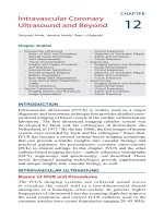
The relationship between PaO2 and SaO2 is depicted
by the oxyhemoglobin dissociation curve (Fig. 15-1).
At a PaO2 greater than 60 mm Hg, large changes in
the PaO2 result in only small changes in the SaO2.
However, at a PaO2 of less than 60 mm Hg, the curve
drops sharply, signifying that a small decrease in PaO2
is associated with a large decrease in SaO2. Factors
such as pH, carbon dioxide concentration, temperature, and levels of 2,3-diphosphoglycerate (2,3-DPG)
influence hemoglobin’s affinity for oxygen and can
cause the curve to shift to the left or to the right (see
Fig. 15-1). When the curve shifts to the right, there is
a reduced capacity for hemoglobin to hold onto oxygen, resulting in more oxygen released to the tissues.
When the curve shifts to the left, there is an increased
capacity for hemoglobin to hold oxygen, resulting in
less oxygen released to the tissues.
100
Shift to the right
Acidosis ( pH)
PaCO2
Temperature
2, 3 DPG
90
SaO2 (%)
Shift to the left
Alkalosis ( pH)
PaCO2
Temperature
2, 3 DPG
75
50
F I G U R E 1 5 - 1 The oxyhemoglobin dis-
25
0
Morton_Chap15.indd 212
20
40
60
PaO2 (mm Hg)
80
100
sociation curve is a graphic depiction
of the relationship between oxyhemoglobin saturation (the percentage of
hemoglobin combined with oxygen, or
the SaO2) and the arterial oxygen tension (PaO2) to which it is exposed.
2/4/2012 3:12:21 PM
Patient Assessment: Respiratory System C H A P T E R 1 5
BOX 15-5
Acid–Base Terminology
Acid: A substance that can donate hydrogen ions (H+).
Example: H2CO3 (an acid) → H+ + HCO3
Base: A substance that can accept hydrogen ions (H+).
Example: HCO3 (a base) + H+ → H2CO3
Acidemia: Acid condition of the blood in which the
pH is less than 7.35
Alkalemia: Alkaline condition of the blood in which
the pH is greater than 7.45
Acidosis: The process causing acidemia
Alkalosis: The process causing alkalemia
BOX 15-6
213
Arterial Blood Gases (ABGs) in
Mixed Respiratory and Metabolic
Disorders
Mixed Acidosis
Mixed Alkalosis
pH: 7.25
PaCO2: 56 mm Hg
HCO3: 15 mEq/L
pH: 7.55
PaCO2: 26 mm Hg
HCO3: 28 mEq/L
TA B LE 15- 4 Possible Causes and Signs and Symptoms of Acid–Base Disorders
Condition
Possible Causes
Respiratory Acidosis
PaCO2 greater than 45 mm Hg
pH less than 7.35
Inadequate elimination of CO2 by lungs
Central nervous system (CNS) depression
Head trauma
Oversedation
Anesthesia
High cord injury
Pneumothorax
Hypoventilation
Bronchial obstruction and atelectasis
Severe pulmonary infections
Heart failure and pulmonary edema
Massive pulmonary embolus
Myasthenia gravis
Multiple sclerosis
Excessive elimination of CO2 by the lungs
Anxiety and nervousness
Fear
Pain
Hyperventilation
Fever
Thyrotoxicosis
CNS lesions
Salicylates
Gram-negative septicemia
Pregnancy
Increased acids
Renal failure
Ketoacidosis
Anaerobic metabolism
Starvation
Salicylate intoxication
Loss of base
Diarrhea
Intestinal fistulas
Respiratory Alkalosis
PaCO2 less than 35 mm Hg
pH greater than 7.45
Metabolic Acidosis
HCO3 less than 22 mEq/L
pH less than 7.35
Metabolic Alkalosis
HCO3 greater than 26 mEq/L
pH greater than 7.45
Morton_Chap15.indd 213
Gain of base
Muscle twitching and cramps
Excess use of bicarbonate
Lactate administration in dialysis
Excess ingestion of antacids
Loss of acids
Vomiting
Nasogastric suctioning
Hypokalemia
Hypochloremia
Administration of diuretics
Increased levels of aldosterone
Signs and Symptoms
Dyspnea
Restlessness
Headache
Tachycardia
Confusion
Lethargy
Dysrhythmias
Respiratory distress
Drowsiness
Decreased responsiveness
Light-headedness
Confusion
Decreased concentration
Paresthesias
Tetanic spasms in the arms and legs
Cardiac dysrhythmias
Palpitations
Sweating
Dry mouth
Blurred vision
Headache
Confusion
Restlessness
Lethargy
Weakness
Stupor/coma
Kussmaul’s respirations
Nausea and vomiting
Dysrhythmias
Warm, flushed skin
Tetany
Dizziness
Lethargy
Weakness
Disorientation
Convulsions
Coma
Nausea and vomiting
Depressed respiration
2/4/2012 3:12:22 PM
214
P A R T F O U R Respiratory System
BOX 15-7
Interpretation of Arterial Blood Gas (ABG) Results
Approach
Sample blood gas
1. Evaluate oxygenation by examining the PaO2 and the
SaO2.
2. Evaluate the pH. Is it acidotic, alkalotic, or normal?
3. Evaluate the PaCO2. Is it high, low, or normal?
4. Evaluate the HCO3. Is it high, low, or normal?
5. Determine whether compensation is occurring. Is it
complete, partial, or uncompensated?
PaO2
SaO2
pH
PaCO2
HCO3
85 mm Hg
90%
7.49
40
29 mEq/L
Normal
Low
Alkalemia
Normal
Increased (metabolic
cause)
Conclusion: Metabolic alkalosis with a low saturation
(uncompensated)
Examples
Sample blood gas
PaO2
SaO2
Ph
PaCO2
80 mm Hg
95%
7.30
55 mm Hg
HCO3
25 mEq/L
Normal
Normal
Acidemia
Increased (respiratory
cause)
Normal
Conclusion: Respiratory acidosis (uncompensated)
Pulse Oximetry
The SpO2 is the arterial oxygen saturation of hemoglobin as measured by pulse oximetry. In pulse
oximetry, light-emitting and light-receiving sensors quantify the amount of light absorbed by oxygenated/deoxygenated hemoglobin in the arterial
blood. Usually, the sensors are in a clip placed on
BOX 15-8
Compensatory Status of Arterial Blood Gases (ABGs)
Uncompensated: pH is abnormal, and either the CO2 or
HCO3 is also abnormal. There is no indication that the
opposite system has tried to correct for the other.
In the example below, the patient’s pH is alkalotic
as a result of the low (below the normal range of 35 to
45 mm Hg) CO2 concentration. The renal system value
(HCO3) has not moved out its normal range (22 to 26
mEq/L) to compensate for the primary respiratory
disorder.
PaO2
pH
PaCO2
HCO3
94 mm Hg
7.52
25 mm Hg
24 mEq/L
Normal
Alkalotic
Decreased
Normal
Partially compensated: pH is abnormal, and both the
CO2 and HCO3 are also abnormal; this indicates that
one system has attempted to correct for the other but
has not been completely successful.
In the example below, the patient’s pH remains alkalotic as a result of the low CO2 concentration. The renal
system value (HCO3) has moved out its normal range
(22 to 26 mEq/L) to compensate for the primary respiratory disorder but has not been able to bring the pH
back within the normal range.
Morton_Chap15.indd 214
a finger, ear lobe, or forehead. The value displayed
by the oximeter is an average of numerous readings taken over a 3- to 10-second period. Oximetry
is not used in place of ABG monitoring. Rather,
pulse oximetry is used to assess trends in oxygen
saturation when the correlation between arterial blood and pulse oximetry readings has been
established.
PaO2
pH
PaCO2
HCO3
94 mm Hg
7.48
25 mm Hg
20 mEq/L
Normal
Alkalotic
Decreased
Decreased
Completely compensated: pH is normal and both the
CO2 and HCO3 are abnormal; the normal pH indicates
that one system has been able to compensate for the
other.
In the example below, the patient’s pH is normal
but is tending toward alkalosis (greater than 7.40). The
primary abnormality is respiratory because the PaCO2
is low (decreased acid concentration). The bicarbonate value of 18 mEq/L reflects decreased concentration
of base and is associated with acidosis, not alkalosis.
In this case, the decreased bicarbonate has completely
compensated for the respiratory alkalosis.
PaO2
pH
94 mm Hg
7.44
PaCO2
HcO3
25 mm Hg
18 mEq/L
Normal
Normal, tending toward
alkalosis
Decreased, primary problem
Decreased, compensatory
response
2/4/2012 3:12:22 PM
Patient Assessment: Respiratory System C H A P T E R 1 5
RED FLAG! Values obtained by pulse oximetry
are unreliable in the presence of
vasoconstricting medications, IV dyes, shock,
cardiac arrest, severe anemia, and dyshemoglobins
(eg, carboxyhemoglobin, methemoglobin).2
End-Tidal Carbon Dioxide Monitoring
End-tidal carbon dioxide (ETCO2) monitoring and
capnography measures the level of carbon dioxide at
the end of exhalation, when the percentage of carbon dioxide dissolved in the arterial blood (PaCO2)
approximates the percentage of alveolar carbon dioxide (PACO2). Therefore, ETCO2 can be used to estimate PaCO2. Although PaCO2 and ETCO2 values are
similar, ETCO2 is usually lower than PaCO2 by 2 to 5
mm Hg.3 The difference between PaCO2 and ETCO2
(PaCO2–ETCO2 gradient) may be attributed to several factors; pulmonary blood flow is the primary
determinant.
ETCO2 values are obtained by analyzing samples
of expired gas from an endotracheal tube, an oral
airway, a nasopharyngeal airway, or a nasal cannula.
Because ETCO2 provides continuous estimates of
alveolar ventilation, it is useful for monitoring the
patient during weaning from a ventilator, in cardiopulmonary resuscitation (CPR), and in endotracheal
intubation.
On a capnogram, the waveform is composed of
four phases, each one representing a specific part of
the respiratory cycle (Fig. 15-2):
1. The first phase is the baseline phase, which represents both the inspiratory phase and the very
beginning of the expiratory phase, when carbon
dioxide–free air in the anatomical dead space is
exhaled. This value should be zero in a healthy adult.
2. The second phase is the expiratory upstroke, which
represents the exhalation of carbon dioxide from
the lungs. Any process that delays the delivery
of carbon dioxide from the patient’s lungs to the
detector (eg, COPD, bronchospasm, kinked ventilator tubing) prolongs the expiratory upstroke.
3. The third phase, the plateau phase, begins as carbon dioxide elimination rapidly continues and
indicates the exhalation of alveolar gases. The
End-tidal carbon dioxide
(ET CO2 ) level
mm Hg
Plateau phase
32
0
Expiration starts;
Inspiration starts;
indicated by CO2 rise
indicated by CO2 fall
(expiratory upstroke
(inspiratory downstroke
phase)
phase)
Baseline
phase
F I G U R E 1 5 - 2 Capnogram tracing.
Morton_Chap15.indd 215
215
ETCO2 is the value generated at the very end of
exhalation, indicating the amount of carbon dioxide exhaled from the least ventilated alveoli.
4. The fourth phase is the inspiratory downstroke. The
downward deflection of the waveform is caused by
the washout of carbon dioxide that occurs in the
presence of the oxygen influx during inspiration.
Mixed Venous Oxygen Saturation
Mixed venous oxygen saturation (SvO2) is a parameter that is measured to evaluate the balance between
oxygen supply and oxygen demand. SvO2 indicates
the adequacy of the supply of oxygen relative to the
demand for oxygen at the tissue levels. Normal SvO2
is 60% to 80%; this means that supply of oxygen to
the tissues is adequate to meet the tissue’s demand.
However, a normal value does not indicate whether
compensatory mechanisms were needed to maintain the balance. For example, in some patients, an
increase in cardiac output is needed to compensate
for a low supply of oxygen.
A pulmonary artery catheter (PAC) with an oximeter built into its tip that allows continuous monitoring of SvO2 provides ongoing assessment of oxygen
supply and demand imbalances. If a catheter with
a built-in oximeter is not available, a blood sample
drawn from the pulmonary artery port of a PAC can
be sent to the laboratory for blood gas and SvO2
analysis.
A low SvO2 value may be caused by a decrease in
oxygen supply to the tissues or an increase in oxygen
use due to a high demand (Table 15-5). A decrease
in SvO2 often occurs before other hemodynamic
changes and therefore is an excellent clinical tool
in the assessment and management of critically ill
patients. Elevated SvO2 values are associated with
increased delivery of oxygen or with decreased
demand (see Table 15-5).
Respiratory Diagnostic Studies
Pulmonary function tests measure the ability of the
chest and lungs to move air into and out of the alveoli. Pulmonary function tests include volume measurements, capacity measurements, and dynamic
measurements (Table 15-6):
• Volume measurements show the amount of air
contained in the lungs during various parts of the
respiratory cycle.
• Capacity measurements quantify part of the pulmonary cycle.
• Dynamic measurements provide data about airway resistance and the energy expended in breathing (work of breathing).
These measurements are influenced by exercise, disease, age, gender, body size, and posture.
Other diagnostic studies that are often used to
evaluate the respiratory system are summarized in
Table 15-7.
2/4/2012 3:12:22 PM
216
P A R T F O U R Respiratory System
TA B L E 1 5- 5 Possible Causes of Abnormalities in Mixed Venous Oxygen Saturation (SvO2)
Abnormality
Possible Cause
Low SvO2 (less than 60%)
Decreased oxygen supply
Low hematocrit from anemia or hemorrhage
Low arterial saturation and hypoxemia from lung disease, ventilation–perfusion
mismatches
Low cardiac output from hypovolemia, heart failure, cardiogenic shock, myocardial
infarction
Increased oxygen demand
Increased metabolic demand, such as hyperthermia, seizures, shivering, pain, anxiety,
stress, strenuous exercise
Increased oxygen supply
Supplemental oxygen
Decreased oxygen demand
Anesthesia, hypothermia
Technical problems
False high reading because of wedged PAC
Fibrin clot at end of catheter
Decreased oxygen consumption
Sepsis
High SvO2 (greater than 80%)
TA B L E 1 5- 6 Volume Measurements, Capacity Measurements, and Dynamic Measurements
Term Used
Symbol Description
Remarks
Tidal volume
VT
Tidal volume may vary with
severe disease.
Inspiratory reserve
volume
Expiratory reserve
volume
IRV
Normal
Values
Volume Measurements
Residual volume
ERV
Volume of air inhaled and exhaled
with each breath
Maximum volume of air that can be
inhaled after a normal inhalation
Maximum volume of air that can be
exhaled forcibly after a normal
exhalation
RV
Volume of air remaining in the lungs
after a maximum exhalation
Vital capacity
VC
Maximum volume of air exhaled from
the point of maximum inspiration
Inspiratory capacity
IC
Maximum volume of air inhaled after
normal expiration
Functional residual
capacity
FRC
Volume of air remaining in lungs after
a normal expiration
Total lung capacity
TLC
Volume of air in lungs after a
maximum inspiration and equal to
the sum of all four volumes (VT,
IRV, ERV, RV)
500 mL
3000 mL
Expiratory reserve volume is
decreased with restrictive
disorders, such as obesity,
ascites, and pregnancy.
Residual volume may be
increased with obstructive
diseases.
1100 mL
1200 mL
Capacity Measurements
Morton_Chap15.indd 216
Decrease in vital capacity
may be found in
neuromuscular disease,
generalized fatigue,
atelectasis, pulmonary
edema, and chronic
obstructive pulmonary
disease (COPD), asthma.
Decrease in inspiratory
capacity may indicate
restrictive disease.
Functional residual capacity
may be increased with
COPD and decreased in
acute respiratory distress
syndrome (ARDS).
Total lung capacity may be
decreased with restrictive
disease (atelectasis,
pneumonia) and
increased in COPD.
4600 mL
3500 mL
2300 mL
5800 mL
2/4/2012 3:12:22 PM
Patient Assessment: Respiratory System C H A P T E R 1 5
217
TA B LE 15- 6 Volume Measurements, Capacity Measurements, and Dynamic Measurements (continued)
Term Used
Symbol Description
Normal
Values
Remarks
Dynamic Measurements
Respiratory rate
(frequency)
Minute volume
(minute ventilation)
Dead space
f
Alveolar ventilation
V˙A
VD
Number of breaths per minute
15 breaths/min
Volume of air inhaled and exhaled per
minute; equal to VT × f
The part of the tidal volume that does
Alveolar dead space occurs
not participate in alveolar gas
only in disease states
exchange; equal to the air contained
(eg, pulmonary embolism,
in the airways (anatomical dead
pulmonary hypertension)
space) plus the alveolar air that
Anatomic plus alveolar dead
is not involved in gas exchange
space is physiologic dead
(alveolar dead space); calculated as
space
PACO2 − PaCO2
The part of the tidal volume that does
A measure of ventilatory
participate in alveolar gas exchange;
effectiveness
calculated as (VT − VD) × f
7500 mL/min
Less than 40%
of the VT
4500 mL/min
TA B LE 15- 7 Respiratory Diagnostic Studies
Test and Purpose
Method of Testing
Nursing Implications
X-rays pass through chest wall, making
it possible to visualize structures.
Bones appear as opaque or white;
heart and blood vessels appear as
gray; lungs filled with air appear
black; lungs with fluid appear white.
• Test can be done at the bedside or in the
diagnostic center.
• Nurse may be asked to help position the patient
and ensure that the patient takes a deep breath
during the test.
To test ventilation, the patient inhales
radioactive gas. Diminished areas of
ventilation are visible on the scan.
To test perfusion, a radioisotope
is injected intravenously, enabling
visualization of the blood supply
to the lungs. When a pulmonary
embolus is present, the blood supply
beyond the embolus is restricted.
• Test is done in a diagnostic center.
• The nurse may need to calm the patient’s
feeling of claustrophobia due to face mask.
• Check for post–procedure allergic reaction.
The larynx, trachea, and bronchi are
visualized through a fiberoptic
bronchoscope.
• The patient often receives sedation or
analgesia before the procedure.
• Postprocedure complications may include
laryngospasm, fever, hemodynamic changes,
cardiac dysrhythmias, pneumothorax,
hemorrhage, or cardiopulmonary arrest.
With the patient placed in an upright or
sitting position, a needle is placed
into the pleural space. A local
anesthetic is used at the site to
reduce pain.
• Before the test, chest radiograph, coagulation
studies, and patient education are done;
antianxiety medication may be given.
• During the procedure, the nurse helps the
patient remain in a position with the arms and
shoulders raised (to facilitate needle insertion
between the ribs) and monitors the patient’s
comfort, anxiety, and respiratory status.
• Postprocedure complications may include
pneumothorax, pain, hypotension, and
pulmonary edema.
Chest Radiography
Used to assess anatomical
and physiological
features of the
chest and to detect
pathological processes.
Ventilation–Perfusion
Scanning
A nuclear imaging test
used to evaluate a
suspected alteration
in the ventilation–
perfusion relationship in
the lung.
Bronchoscopy
Used to examine
lung tissue, collect
secretions, determine
the extent and location
of a pathologic process,
and obtain a biopsy.
Thoracentesis
Used to remove air, fluid,
or both from the chest;
to obtain specimens for
diagnostic evaluation; or
instill medications.
(continued on page 218)
Morton_Chap15.indd 217
2/4/2012 3:12:22 PM
218
P A R T F O U R Respiratory System
TA B L E 1 5- 7 Respiratory Diagnostic Studies (continued)
Test and Purpose
Method of Testing
Nursing Implications
The patient is asked to cough up
sputum from the lungs.
• The nurse instructs the patient not to place
saliva in the container but instead cough up
sputum from the lungs.
A radiopaque contrast material is
injected into one or both arms, the
femoral vein, or a catheter placed
in the pulmonary artery. Positive
test is indicated by impaired flow of
substance through narrowed vessel
or by abrupt cessation of flow.
• The nurse monitors the patient’s pulse, blood
pressure, and breathing during test.
• Possible complications include allergic reaction
to dye, pulmonary embolus, and abnormal
cardiac rhythm.
Continuously rotating x-rays send
images to a computer to create a 3D
composite image.
• Test is done in a diagnostic center.
• The nurse monitors for claustrophobia and
administers a mild sedative if necessary.
Sputum Culture
Used to identify specific
microorganisms and
their corresponding
drug sensitivity.
Pulmonary Angiography
Used to visualize the
pulmonary vasculature.
Spiral Computed
Tomography (CT)
Used to screen for tumors,
pulmonary embolism,
and abdominal aortic
aneurysm.
CA S E STUDY
M
r. J. is a 75-year-old man who has been
admitted to the cardiac care unit with a diagnosis of
exacerbated heart failure. He has a history of two
myocardial infarctions and underwent a triple coronary artery bypass graft 4 years ago.
On admission to the unit, Mr. J. is profoundly short
of breath, restless, and tachycardic. His daughter, who
accompanied him to the hospital, reports that Mr. J. is
uncharacteristically confused. On physical examination, his vital signs are as follows: RR, 32 breaths/min;
HR, 126 beats/min; and BP, 100/64 mm Hg. The nurse
notes that Mr. J. is using accessory muscles for breathing, and his jugular veins are visibly distended at 45
degrees. Mr. J.’s mucous membranes are pale, and he
has a Glasgow Coma Scale score of 14. On auscultation, the nurse hears coarse crackles in both bases
with some audible expiratory wheezing. During assessment of breath sounds, the nurse is able to clearly hear
whispered sounds through the stethoscope. Arterial
blood gases (ABGs) are PaO2, 68 mm Hg; PaCO2, 49
mm Hg; HCO3, 29 mEq/L; and pH, 7.31.
1. What three findings from Mr. J.’s assessment are
consistent with a diagnosis of heart failure?
2. Describe some of the differences in respiratory
assessment of the older patient.
Morton_Chap15.indd 218
3. What signs of respiratory distress are apparent,
even before auscultating the lungs or obtaining
arterial blood gas (ABG) results?
4. Why is Mr. J. tachypneic?
5. Why is the nurse able to hear whispered sounds
clearly with the stethoscope? What is this condition called?
6. Interpret the ABG results. Is Mr. J.
compensating?
References
1. Miller RD, et al: Chapter 71: Geriatrics: Pulmonary changes.
In Miller’s Anesthesia, 7th edition. Churchill Livingstone,
2009
2. Wilson B, et al: The accuracy of pulse oximetry in emergency department: patients with severe sepsis and septic
shock. BMC Emerg Med 10:9, 2010
3. Respiratory Care. In Best Practices: Evidence-Based Nursing
Procedures, 2nd ed. Lippincott Williams & Wilkins, 2007,
p. 298–302
Want to know more? A wide variety of resources to enhance your learning and understanding of this chapter are available on
. Visit
to access chapter review
questions and more!
2/4/2012 3:12:23 PM
CHAPTER
Patient Management:
Respiratory System
16
OBJECTIVES
Based on the content in this chapter, the reader should be able to:
1 Describe various bronchial hygiene therapy (BHT) techniques and explain their
role in preventing and treating pulmonary complications.
2 Describe the nursing assessment of patients on oxygen therapy.
3 Discuss nursing interventions necessary to prevent complications in a patient
with a chest tube drainage system.
4 Describe nursing considerations specific to the major classes of drugs used to
treat respiratory disorders.
5 List and define types of surgeries that may be used to treat respiratory system
disorders.
Bronchial Hygiene Therapy
Hospitalized patients are often not able to deep
breathe, cough, or clear mucus effectively because
of weakness, sedation, pain, or an artificial airway.
Bronchial hygiene therapy (BHT) aims to improve
ventilation and diffusion through secretion mobilization and removal and through improved gas
exchange.
BHT methods include coughing and deep breathing, airway clearance adjunct therapies, chest physiotherapy (CPT), and bronchodilator therapy. BHT
methods are used individually or in combination,
depending on the patient’s needs. Physical assessment, chest radiography, and arterial blood gases
(ABGs) are used to determine the need for BHT, the
appropriate methods to use, and the effectiveness
of these interventions. Incentive spirometry may be
given before any of the BHT methods to promote
mucus removal.
Coughing and Deep Breathing
The objectives of coughing and deep breathing are
to promote lung expansion, mobilize secretions, and
prevent the complications of retained secretions
(atelectasis and pneumonia). Even if crackles or
rhonchi are not auscultated, the nurse encourages
the high risk patient to cough and deep breathe as a
prophylactic measure every hour. These techniques
are effective only if the patient is able to cooperate
and has the strength to cough productively.
The nurse instructs the patient to sit upright,
inhale maximally and cough, and then take a slow,
deep breath and hold it for 2 to 3 seconds. Use
of incentive spirometry along with coughing and
219
Morton_Chap16.indd 219
2/4/2012 3:14:30 PM
220
P A R T F O U R Respiratory System
deep-breathing exercises improves inhaled volumes and prevents atelectasis. Effective incentive
spirometry provides the patient with immediate
visual feedback on the breath depth and encourages the patient to increase breath volume. Ideally,
the patient uses the incentive spirometer hourly
while awake, completing 10 breaths each session
followed by coughing and striving to progressively
increase breath volumes.
Airway Clearance Adjunct Therapies
Airway clearance adjunct therapies may be useful for patients who require mucus removal when
coughing efforts are limited by a disease process,
injury, or surgery.
• Autogenic drainage (“huff cough”). It is a breathing technique frequently used by patients with cystic fibrosis and other chronic pulmonary diseases
associated with the production of large amounts
of thick mucus. To practice the technique, the
patient takes a series of controlled breaths, exhaling with gentle huffs to unstick the mucus while at
the same time suppressing the urge to cough.
• Oscillating positive expiratory pressure (PEP).
An oscillating PEP device (eg, Acapella valve, Flutter
valve) loosens mucus by producing PEP and oscillatory vibrations in the airways so that the mucus
can then be cleared with a cough. The nurse manually assists the patient’s cough by exerting positive
pressure on the abdominal costal margin during
exhalation, thus increasing the cough’s force.
• High-frequency chest wall oscillation. The
patient wears a vest-like device that uses air pulses
to compress the chest wall, loosening secretions.
High-frequency chest wall oscillation has been
shown to improve mucus removal and pulmonary
function, is well tolerated by surgical patients, and
can be self-administered at home.
• Positive airway pressure (PAP). PAP devices
enable airway recruitment and reduce atelectasis
by delivering pressures between 5 and 20 cm H2O
with variable flow of oxygen during therapy. They
are used in patients when other airway clearance
therapies are not sufficient to reduce or prevent
atelectasis.
Chest Physiotherapy
CPT techniques include postural drainage, chest
percussion and vibration, and patient positioning.
CPT is preceded by bronchodilator therapy and
followed by deep breathing and coughing or other
BHT techniques. Patients with an artificial airway
or an ineffective cough may require suctioning after
CPT. No single method of CPT has been shown to be
superior, and there are many contraindications to
using these techniques.
Studies have questioned the efficacy of CPT,
except in segmental atelectasis caused by mucus
obstruction and diseases that result in increased
Morton_Chap16.indd 220
sputum production.1 Bronchoscopy with bronchoalveolar lavage (BAL) is an alternative to CPT for
removing mucus plugs that result in atelectasis. The
inclusion of CPT in the plan of care must be individualized and evaluated in terms of derived benefit
versus potential risks.
Postural Drainage
In postural drainage, gravity facilitates drainage of
pulmonary secretions. The positions used depend
on the lobes affected by atelectasis or accumulations
of fluid or mucus (Fig. 16-1). Postural drainage in all
positions is not indicated for all critically ill patients.
The nurse must closely monitor the patient who is in
a head-down position for aspiration, respiratory distress, and dysrhythmias. Alternate techniques may
include gentle chest percussion and vibration.
RED FLAG! Contraindications to postural
drainage include increased intracranial pressure
(ICP), tube feeding, inability to cough, hypoxia or
respiratory instability, hemodynamic instability,
decreased mental status, recent eye surgery, hiatal
hernia, and obesity.
Chest Percussion and Vibration
Chest percussion and vibration are used to dislodge
secretions. Percussion involves striking the chest
wall with the hands formed into a cupped shape.
The patient’s position depends on the segment of
lung to be percussed. Vibration involves manually compressing the chest wall while the patient
exhales through pursed lips to increase the velocity
and turbulence of exhaled air to loosen secretions.
Vibration is used instead of percussion if the chest
wall is extremely painful. Critical care unit beds have
options to percuss or vibrate, with variable settings
for high to low frequency of percussion or vibration.
The nurse assesses the patient for tolerance to the
level of therapy.
RED FLAG! Contraindications to percussion
and vibration include fractured ribs, osteoporosis,
chest or abdominal trauma or surgery, pulmonary
hemorrhage or embolus, chest malignancy,
mastectomy, pneumothorax, subcutaneous
emphysema, cervical cord trauma, tuberculosis,
pleural effusions or empyema, and asthma.
Patient Positioning
Turning the patient laterally every 2 hours (at minimum) aids in mobilizing secretions for removal with
cough or suctioning. Changing the patient’s position
affects gas exchange, and positioning the patient
with the “good” lung down improves oxygenation by
improving ventilation to perfusion match.2
RED FLAG! Positioning is altered if the patient
has a lung abscess. In this case, the preferred
position is with the diseased lung down, because
otherwise gravity can cause the abscessed lung’s
purulent contents to drain into the opposite lung.
2/4/2012 3:14:33 PM
Patient Management: Respiratory System C H A P T E R 1 6
221
A. Face-lying hips elevated 16–18 inches on
pillows, making a 30°–45° angle.
Purpose: to drain the posterior lower lobes.
B. Lying on the left side—hips elevated
16–18 inches on pillows.
Purpose: to drain the right lateral lower lung
segments.
C. Back lying—hips elevated 16–18 inches on
pillows.
Purpose: to drain the anterior lower lung
segments.
D. Sitting upright or semireclining.
Purpose: to drain the upper lung field and
allow more forceful coughing.
E. Lying on the right side—hips elevated on pillows
forming a 30°–45° angle.
Purpose: to drain the left lower lobes.
F I G U R E 1 6 - 1 Positions used in lung drainage.
Continuous lateral rotation therapy (CLRT),
defined as continuous lateral positioning of less
than 40 degrees for 18 of 24 hours daily, improves
oxygenation and blood flow to the lung tissue in
affected regions and promotes secretion removal
and airway patency.2 Using lateral rotation therapy
beds is more effective than the inconsistent nursing
care of turning every 2 hours at minimum.3 CLRT
beds rotate to less than 40 degrees, while kinetic
therapy beds rotate to 40 degrees or more. The best
evidence-based research involves kinetic therapy
beds. The nurse assesses the patient for tolerance
to position changes when a CLRT or kinetic therapy
bed is in use.
Patients who are ventilated benefit from having
the head of the bed elevated 30 degrees at all times.4
The rationale is to promote lung expansion, prevent the aspiration that can occur in the recumbent
position in intubated patients, and prevent ventilator-associated pneumonia (VAP). Rotation therapy
may also help reduce pneumonia, although it may
not reduce days on the ventilator or the length of
Morton_Chap16.indd 221
hospital stay. For best outcomes, rotation must be
continuous and at the maximum for each side.
Prone positioning is an advanced technique used
with critically ill ventilated patients who have acute
lung injury (ALI) or acute respiratory distress syndrome (ARDS) with a low PaO2/FiO2 ratio. Studies
have demonstrated improved oxygenation in these
patients when placed in the prone position, although
this maneuver may not ultimately improve survival.5
Prone positioning involves multiple personnel and
specialized equipment, and must be performed only
by specially trained staff to prevent complications.
Progressive mobility, from sitting up in a chair to
ambulation, is also used as part of pulmonary hygiene.
Oxygen Therapy
Oxygen therapy is used to correct hypoxemia,
decrease the work of breathing, and decrease myocardial work. The goals for all patients on oxygen
therapy are a stable arterial oxygen saturation (SaO2)
2/4/2012 3:14:33 PM
222
P A R T F O U R Respiratory System
level, eupneic respirations, and a decrease in anxiety and shortness of breath. These goals should be
accomplished through delivery of the least amount
of supplemental oxygen needed, so the nurse continuously monitors the patient on oxygen for desired
results, as well as for complications.
RED FLAG! Complications of oxygen therapy
include respiratory arrest; skin breakdown from
straps and masks; dry nasal mucous membranes;
epistaxis, infection in the nares; oxygen toxicity;
absorptive atelectasis; and carbon dioxide narcosis
(manifested by altered mental status, confusion,
headache, and somnolence).
Several methods of oxygen delivery are available
(Box 16-1). The choice of delivery method depends
on the patient’s condition. Low-flow oxygen devices
are suitable for patients with normal respiratory
patterns, rates, and ventilation volumes. High-flow
BOX 16-1
oxygen devices are suitable for patients with high
oxygen requirements because high-flow devices
deliver up to 100% FiO2 and maintain humidification, which is essential to prevent drying of the nasal
mucosa. The nurse monitors the SaO2 closely for at
least 30 to 60 minutes when switching from a lowflow to a high-flow oxygen delivery device, evaluates
ABGs as needed, and assesses patient tolerance. If
increased distress, desaturation, or both are noted,
more extreme interventions (eg, intubation) may be
necessary.
Oxygen toxicity starts to occur in patients breathing an FiO2 of more than 50% for longer than
24 hours. The FiO2 should be decreased as tolerated
to the lowest possible setting as long as the SaO2
remains greater than 90%. The pathophysiological
changes that occur with oxygen toxicity may progress from capillary leaking to pulmonary edema and
possibly to ALI or ARDS with prolonged high FiO2
continues for several days. Patients on a high FiO2
Oxygen Delivery Methods With Delivered Fraction of Inspired Oxygen (FiO2)
High-Flow Devices
Venturi Mask
High-Flow Nasal Cannula
Oxygen Flow
(Minimal Rate) (L/min)
Flow (L/min)
1–35
FiO2 (%)
21–100
Low-Flow Devices
Nasal Cannula
Flow (L/min)
1
2
3
4
5
6
FiO2 (%)
21–25
25–28
28–32
32–36
36–40
40–44
Facemask
Flow (L/min)
5–6
6–7
7–10
FiO2 (%)
40
50
60
Face Tent
Air is mixed with the oxygen flow in the mask, resulting in variable delivery with humidification (21%
delivered with compressed air and up to 50% delivered with 10 L/min oxygen flow attached). A face tent
is often used for patients who cannot tolerate the
claustrophobic feeling associated with more traditional masks.
Morton_Chap16.indd 222
FiO2 Settinga (%)
4
4
6
8
8
10
25
28
31
35
40
50
a
FiO2 setting is based on venturi setting/adapter used and
oxygen flow.
Nonrebreather Mask
The nonrebreather mask is used in severe hypoxemia
to deliver the highest oxygen concentration. The oneway valve on one side allows for the exhalation of carbon dioxide. The mask delivers 80% to 95% FiO2 at a
flow rate of 10 L/min depending on the patient’s rate
and depth of breathing, with some room air entrained
through the open port on the mask. The mask should fit
snugly to prevent additional entrainment of room air.
Tracheostomy Collar and T-Piece
The T-piece is a T-shaped adapter used to provide oxygen
to either an endotracheal or a tracheostomy tube. The
tracheostomy collar may also be used and is generally preferred because it is more comfortable than the T-piece. The
strap on the tracheostomy collar is adjusted to keep the
collar on top of the tracheostomy. With both the T-piece
and tracheostomy collar, the goal is to provide a high
enough flow rate (at least 10 L/min with humidification) to
ensure that there is a minimal amount of entrained room
air. Flow can also be provided by a ventilator.
2/4/2012 3:14:34 PM
Patient Management: Respiratory System C H A P T E R 1 6
223
TA B LE 16- 1 Indications for Chest Tube Placement
Indication
Potential Causes
Hemothorax
Chest trauma, neoplasms, pleural tears, excessive anticoagulation, postthoracic
surgery, post–open lung biopsy
Pneumothorax
Spontaneous (greater than
20%)
Tension
Bronchopleural fistula
Pleural effusion
Chylothorax
Bleb rupture, lung disease
Mechanical ventilation, penetrating puncture wound, prolonged clamping of chest
tubes, lack of seal in chest tube drainage system
Tissue damage, esophageal cancer, aspiration of toxic chemicals, Boerhaave’s
syndrome (spontaneous esophageal rupture)
Neoplasms, cardiopulmonary disease, inflammatory conditions, recurrent infections,
pneumonia
Trauma or thoracic surgery, malignancy, congenital abnormalities
may also develop absorptive atelectasis as a result
of less nitrogen in the delivered gas mixture.
Because nitrogen is not absorbed, it exerts pressure within the alveoli, keeping the alveoli open.
When nitrogen is “washed out,” the oxygen replacing it is absorbed, resulting in alveolar collapse
(atelectasis).
Chest Tubes
Chest tubes are used to remove air or fluid from the
pleural space, restore intrapleural negative pressure,
reexpand a collapsed or partially collapsed lung,
and prevent reflux of drainage back into the chest.
Indications for chest tube placement are listed in
Table 16-1.
Equipment
Most chest tubes are multifenestrated transparent
tubes with distance and radiopaque markers that
facilitate visualization of the tube on chest radiographs (necessary for verifying correct positioning
in the pleural space). Larger tubes (20 to 36 French)
are used to drain blood or thick pleural drainage.
They are placed at about the fifth to sixth intercostal space (ICS) midaxillary. Smaller tubes (16 to
20 French) are used to remove air and are placed at
the second to third ICS midclavicular.
Chest tubes are attached to a drainage system.
Modern systems are disposable and have three
chambers (Fig. 16-2). The first chamber is the collection receptacle, the second chamber is the water
seal, and the third chamber is suction. The water
Parietal pleura
Visceral
pleura
To suction source
(or air)
From patient
Vent to
room air
Lung
Pleural cavity
20 mm
250 mm
Drainage
collection
chambers
2 mm
1
2
3
Water seal
F I G U R E 1 6 - 2 A disposable chest tube drainage system.
Morton_Chap16.indd 223
2/4/2012 3:14:34 PM
224
P A R T F O U R Respiratory System
seal chamber acts as a one-way valve, allowing
air to escape while preventing air from reentering the pleural space. The fluid level in the water
seal chamber fluctuates during respiration. During
inspiration, pleural pressures become more negative, causing the fluid level in the water seal chamber to rise. During expiration, pleural pressures
become more positive, causing the fluid level to
descend. If the patient is being mechanically ventilated, this process is reversed. Intermittent bubbling is seen in the water seal chamber as air and
fluid drain from the pleural cavity. Constant bubbling indicates either an air leak in the system or a
bronchopleural fistula.
In a disposable system that requires water
suction, it is achieved by adding water up to the
prescribed level in the suction chamber, usually
−20 cm H2O. It is the height of the water column
in the suction chamber, not the amount of wall
suction, that determines the amount of suction
applied to the chest tube, most commonly −20 cm
H2O. Once the wall suction exceeds the force necessary to “lift” this column of fluid, any additional
suction simply pulls air from a vented cap atop
the chamber up through the water. The amount
of wall suction applied should be sufficient to create a “gently rolling” bubble in the suction control
chamber. Vigorous bubbling results in water loss
through evaporation, changing suction pressure
and increasing the noise level in the patient’s room.
It is important to assess the system for water loss
and to add sterile water as necessary to maintain
the prescribed level of suction.
Dry suction (waterless) systems use a spring
mechanism to control the suction level and can provide levels of suction ranging from −10 to −40 cm
H2O. The amount of negative pressure is dialed in,
again, it is the amount dialed in not the wall suction
which determines the amount of suction. Dry suction systems that can deliver higher levels of suction
may be necessary in patients with large bronchopleural fistulas, hemorrhage, or obesity. They also
afford the patient a quieter environment.
RED FLAG! The chest tube drainage system
should never be raised above the chest, or the
drainage will back up into the chest.
Chest Tube Placement
The patient is placed in Fowler’s or semi-Fowler’s
position for the procedure. Because the parietal
pleura is innervated by the intercostal and phrenic
nerves, chest tube insertion is a painful procedure
and administration of analgesics is indicated. After
insertion, bacteriostatic ointment or petroleum
gauze can be applied to the incision site. Petroleum
gauze is thought to prevent air leaks; however, it also
has the potential to macerate the skin and predispose
the site to infection. A 4 × 4 gauze pad with a split
is positioned over the tube and taped occlusively to
the chest. All connections from the insertion site to
Morton_Chap16.indd 224
BOX 16-2
Chest Tube Drainage System
Assessment and Management
1. Assess cardiopulmonary status and vital signs
every 2 hours and as needed.
2. Check and maintain tube patency every 2 hours
and as needed.
3. Monitor and document the type, color, consistency,
and amount of drainage.
4. Mark the amount of drainage on the collection
chamber in hourly or shift increments,
depending on drainage, and document in output
record.
5. Prevent dependent loops from forming in tubing;
ensure that the patient does not inadvertently lie
on the tubing.
6. Assess for fluctuation of the water level
(“tidaling”) in the water seal chamber with
respiration or mechanical ventilation breaths.
7. Assess for the air leaks, manifested as constant
bubbling in the water seal chamber. If constant
bubbling is noted, identify the location of the
leak by first turning off the suction. Then,
beginning at the insertion site, briefly occlude
the chest tube or drainage tube below each
connection point until the drainage unit is
reached.
8. Check that all tubing connections are securely
sealed and taped.
9. Ensure water seal chambers are filled to the 2-cm
water line. Relieve negative pressure if the water
level is above the 2-cm water line.
10. Assess the patient for pain, intervene as needed,
and reassess appropriately. Pain management may
include the use of analgesics, a lidocaine patch, or
nonsteroidal anti-inflammatory drugs (NSAIDs).
11. Assess the actual chest tube insertion site for signs
of infection and subcutaneous emphysema.
12. Change the dressing per unit guidelines, when
soiled, and when ordered.
the drainage collection system are securely taped to
prevent air leaks as well as inadvertent disconnection. The proximal portion of the tube is taped to the
chest to prevent traction on the tube and sutures if
the patient moves. A postinsertion chest radiograph
is always ordered to confirm proper positioning.
The lungs are auscultated, and the condition of the
tissue around the insertion site is evaluated for the
presence of subcutaneous air. Ongoing assessment
and management of a patient with a chest tube is
summarized in Box 16-2.
RED FLAG! Occasionally, the chest tube may
fall out or be accidentally pulled out. If this occurs,
the insertion site should be quickly sealed off using
petroleum gauze covered with dry gauze and an
occlusive tape dressing to prevent air from entering
the pleural cavity.
2/4/2012 3:14:35 PM
Patient Management: Respiratory System C H A P T E R 1 6
RED FLAG! The most serious complication
associated with chest tube placement is tension
pneumothorax, which can develop if there is an
obstruction in the chest tube that prevents air from
leaving (thus allowing it to accumulate in the pleural
space.) Clamping chest tubes predisposes patients
to this complication and is only recommended as a
momentary measure, such as when it is necessary
to locate the source of an air leak or replace the
chest tube drainage unit.
Chest Tube Removal
Chest tubes are removed after drainage is minimal.
Prior to chest tube removal (12 to 24 hours before),
the wall suction is disconnected (ie, the chest tube
is placed on water seal). Premature removal of the
chest tube may cause reaccumulation of the pneumothorax. Before the chest tube is removed, the
patient is premedicated to alleviate pain. The tube is
removed in one quick movement during expiration
to prevent entraining air back into the pleural cavity. Immediately after tube removal, the lung fields
are auscultated for any change in breath sounds,
and an occlusive sterile dressing with petroleum
gauze is applied over the site. A chest radiograph
is obtained to look for the presence of residual air
or fluid.
Pharmacotherapy
Bronchodilators
Bronchodilators dilate the airways by relaxing
bronchial smooth muscle. Bronchodilator therapy
can be delivered through metered-dose inhalers
(MDIs) or nebulization. Patient inhalation ensures
delivery into the lungs. Assessment before, during, and after the therapy is essential and includes
breath sounds, pulse, respiratory rate, and pulmonary function tests to measure improvement in
severity of airway obstruction. ABGs also may be
indicated.
• b2-Adrenergic blockers. Because of their rapid
onset of action, β-adrenergic blockers are the
bronchodilators of choice for the treatment of
acute exacerbation of asthma or severe bronchial constriction. The bronchodilator effects of
β-adrenergic blockers result from stimulation
of β2-adrenergic receptors in the lung bronchial
smooth muscle. These agents may also stimulate
β1-adrenergic receptors in the heart, leading to
undesired cardiac effects. β2-selective drugs are
more specific for the β2-receptor, although they
retain some β1 activity. β2-Adrenergic blockers may
be administered orally or inhaled. Inhaled therapy
has been shown to produce bronchodilation comparable to that of oral administration, with fewer
adverse systemic effects.
• Anticholinergic agents. These drugs produce
bronchodilation by reducing intrinsic vagal tone
Morton_Chap16.indd 225
225
to the airways. They also block reflex bronchoconstriction caused by inhaled irritants.
• Methylxanthines. The use of methylxanthines
in the treatment of bronchospastic airway disease is controversial. Theophylline, the prototype
methylxanthine, may be used chronically in the
treatment of bronchospastic disease but is usually
considered third- or fourth-line therapy. Some
patients with severe disease that is not controlled
with β-adrenergic blockers, anticholinergics, or
anti-inflammatory agents may benefit from theophylline. Aminophylline, the IV form of theophylline, is rarely used in acute exacerbations
because of the lack of evidence that it is beneficial in this situation and it produces significant
tachycardia.
Anti-Inflammatory Agents
Anti-inflammatory agents may be used prophylactically to interrupt the development of bronchial
inflammation. They may also be used to reduce or
terminate ongoing inflammation in the airway.
• Corticosteroids are the most effective antiinflammatory agents for the treatment of reversible airflow obstruction. Corticosteroid therapy
should be initiated simultaneously with bronchodilator therapy because the onset of action may be
6 to 12 hours. Corticosteroids may be administered
parenterally, orally, or as aerosols. In acute exacerbations, high-dose parenteral steroids (eg, IV
methylprednisolone) are used and then tapered as
the patient tolerates. Short courses of oral therapy
may be used to prevent the progression of acute
attacks. Long-term oral therapy is associated with
systemic adverse effects and should be avoided if
possible.
• Mast cell stabilizers are thought to stabilize the
cell membrane and prevent the release of mediators from mast cells. These agents are not indicated
for acute exacerbations of asthma. Rather, they are
used prophylactically to prevent acute airway narrowing after exposure to allergens (eg, exercise,
cold air). A 4- to 6-week trial may be required to
determine efficacy in individual patients. The goal
is to reduce the frequency and severity of asthma
attacks and enhance the effects of concomitantly
administered bronchodilator and steroid therapy.
It may be possible to decrease the dose of bronchodilators or corticosteroids in patients who respond
to mast cell stabilizers.
• Leukotriene receptor antagonists may be used
in the management of exercise-induced bronchospasm, asthma, allergic rhinitis, and urticaria.
These agents block the activity of endogenous
inflammatory mediators, particularly leukotrienes, which cause increased vascular permeability,
mucus secretion, airway edema, bronchoconstriction, and other inflammatory cell process activities.
Leukotriene receptor antagonists are administered
once daily and are usually well tolerated. They are
2/4/2012 3:14:35 PM
226
P A R T F O U R Respiratory System
not administered for acute conditions; rather,
they are used as a part of an ongoing program of
therapy.6
Neuromuscular Blocking Agents
Critically ill patients frequently require pharmacological intervention for analgesia, sedation, anxiety
control, and facilitation of mechanical ventilation.
If metabolic demands and work of breathing continue to compromise ventilatory or hemodynamic
stability after maximization of sedation, neuromuscular blocking (NMB) agents may be required. NMB
agents induce muscular paralysis by blocking acetylcholine at the motor endplate. The paralysis prevents the patient from “fighting” the ventilator and
increasing the work of breathing. The goal of therapy with NMB agents is to maximize oxygenation
and prevent complications such as barotrauma.
NMB drugs do not possess analgesic or sedative
properties. The patient is awake and aware, but
unable to move. When NMB agents are used, sedation and analgesia are required, along with patient
and family education. Numerous reports of prolonged paralysis following the use of NMB agents
have prompted many facilities to initiate protocols
for monitoring with the use of peripheral nerve
stimulators.
Thoracic Surgery
Thoracic surgery is indicated as part of the management plan for many disorders involving the lungs
and associated structures.
• Wedge resection is performed for the removal of
benign or malignant lesions.
• Segmentectomy is the preferred method when
patients are a poor risk with limited pulmonary
reserve. Bleeding may be extensive following the
surgery, and two chest drains are usually in place
to drain air or blood.
• Lobectomy may be performed as a treatment for
malignant or benign tumors and for infections
such as bronchiectasis, tuberculosis, or fungal
infection.
• Pneumonectomy is performed to remove one lung,
usually because of primary carcinoma or significant infection.
• Lung volume reduction surgery (LVRS) involves
resecting parts of the lung to reduce hyperinflation
(eg, as part of the treatment for emphysema).
• Lung transplantation may involve one lung or
both lungs, and it may be done along with heart
transplantation. To be considered a viable candidate for lung transplantation, a patient must have
minimal comorbidities and advanced lung disease
that is unresponsive to other therapies.
Morton_Chap16.indd 226
CAS E S T U DY
M
r. B. is admitted to the critical care unit for
the diagnosis of pancreatitis. The physician places
a right subclavian central line. Immediately after
the line placement, the nurse notes that Mr. B.
has increasing dyspnea and tachycardia. Further
assessment reveals diminished breath sounds on
the right and unequal chest wall expansion. A chest
radiograph is obtained and the physician is notified.
Mr. B. is diagnosed with a right pneumothorax, and
a chest tube connected to a drainage system and
−20 cm H2O suction is placed.
Two days later, the nurse is assessing Mr. B. and
notes intermittent bubbling in the water seal chamber, fluctuation of the fluid in the tubing, a small
amount of subcutaneous air, and a dry and occlusive
dressing. Chest radiographs, obtained daily, demonstrate the presence of a small pneumothorax. The
chest tube is still connected to −20 cm H2O suction.
Five days later, the chest radiograph demonstrates complete resolution of the pneumothorax.
The chest tube is taken off of suction and left to
water seal for 8 hours. Mr. B. tolerates this without any signs of dyspnea and the chest tube is
removed by the physician. The incision site is
covered with petroleum gauze and occlusive tape
is applied to secure the dressing.
1. What was the cause of the pneumothorax?
2. What is the clinical difference between finding
intermittent bubbling and constant bubbling in the
water seal chamber?
3. What would be the reasoning for applying a
petroleum gauze dressing after removal of the
chest tube?
References
1. Nettina SM: Respiratory disorders. In Mills EJ (ed):
Lippincott Manual of Nursing Practice, 9th ed. Philadelphia,
PA: Lippincott Williams & Wilkins, 2009
2. Staudinger T, et al.: Continuous lateral rotation therapy to
prevent ventilator-associated pneumonia. Crit Care Med
38(2):706–707, 2010
3. Swadener-Culpepper, L. Continuous lateral rotation therapy.
Critical Care Nurse 30(2):S5–S7, 2010
4. Tolentino-DelosReyes AF, et al.: Am J Crit Care 16(1):20–27,
2007
5. Kopterides P, Siempos I, Armagaidis A, et al.: Prone positioning in hypoxemix respiratory failure: Meta analysis of
randomized controlled trials. J Crit Care 24:89–100, 2009
6. Karch AM (ed): Lippincott’s Nursing Drug Guide, 2007 ed.
Philadelphia, PA: Lippincott Williams & Wilkins, 2007
Want to know more? A wide variety of resources to enhance your learning and understanding of this chapter are available on
. Visit
to access chapter review
questions and more!
2/4/2012 3:14:35 PM
CHAPTER
Common Respiratory Disorders
17
OBJECTIVES
Based on the content in this chapter, the reader should be able to:
1 Describe the pathophysiology, assessment, and management of pneumonia in
the critically ill patient.
2 Describe the pathophysiology, assessment, and management of acute
respiratory failure.
3 Differentiate between hypoxemic (type I) acute respiratory failure and
hypercapnic (type II) acute respiratory failure.
4 Describe the pathophysiology, assessment, and management of acute
respiratory distress syndrome (ARDS).
5 Discuss the pathophysiology, assessment, and management of pleural effusion.
6 Describe the pathophysiology, assessment, and management of
pneumothorax.
7 Discuss the pathophysiology, assessment, management, and prevention of
pulmonary embolism.
8 Explain the pathophysiology, assessment, and management of an acute
exacerbation of chronic obstructive pulmonary disease (COPD).
9 Describe the pathophysiology, assessment, and management of an acute
exacerbation of asthma and status asthmaticus.
Pneumonia
Pneumonia is a common infection in both the community and hospital. In the United States, pneumonia is the leading cause of death from infectious
disease, the second most common hospital-acquired
infection, and the seventh leading cause of death.1
Critical care nurses encounter pneumonia when it
complicates the course of a serious illness or leads
to acute respiratory distress. According to guidelines developed by the American Thoracic Society
(ATS), patients with severe community-acquired
pneumonia (CAP) require admission to the critical
care unit. Severe CAP is defined as the presence of
one of two major criteria or the presence of two
of three minor criteria (Box 17-1).2 Streptococcus
pneumoniae (pneumococcus) is the predominant pathogen in patients with CAP who require
hospitalization.
The Older Patient. The incidence of CAP requiring
hospitalization is four times higher in patients older
than 65 years than it is in those 45 to 64 years of
age.3 In addition, the cause of CAP in patients older
than 65 years is frequently a drug-resistant strain of
S. pneumoniae.2
227
Morton_Chap17.indd 227
2/4/2012 3:15:37 PM
228
P A R T F O U R Respiratory System
BOX 17-1
American Thoracic Society (ATS)
Criteria for Diagnosis of Severe
Community-Acquired Pneumonia
(CAP)
Major Criteria
• Need for mechanical ventilation
• Need for vasopressors for greater than 4 hours
(septic shock)
• Acute renal failure (urine output less than 80 mL in
4 hours or serum creatinine greater than 2 mg/dL in
the absence of chronic renal failure)
• Increase in size of infiltrates by more than 50% in
presence of clinical nonresponse to treatment or
deterioration
Minor Criteria
• Respiratory rate greater than 30 breaths/min
• Systolic blood pressure less than or equal to
90 mm Hg
• Diastolic blood pressure less than 60 mm Hg, multilobar disease
• PaO2/FiO2 ratio less than 250
Adapted from American Thoracic Society: Guidelines for the
management of adults with community-acquired pneumonia. Am
J Respir Crit Care Med 163:1730–1754, 2001.
Hospital-acquired pneumonia (HAP) is pneumonia occurring more than 48 hours after admission to
a hospital, which excludes infection that is incubating at the time of admission.4 Ventilator-associated
pneumonia (VAP) is the occurrence of pneumonia
more than 48 to 72 hours after intubation. HAP
and VAP continue to cause morbidity and mortality despite advances in antimicrobial therapy and
advanced supportive measures.4
Bacteria, viruses, mycoplasmas, fungi, and aspiration of foreign material can cause pneumonia.
Etiology varies greatly depending on whether the
pneumonia is community acquired or hospital
acquired.5 HAP and VAP may be polymicrobial and
multidrug resistant.
Pathophysiology
Pneumonia is an inflammatory response to inhaled
or aspirated foreign material or the uncontrolled
multiplication of microorganisms invading the
lower respiratory tract. This response results in the
accumulation of neutrophils and other proinflammatory cytokines in the peripheral bronchi and alveolar spaces.6 The severity of pneumonia depends on
the amount of material aspirated, the virulence of
the organism, the amount of bacteria in the aspirate, and the host defenses.6
The means by which pathogens enter the lower
respiratory tract include aspiration, inhalation,
hematogenous spread from a distant site, and translocation. Risk factors that predispose a patient to
one of these mechanisms include conditions that
enhance colonization of the oropharynx, conditions
Morton_Chap17.indd 228
favoring aspiration, conditions requiring prolonged
intubation, and host factors.6 The risk for clinically
significant aspiration is increased in patients who
are unable to protect their airways.
Colonization of the oropharynx has been identified
as an independent factor in the development of HAP
and VAP. Gram-positive bacteria and anaerobic bacteria normally live in the oropharynx. When normal
oropharyngeal flora are destroyed, the oropharynx is
susceptible to colonization by pathogenic bacteria.
Pathogenic organisms that have colonized the oropharynx are readily available for aspiration into the
tracheobronchial tree. Gastric colonization may also
lead to retrograde colonization of the oropharynx,
although the role the stomach plays in the development of pneumonia is controversial. The stomach
is normally sterile because of the bactericidal activity of hydrochloric acid. However, when gastric pH
increases above normal (eg, with the use of histamine
type 2 antagonists or antacids), microorganisms are
able to multiply, increasing the risk for retrograde
colonization of the oropharynx and pneumonia.4
Inhalation of bacteria-laden aerosols from
contaminated respiratory equipment is another
potential source of pneumonia-causing bacteria.
Condensate collection in the ventilator tubing can
become contaminated with secretions and serve as
a reservoir for bacterial growth.
Assessment
Knowledge of risk factors and symptoms assists in
making the diagnosis and identifying the causative
organism. A comprehensive cardiovascular and pulmonary assessment is completed, with a focus on
the ATS major and minor criteria (see Box 17-1). The
nurse assesses for signs of hypoxemia and dyspnea.
Patients presenting with new-onset respiratory
symptoms (eg, cough, sputum production, dyspnea,
pleuritic chest pain) usually have an accompanying
fever and chills. Decreased breath sounds and crackles or bronchial breath sounds are heard over the
area of consolidation.
The Older Patient. Confusion and tachypnea are
common presenting symptoms in older patients
with pneumonia. The usual symptoms (fever, chills,
increased white blood cell (WBC) count) may be
absent. Other symptoms include weakness, lethargy,
failure to thrive, anorexia, abdominal pain, episodes
of falling, incontinence, headache, delirium, and
nonspecific deterioration.
RED FLAG! Disorders that may mimic pneumonia
clinically include heart failure, atelectasis, pulmonary
thromboembolism, drug reactions, pulmonary
hemorrhage, and ARDS.
Diagnostic tests are ordered to determine whether
pneumonia is the cause of the patient’s symptoms
and to identify the pathogen when pneumonia is
present. Table 17-1 summarizes the current ATS
2/4/2012 3:15:39 PM
Common Respiratory Disorders C H A P T E R 1 7
229
Studies in Patients With Severe Community-Acquired Pneumonia (CAP) or Severe
TA B LE 17- 1 Diagnostic
Hospital-Acquired Pneumonia (HAP)
Study
Rationale
Chest radiograph (anterior–posterior and lateral)
Identifies the presence, location, and severity of infiltrates
(multilobar, rapidly spreading, or cavitary infiltrates indicate
severe pneumonia)
Facilitates assessment for pleural effusions
Differentiates pneumonia from other conditions
Isolates the etiologic pathogen in 8%–20% of cases
Documents the presence of multiple-organ dysfunction
Helps define severity of illness
Two sets of blood cultures from separate sites
Complete blood count
Serum electrolyte panel, renal and liver function
tests
Arterial blood gases (ABGs)
Thoracentesis (if pleural effusion greater than 10 mm
identified on lateral decubitus film)
Pleural fluid studies, including
WBC count with differential
Protein
Glucose
Lactate dehydrogenase (LDH)
pH
Gram stain and acid-fast stain
Culture for bacteria, fungi, and mycobacteria
Defines severity of illness
Determines need for supplemental oxygen and mechanical
ventilation
Rules out empyema
From data in American Thoracic Society: Guidelines for the management of adults with community-acquired
pneumonia. Am J Respir Crit Care Med 163:1730–1754, 2001.
recommendations. Lower respiratory secretions
can be easily obtained in intubated patients using
endotracheal aspiration and may assist in excluding certain pathogens and modifying initial empirical treatment. Invasive diagnostic techniques, such
as bronchoalveolar lavage (BAL) or bronchoscopy
with protected specimen brush (PSB), may be
used in selected circumstances (eg, nonresponse
to antimicrobial therapy, immunosuppression, suspected tuberculosis in the absence of a productive
cough, pneumonia with suspected neoplasm or foreign body, or conditions that require lung biopsy).4
Pneumococcal urinary antigen assay, which returns
results within 15 minutes, is recommended as an
addition to blood culture testing.7 The IDSA recommends HIV testing for people between the ages of
15 and 54 years as well.7
need to modify therapy.4 The duration of therapy
depends on many factors, including the presence
of concurrent illness or bacteremia, the severity of
pneumonia at the onset of antibiotic therapy, the
causative organism, the risk for multidrug resistance, and the rapidity of clinical response.4
Management
Acute Respiratory Failure
Antibiotic Therapy
Acute respiratory failure is a sudden and life-threatening deterioration in pulmonary gas exchange,
resulting in carbon dioxide retention and inadequate
oxygenation. Acute respiratory failure is defined as
an arterial oxygen tension (PaO2) of 50 mm Hg or less,
an arterial carbon dioxide tension (PaCO2) greater
than 50 mm Hg, and an arterial pH less than 7.35.
Patients with advanced COPD and chronic hypercapnia may exhibit an acute increase in PaCO2 to a
high level, a decrease in blood pH, and a significant
increase in serum bicarbonate during the onset of
acute respiratory failure. Acute respiratory failure
Patients are initially treated empirically, based on
the severity of disease and the likely pathogens.4
Because data show that hospitalized patients with
CAP who receive their first dose of antibiotic therapy within 8 hours of arrival at the hospital have
reduced mortality at 30 days, initial therapy should
be instituted rapidly.2 Double antibiotic coverage
is necessary for patients with severe CAP.7 Initial
therapy should not be changed within the first 48 to
72 hours unless progressive deterioration is evident
or initial blood or respiratory cultures indicate a
Morton_Chap17.indd 229
Supportive Therapy
Oxygen therapy may be required to maintain adequate gas exchange. Humidified oxygen should be
administered by mask or endotracheal tube to promote adequate ventilation. Mechanical ventilation
to correct hypoxemia is frequently required in both
severe CAP and HAP. Aggressive bronchial hygiene
therapy (BHT) and adequate nutritional support are
critical.
2/4/2012 3:15:40 PM
230
P A R T F O U R Respiratory System
BOX 17-2
Causes of Acute Respiratory Failure
Intrinsic Lung and Airway Diseases
Large Airway Obstruction
• Congenital deformities
• Acute laryngitis, epiglottitis
• Foreign bodies
• Intrinsic tumors
• Extrinsic pressure
• Traumatic injury
• Enlarged tonsils and adenoids
• Obstructive sleep apnea
Bronchial Diseases
• Chronic bronchitis
• Asthma
• Acute bronchiolitis
Parenchymal Diseases
• Pulmonary emphysema
• Pulmonary fibrosis and other chronic diffuse infiltrative diseases
• Severe pneumonia
• Acute lung injury (ALI), acute respiratory distress
syndrome (ARDS)
Vascular Disease
• Cardiac pulmonary edema
• Massive or recurrent pulmonary embolism
• Pulmonary vasculitis
Extrapulmonary Disorders
Diseases of the Pleura and the Chest Wall
• Pneumothorax
• Pleural effusion
• Fibrothorax
may be caused by a variety of pulmonary and nonpulmonary diseases (Box 17-2). Many factors may
precipitate or exacerbate acute respiratory failure
(Box 17-3).
BOX 17-3
Precipitating and Exacerbating
Factors in Acute Respiratory
Failure
• Changes in tracheobronchial secretions
• Disturbances in tracheobronchial clearance
• Viral or bacterial pneumonia
• Drugs: sedatives, narcotics, anesthesia, oxygen
• Inhalation or aspiration of irritants, vomitus, or
foreign body
• Cardiovascular disorders: heart failure, pulmonary
embolism, shock
• Mechanical factors: pneumothorax, pleural effusion,
abdominal distention
• Trauma, including surgery
• Neuromuscular abnormalities
• Allergic disorders: bronchospasm
• Increased oxygen demand: fever, infection
• Inspiratory muscle fatigue
Morton_Chap17.indd 230
• Thoracic wall deformity
• Traumatic injury to the chest wall (flail chest)
• Obesity
Disorders of the Respiratory Muscles and the
Neuromuscular Junction
• Myasthenia gravis and myasthenia-like disorders
• Muscular dystrophies
• Polymyositis
• Botulism
• Muscle-paralyzing drugs
• Severe hypokalemia and hypophosphatemia
Disorders of the Peripheral Nerves and Spinal
Cord
• Poliomyelitis
• Guillain–Barré syndrome
• Spinal cord trauma (quadriplegia)
• Amyotrophic lateral sclerosis
• Tetanus
• Multiple sclerosis
Disorders of the Central Nervous System
• Sedative and narcotic drug overdose
• Head trauma
• Cerebral hypoxia
• Stroke
• CNS infection
• Epileptic seizure: status epilepticus
• Metabolic and endocrine disorders
• Bulbar poliomyelitis
• Primary alveolar hypoventilation
• Sleep apnea syndrome
There are three main types of acute respiratory
failure:
• Acute hypoxemic respiratory failure (type I).
Type I acute respiratory failure is the result of
abnormal oxygen transport secondary to pulmonary parenchymal disease, with increased
alveolar ventilation resulting in a low PaCO2.8
The principal problem in type I acute respiratory failure is the inability to achieve adequate
oxygenation, as evidenced by a PaO2 of 50 mm
Hg or less and a PaCO2 of 40 mm Hg or less.
Right-to-left shunt and alveolar hypoventilation
are the most clinically significant causes of type I
failure.8
• Acute hypercapnic respiratory failure (type II).
Type II acute respiratory failure (ventilatory failure) is the result of inadequate alveolar ventilation
secondary to decreased ventilatory drive, respiratory muscle fatigue or failure, and increased work
of breathing.8 Type II acute respiratory failure is
characterized by marked elevation of carbon dioxide levels with relative preservation of oxygenation. Hypoxemia results from reduced alveolar
pressure of oxygen (PAO2) and is proportionate to
hypercapnia.8
2/4/2012 3:15:40 PM
Common Respiratory Disorders C H A P T E R 1 7
• Combined hypoxemic and hypercapnic respiratory failure (type I and type II). The combined
type of acute respiratory failure develops as a consequence of inadequate alveolar ventilation and
abnormal gas transport. Any cause of type I failure
may lead to combined failure, especially if increased
work of breathing and hypercapnia are involved.
Pathophysiology
A vicious positive feedback mechanism characterizes
the deleterious effects of continued hypoxemia and
hypercapnia. Mechanisms of hypoxemia in acute
respiratory failure are summarized in Table 17-2.
Effects of prolonged hypoxemia and hypercapnia
include
•
•
•
•
•
•
•
Increased pulmonary vascular resistance
Right ventricular failure (cor pulmonale)
Right ventricular hypertrophy
Impaired left ventricular function
Reduced cardiac output
Cardiogenic pulmonary edema
Diaphragmatic fatigue from increased workload
of respiratory muscles
Assessment
Presentation of acute respiratory failure varies,
depending on the underlying disease, precipitating
factors, and degree of hypoxemia, hypercapnia, or
acidosis. The classic symptom of hypoxemia is dyspnea,9 although dyspnea may be completely absent
in ventilatory failure resulting from depression of
the respiratory center. Other presenting symptoms
of hypoxemia include cyanosis, restlessness, confusion, anxiety, delirium, tachypnea, tachycardia,
hypertension, cardiac dysrhythmias, and tremor.9
231
The cardinal symptoms of hypercapnia are dyspnea and headache. Other clinical manifestations
of hypercapnia include peripheral and conjunctival
hyperemia, hypertension, tachycardia, tachypnea,
impaired consciousness, papilledema, and asterixis
(wrist tremor).9 Uncorrected carbon dioxide narcosis leads to diminished alertness, disorientation,
increased intracranial pressure (ICP), and loss of
consciousness. Associated findings in acute respiratory failure may include use of accessory muscles
for respiration, intercostal or supraclavicular retraction, and paradoxical abdominal movement if diaphragmatic weakness or fatigue is present.
Arterial blood gas (ABG) analysis is needed to
determine PaO2, PaCO2, and blood pH levels and
confirm the diagnosis of acute respiratory failure.
Other diagnostic tests that may be ordered to aid
in determining the underlying cause may include
chest radiography, sputum examination, pulmonary
function testing, angiography, ventilation–perfusion
scanning, computed tomography (CT), toxicology
screening, complete blood count, serum electrolytes, cytology, urinalysis, bronchogram, bronchoscopy, electrocardiography, echocardiography, and
thoracentesis.8 Table 17-3 summarizes key clinical
findings and diagnostic tests according to the underlying cause of the respiratory failure.
Management
Treatment of acute respiratory failure warrants
immediate intervention to correct or compensate
for the gas exchange abnormality and identify the
cause. Therapy is directed toward correcting the
cause and alleviating the hypoxia and hypercapnia
(see Table 17-3).
If alveolar ventilation is inadequate to maintain
PaO2 or PaCO2 levels (due to respiratory or neurological
TA B LE 17- 2 Mechanisms of Hypoxemia in Acute Respiratory Failure
Mechanism
Comments
Ventilation–perfusion mismatching (“dead space”)
Resultant hypoxemia is reversible with supplemental
oxygen
Oxygen content of inhaled gas is decreased
Inhalation of a hypoxic gas mixture or severe reduction
of barometric pressure (eg, toxic inhalation, oxygen
consumption in fire, high altitudes)
Alveolar hypoventilation
Impaired diffusion (eg, emphysema, diffuse lung injury)
Right-to-left shunt
Abnormal pulmonary gas exchange, cardiac output that is
too high or too low, high metabolic rate
Morton_Chap17.indd 231
Alveolar partial pressure of oxygen (PaO2) is decreased
while alveolar partial pressure of carbon dioxide
(PaCO2) is increased
Prevents complete equilibration of alveolar gas with
pulmonary capillary blood; small effect is usually easily
compensated by a small increase in the fraction of
inspired oxygen (FiO2)
Indicates closure of air passages, especially the distal
airways and alveoli
Changes in FiO2 have little effect on the arterial carbon
dioxide tension (PaO2) when the shunt exceeds 30%
Increased oxygen extraction from arterial blood results in
decreased PaO2
Oxygen content of mixed venous blood is reduced
2/4/2012 3:15:40 PM