CHỈ ĐỊNH CHỤP ĐM VÀNH TRONG BỆNH ĐỘNG MẠCH VÀNH
Bạn đang xem bản rút gọn của tài liệu. Xem và tải ngay bản đầy đủ của tài liệu tại đây (1.75 MB, 24 trang )
CHỈ ĐỊNH CHỤP ĐM
VÀNH TRONG BỆNH
ĐỘNG MẠCH VÀNH
A\Prof. Tröông Quang Bình MD, PhD,
FSCAI
University Medical Center
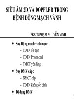
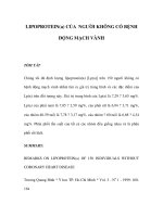
Hội chứng mạch vành cấp
CHỈ ĐỊNH CHỤP
VÀ CAN THIỆP
ĐM VÀNH RÕ
RÀNG
STEMI
Pts presenting in a
hospital with PCI
< 12 h after onset of chest
pain
Pts presenting in a
hospital without PCI
≥ 3 – 12 hours
Immediate
transfer
< 3 hours
Thrombolysis
Failed
Successful
PCI within up to 24
hours available
PCI within up to 24
hours not available
Predischarge
ischemia
Primary PCI
Rescue PCI
Post thrombolysis
PCI
Ischemia-driven
PCI
Pts presenting with NSTE-ACS
ASA/Clopidogrel/UFH
Nitrate, Betablocker
High risk
Low risk
Initally planned
invasive stratey
Immediate (< 2.5 hrs)
angio planned: GPI can
be postponed
PCI +
Abciximab
Initally planned
conservative
stratey
Early (< 48 hrs) angio
planned: upstream GPI
(Tirofiban, Eptifibatide)
PCI +
continuing
Abciximab or
Eptifibatide
Early noninvasive
stress testing
PCI + provisional
Abciximab or
Eptifibatide
Medical
treatment
CHỈ ĐỊNH CHỤP ĐM
VÀNH TRONG BỆNH ĐM
VÀNH MẠN ???
Boden WE et al. N Engl J Med. 2007;356:1503-16.
Boden WE et al. Am Heart J. 2006;151:1173-9.
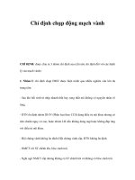
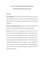
COURAGE
Clinical Outcomes Utilizing Revascularization
and Aggressive Drug Evaluation
COURAGE: Summary and implications
PCI added to OMT did not reduce
risk of death, MI, or other major CV
events compared with OMT alone
Boden WE et al. N Engl J Med. 2007;356:1503-16.
CHẨN ĐOÁN?
2013 ESC guidelines on the management of stable coronary artery disease
Uptodate 2012:
Thomas Levin, MD, Julian M Aroes
ty, MD
• We agree with the 2005
ACC/AHA/SCAI guideline update for
percutaneous coronary intervention,
which recommends that all patients
with stable angina undergo testing
for risk stratification
Exercise
ECG
Stress
gaéng
Test
söùc
Stress
echocardiography
Nuclear cardiology
MRI tim
Khaỷo saựt vaọn ủoọng vuứng
MSCT
Mức độ khuyến cáo
Mức độ bằng chứng
2014 ESC/EACTS Guidelines on myocardial revascularization
I
2014 ESC/EACTS Guidelines on myocardial revascularization
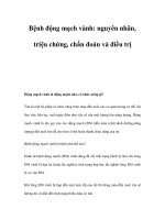
CÁC NGHIỆM PHÁP
Nghiệm pháp gắng sức
nguy
cơ
cao
Chỉ đònh nhóm I
Nghiệm pháp gắng sức dương tính sớm ( < 6.5 METS)
Những bệnh nhân được xếp vào nhóm nguy cơ cao:
ST chênh xuống >2mm ở nhiều chuyển đạo hoặc
kéo dài >6 phút khi nghỉ.
ST chênh lên ở các chuyển đạo không có sóng Q.
HA tụt > 10mmHg khi gắng sức.
Một kỹ thuật chẩn đoán hình ảnh cùng lúc với
nghiệm phát gắng sức cho thấy thất trái và EF
giảm >10% hoặc có nhiều vùng thiếu máu cục bộ.
KẾT LUẬN: CĐ CHỤP MẠCH VÀNH QUA DA
TRONG BỆNH MẠCH VÀNH MẠN
1. ĐAU NGỰC với EF thấp CAG
2. ĐAU NGỰC với PTP > 85% CAG
3. ĐAU NGỰC với PTP 16-85%:
- Stress ECG
- Stress Echo
- Stress MRI
- Nuclear imaging
- PET perfusion
- CTA
có KQ nguy cơ cao CAG
Cám ơn sự chú ý
của quý đồng nghiệp