Bs tai gay xuong suon, sun suon
Bạn đang xem bản rút gọn của tài liệu. Xem và tải ngay bản đầy đủ của tài liệu tại đây (266.64 KB, 11 trang )
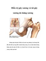
GÃY XƯƠNG SƯỜN, SỤN SƯỜN
BS Lê Văn Tài
Trung Tâm Y Khoa MEDIC
• A bone fracture is a medical condition in
which there is a damage in the continuity of
the bone.
• A bone fracture can be the result of high force
impact or stress, or a minimal trauma injury as
a result of certain medical conditions that
weaken the bones, such as osteoporosis,
bone cancer, or osteogenesis imperfecta,
where the fracture is then properly termed a
pathologic fracture.
GẢY XƯƠNG SƯỜN
• High-resolution sonography was performed with a 5-12–
MHz linear array sonography unit.
• The high-resolution sonography of the ribs and costal
cartilage in the most painful area was performed with the
transducer aligned transversely (i.e., parallel to the long
axis of the rib).
• The patients were then turned to a lateral decubitus
position. Thereafter, the entire length of each rib in the
affected area and above and below the rib was scanned
from the costosternal to the costovertebral junction.
• The high-resolution sonograms were reviewed for the
presence of representative findings: cortical disruption
(focal interruption of the echogenic cortical line), callus
formation (echogenic dump with posterior shadow),
cortical deformity (changed shape of echogenic cortical
line such as angulation or stepping.
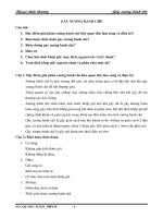
43-year-old man with trauma 10 days
previously and confirmed traumatic
rib fracture. Radiograph shows
cortical disruption with soft-tissue
swelling (arrows).
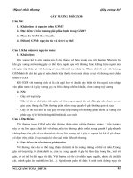
43-year-old man with trauma 10 days
previously and confirmed traumatic
rib fracture. Sonogram shows cortical
disruption (arrow) and hematoma
formation (arrowheads) on
transverse and longitudinal scans.
GÃY SỤN SƯỜN
• Costal cartilage fractures are fractures of the
cartilage connecting the ribs anteriorly to the
sternum.
• There is little published data on costal cartilage
fractures. Most reported cases are in males and
resulted from blunt trauma or a fall.
• In young children a costal cartilage fracture can
present as a chest wall mass associated with
pain (differentials include neoplasm and posttraumatic haematoma). In older patients pain is
the primary complaint.
• Costal cartilages can fracture due to blunt
trauma sustained in high-energy trauma or a
fall. While the more immobile first and second
ribs seem to be more prone to costochondral
separation, the lower ribs can suffer costal
cartilage fracturing more easily.
• Disruptions or fractures of costal cartilage
might result in an unstable rib cage and may
expose thoracic contents, such as the heart,
to injury.
• Plain film: Fractures of the costal
cartilage are challenging to establish on
physical examination and not visible on
plain radiographs, unless there is severe
calcification of the cartilage. Due to their
low incidence they may also be easily
overlooked on additional imaging.
• CT: CT is a reliable method of identifying
costal cartilage fractures. They tend to be
mid-substance (i.e. in the centre of the
costal cartilage) and CT offers the
advantage of demonstrating other injuries,
such as rib fractures and involvement of
lung parenchyma.
• Ultrasound: Ultrasound can be used reliably to
diagnose costal cartilage fractures and can
increase the sensitivity of their detection when
used together with CT scanning Unlike CT it
does not involve radiation and is therefore
preferable in a paediatric population and for
follow-up examinations. Furthermore, it may be
helpful to perform an ultrasound examination
when there is a high clinical suspicion, with or
without a mass, and other modalities have not
demonstrated an injury.
• Signs of a cartilage fracture include a fracture
line, disruption of the anterior echogenic margin,
a step-off deformity or gas located at the
costochondral junction.
• MRI: MRI can demonstrate costal cartilage
fractures as well and, like ultrasound, doesn't
result in radiation exposure.
• Treatment and prognosis: It is unclear if
costal cartilage fractures actually completely
heal, but reported cases state a return to
normal and sports activities when the pain
goes away.