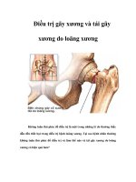
Bs tai gay xuong uc
Bạn đang xem bản rút gọn của tài liệu. Xem và tải ngay bản đầy đủ của tài liệu tại đây (219.46 KB, 9 trang )
GÃY XƯƠNG ỨC
BS Lê Văn Tài
Trung Tâm Ykhoa MEDIC
• A sternal fracture is a very common injury
that occurs after motor vehicle collisions,
particularly in restrained front seat
occupants.
• Sternal fractures are seen in 8% to 10% of
patients with blunt trauma to the chest.
• The most common cause of a sternal
fracture is the direct impact on the sternum
from the steering column or seat belt on
sudden deceleration.
• Indirect mechanisms resulting in a sternal
fracture or dislocation include a blow to the
upper thoracic or cervical segment of the
spine, with transmission of the force through
the upper ribs or clavicle to the sternum, and
stress fractures or collapse of the sternum.
• Sternal fractures can usually be identified
with standard lateral radiography.
• However, this technique is not adequate in
the emergency department because the
exact positioning of severely injured
patients for radiography is difficult.
Therefore, diagnosis of sternal fractures is
often delayed.
• 5- to 10-MHz linear array transducer. The
transducer was placed in the maximum
tenderness area with the patient in a supine
position, and both longitudinal and
transverse images were taken.
• The criteria for a sternal fracture on
sonography were discontinuity and a bony
defect in the sternal cortical margin with or
without soft tissue hemorrhage or swelling.
A, Illustration of A the normal sternum, including the manubrium, sternal body, and xiphoid process.
On this illustration, longitudinal and transverse scanning lines are represented (A–E).
B, Normal contour and no bony defect in the
manubrium (MANU), sternal body, and xiphoid process
(XI) on longitudinal sonography at the A-line.
Two gaps (arrows) are shown in the manubriosternal junction (M-S JUNCT) and sternoxiphoid junction (ST-XIPH JUNCT).
C, Normal transverse contours of the manubrium on B-line scanning (B), the sternal body on C-line scanning between the
third and fourth sternocostal junctions (C) and on D-line scanning of the fourth sternocostal junction (D), and the xiphoid
processon E-line scanning (E).
A
B
Figure 2. Fracture in the upper third of the sternal body in a 39-year-old man.
A, Focal cortical discontinuity (arrow) is shown on longitudinal sonography of the mid
sternal body.
B, Double anterior cortical margins (arrows) are shown on transverse sonography of
the sternal body.
Figure 3. Fractures in the mid sternal body in a 29-year-old man.
A, Lateral sternal view on CR (left) shows a fracture (arrows) with displacement
in the mid sternal body.
C, Transverse sonography shows a large amount of peristernal hemorrhage
(single arrows) and a sternal fracture (double arrow).
• Sonography can be more useful because it is
easy in general to perform, can be performed
easily on a patient who is lying down, and does
not emit radiation. Also, fluids (local hematoma,
and pleural effusion or hemothorax) and
associated rib fractures are better observed on
sonography than on CR.
• However, Hendrich et al reported that
sonography was not suitable for portraying the
grade of displacement, and Engin et al reported
that the grade of displacement found on
sonography was lower than that found on CR
because the posterior side of the sternum could
not be shown well by sonography.