A novel method for the preparation of silver/chitosan-O-methoxy polyethylene glycol core shell nanoparticles
Bạn đang xem bản rút gọn của tài liệu. Xem và tải ngay bản đầy đủ của tài liệu tại đây (466 KB, 7 trang )
Appl Nanosci (2014) 4:113–119
DOI 10.1007/s13204-012-0180-y
ORIGINAL ARTICLE
A novel green one-step synthesis of silver nanoparticles using
chitosan: catalytic activity and antimicrobial studies
Maragoni Venkatesham • Dasari Ayodhya
Alle Madhusudhan • Nagati Veera Babu •
Guttena Veerabhadram
•
Received: 19 October 2012 / Accepted: 22 November 2012 / Published online: 16 December 2012
Ó The Author(s) 2012. This article is published with open access at Springerlink.com
Abstract Stable silver nanoparticles were synthesized
using chitosan acting as both reducing and stabilizing agent
without using any toxic chemicals. This reaction was carried out in an autoclave at a pressure of 15 psi and 120 °C
temperature by varying the time. The influence of different
parameters such as time, change of concentration of silver
nitrate and concentration of chitosan on the formation of
silver nanoparticles were studied. The synthesized silver
nanoparticles were characterized by UV–visible spectroscopy, Fourier transform infrared, X-ray diffraction and
transmission electron microscopy. The results of catalytic
reduction of 4-nitrophenol by sodium borohydride in the
presence of green synthesized silver nanoparticles were
presented. The antimicrobial activity of silver nanoparticles was tested against Escherichia coli and Micrococcus
luteus and was found to be possessing inhibiting property.
Keywords Green synthesis Á Chitosan Á Autoclave Á
Nanosilver Á Catalysis and antimicrobial activity
Introduction
Metallic nanoparticles have been widely studied in recent
years because of their potential use as catalysts (Chimentao
et al. 2004; Gong and Mullins 2009; Campelo et al. 2009;
Li et al. 2010). Silver nanoparticles are of particular
M. Venkatesham Á D. Ayodhya Á A. Madhusudhan Á
G. Veerabhadram (&)
Department of Chemistry, Osmania University,
Hyderabad 500007, India
e-mail:
N. Veera Babu
Department of Biochemistry, Osmania University,
Hyderabad 500007, India
interesting due to their role as substrates in the studies of
catalysis (Tsujino and Matsumura 2005; Shimizu et al.
2010), surface enhancement, Raman spectroscopy (Debarre
et al. 2004; Terekhov et al. 2011) and in the biomedical
field (Xu et al. 2006; Liu et al. 2010; Krishna Rao et al.
2012). Because of their vast applications in various fields,
many techniques of synthesizing silver nanoparticles have
been investigated and some of them are: chemical reduction (Bhui and Misra 2012), electrochemical reduction
(Starowicz et al. 2006; Hosseini and Momeni 2010), photochemical reduction (Kutsenko and Granchak 2009),
microemulsion (Zhang et al. 2011), gamma-ray irradiation
(Huang et al. 2009; Rao et al. 2010), UV irradiation
(Spadaro et al. 2010), ultrasonic method (Byeon and Kim
2012), microwave method (Nadagouda et al. 2011), etc.
Different varieties of stabilizers have been used in silver
nanoparticles preparation, as mentioned above, to achieve
the best control of size, distribution, shape, stability and
solubility of silver nanoparticles. The most commonly used
stabilizers are polyvinyl pyrrolidone (PVP) (Link et al.
1999; Tan et al. 2003), polyvinyl alcohol (PVA) (Abdul
kareem and Anu kaliani 2011), polyaniline (Bouazza et al.
2009) and polyethylene glycol (PEG) (Tan et al. 2003).
Natural polymers have also been used in the preparation of
nanosilver because they are nontoxic and biocompatible.
Starch (Hu et al. 2008) and chitosan (Hettiarachchi and
Wickramarachchi 2011) have been used as stabilizers for
the preparation of metal nanoparticles. Many of the nanoparticle synthesis methods, however, involve use of hazardous chemicals, low material conversions and high
energy requirements. Over past decade, keen interest has
been evinced in green synthesis. Green synthesis is costeffective, environment friendly, easily scaled up for largescale synthesis and also there is no need to use toxic
chemicals.
123
114
Appl Nanosci (2014) 4:113–119
Chitosan is a biodegradable polysaccharide copolymer
of N-acetyl-D-glucosamine and D-glucosamine, obtained by
the alkaline deacetylation of chitin (Onishi and Machida
1999). It is considered as a nontoxic, biodegradable, biocompatible and environmental-friendly material with many
superior properties (Jigar and Sinha 2007). It is the second
most plentiful natural biopolymer and is relatively cheap
(Ma et al. 2008). In this paper, we report the green synthesis of silver nanoparticles using chitosan as both
reducing and stabilizing agent and without using any toxic
chemicals. This reaction is carried out in an autoclave at a
pressure of 15 psi and at 120 °C temperature by varying
the time. The influence of different parameters such as
time, change of concentration of silver nitrate and concentration of chitosan on the formation of silver nanoparticles was studied.
In recent days, nitro aromatic compounds are widely
used in chemical industries. These are xenobiotics, used in
manufacturing of pesticides, dyes, plasticizers, fungicides
and explosives. These anthropogenic compounds are
highly hazardous when released in the environment. In
particular, 4-nitrophenol has been listed as priority pollutant by the US Environmental Protection Agency (EPA)
because of its higher solubility and stability in water.
4-nitrophenol stays a longer time in water and surface soil
without degradation and gets accumulated in deep soil
indefinitely (Pocurull et al. 1996). In the present study, we
report the catalytic activity of green synthesized silver
nanoparticles toward 4-nitrophenol reduction and microbial
activity toward Gram-positive and Gram-negative bacteria.
mixture was kept in autoclave at 15 psi pressure, at 120 °C
for different time intervals. The resulting solution was clear
yellow in color indicating the formation of silver nanoparticles. Studies were made varying the concentration of
silver nitrate (0.1–0.5 %), keeping the concentration of
chitosan (0.5 %) constant, and also varying the concentration of chitosan (0.1–0.5 %), keeping silver nitrate
(0.5 %) concentration constant.
Characterizations
Experimental
The silver nanoparticles, stabilized in chitosan solution,
were analyzed by UV–vis absorbance spectroscopy. UV–
vis spectroscopic measurements were made at room temperature using a Shimadzu dual beam UV–vis spectrophotometer, Japan. Fourier transform infrared (FTIR)
spectra of silver nanoparticles stabilized in chitosan and
chitosan alone were recorded in KBr pellets using an FTIR
spectrophotometer (Bruker Optics, Germany). The scan
was performed in the range 400–4,000 cm-1. X-ray diffraction (XRD) measurement of silver nanoparticles stabilized in chitosan was carried out on X’pert Pro X-ray
diffractometer (Panalytical B.V., Netherlands) operating
at 40 kV and a current of 30 mA at a scan rate of
0.388 min-1. The size distribution and crystallinity of
the silver nanoparticles stabilized in chitosan were obtained
by high-resolution transmission electron microscopy
(HRTEM) measurement, casting nanoparticle dispersion
on carbon-coated copper grids and allowing drying at room
temperature. Measurements were done on Tecnai G2 F30
S-Twin instrument (FEI Company, USA) operated at an
accelerating voltage of 200 kV with a lattice resolution of
0.14 nm and point image resolution of 0.20 nm.
Materials
Catalytic activity
Chitosan, MW & 70,000 and [80 % deacetylated, was
purchased from India Sea Foods, Kochi, India. Acetic acid,
4-nitrophenol, sodium chloride, silver nitrate and sodium
borohydride were obtained from S D Fine-chem Limited,
Mumbai, India. The test strains, Escherichia coli MTCC
1303 and Micrococcus luteus MTCC 2987, were purchased
from IMTECH, Chandigarh, India. Yeast extract, tryptophan and bacterial-grade agar–agar were purchased from
Himedia Laboratories, Mumbai, India.
To a 3 ml cuvette containing freshly prepared sodium
borohydride (1 ml, 0.2 M) solution, 4-nitrophenol (1.9 ml,
0.2 mM) solution was added. The cuvette was then placed
in a UV–vis spectrophotometer and the absorbance against
wavelengths recorded. After adding silver nanoparticles
stabilized in chitosan (0.1 ml, 0.1 %) solution, the cuvette
was shaken vigorously for mixing and kept in a UV–vis
spectrophotometer.
Antibacterial property of samples
Preparation of silver nanoparticles
Chitosan (0.5 %) solution was prepared by dissolving
chitosan (0.5 g) in acetic acid (100 ml, 2 %) solution and
also silver nitrate (0.5 g) in deionized water (100 ml).
Chitosan (5 ml, 0.5 %) solution was mixed with silver
nitrate (5 ml, 0.5 %) solution in a boiling tube. This
123
Luria–Bertani (LB) agar medium was prepared by adding
yeast extract (0.5 g), tryptophan (1 g), sodium chloride (1 g)
and bacterial grade agar (2.5 g) in distilled water (100 ml).
Then the agar medium was sterilized by autoclaving at a
pressure of 15 psi and 120 °C temperature for 30 min. This
medium was transferred into sterilized Petri dishes in a
Appl Nanosci (2014) 4:113–119
115
laminar air flow. After solidification of the media, overnight
culture of E. coli (100 ll) and M. luteus (100 ll) was spread
separately on the solid surface of the media. Sterile discs
were kept on these inoculated plates with the help of sterile
forceps. Sample (10 ll) solutions were placed on these discs
and were incubated at 37 °C for 24 h in a bacterial incubator. The inhibition zone that appeared around the disc was
measured and recorded as the antibacterial effect of chitosan
and the nanosilver stabilized in chitosan.
Results and discussion
UV–visible spectroscopy analysis
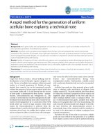
UV–visible absorption spectra recorded is quite sensitive to
the formation of silver nanoparticles because of the fact
that silver nanoparticles exhibit an intense absorption peak
due to surface plasmon resonance (SPR). Figures 1 and 2
show the UV–vis spectra of silver nanoparticles prepared
with different concentrations of chitosan and silver nitrate.
All spectra exhibit an absorption band in the range of
410–430 nm, a typical plasmon resonance band of silver
nanoparticles. There are no peaks located around 335 and
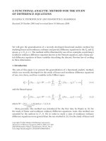
560 nm, indicating the absence of nanoparticle aggregation. The reduction was studied with silver nitrate (0.5 %)
and chitosan (0.5 %) by varying the time (10–60 min). The
absorption against wavelength curves at various times is
given in Fig. 3. It was noticed that the reduction capacity of
chitosan increased with time. As the autoclaving time
increases, possibly more and more of hydroxyl groups are
converted to carbonyl groups by air oxidation, which in
turn reduces the silver ions. A single strong peak with a
maximum around 420 nm was observed in the UV–vis
spectra, which corresponds to typical SPR of conducting
electrons from the surface of silver nanoparticles. Figure 1
shows the UV–vis spectra of the silver nanoparticles prepared with different concentrations of silver nitrate
(0.1–0.5 %) with chitosan (0.5 %) and 50 min of autoclaving. The efficiency of nanoparticle synthesis increases
with increase in the concentration of silver nitrate, due to
an enhancement in the oxidation of hydroxyl groups of
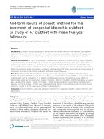
chitosan by silver ions. Further, the production of nanoparticles from silver nitrate (0.5 %) was monitored with
varying concentrations of chitosan (0.1–0.5 %) for 50 min
of reaction time and their respective spectra are depicted in
Fig. 2. The intensity of the absorption of the solutions
increased with increase in the concentration of chitosan.
Fourier transform infrared spectroscopy analysis
Figure 4 shows the FTIR spectrum of chitosan and silver
nanoparticles stabilized in chitosan. The IR spectrum of
Fig. 1 UV–visible absorption spectra of silver nanoparticles stabilized in chitosan using various concentrations of (i) silver nitrate
(0.1 %), (ii) silver nitrate (0.2 %), (iii) silver nitrate (0.3 %), (iv)
silver nitrate (0.4 %) and (v) silver nitrate (0.5 %) at chitosan (0.5 %)
and 50 min of autoclaving
Fig. 2 UV–visible absorption spectra of silver nanoparticles stabilized in chitosan using various concentrations of (i) chitosan (0.1 %),
(ii) chitosan (0.2 %), (iii) chitosan (0.3 %), (iv) chitosan (0.4 %) and
(v) chitosan (0.5 %) at silver nitrate (0.5 %) and 50 min of
autoclaving
chitosan absorption bands at 3,447 and 2,881 cm-1 represent the –NH2, –OH and –CH2, and –CH3 aliphatic groups.
Absorption bands at 1,600 cm-1 represent the amino group
bending vibrations and 1,422 cm-1 of the –OH group of the
primary alcoholic group. The amino group has a characteristic absorption band in the region of 3,400–3,500 cm-1,
which is masked by the broad spectrum band from the –OH
group. The absorption band at 1,657 cm-1 is attributed to
the –CONH2 group of chitosan. In the FTIR spectrum of
silver nanoparticle stabilized in chitosan, the absorption
bands at 1,657 cm-1 and 1,600 cm-1, representing chitosan
123
116
Appl Nanosci (2014) 4:113–119
(111), (200), (220) and (311) sets of lattice planes,
respectively, are observed and shown in Table 1, which
may be indexed as the band for face centered cubic (fcc)
structure of silver nanoparticles. The peak corresponding to
the (111) plane is more intense than the other planes. The
broadening of these peaks is mostly due to the effect of
nano-sized particles.
Transmission electron microscopy (TEM) analysis
of silver nanoparticles stabilized in chitosan
Fig. 3 UV–visible absorption spectra of silver nanoparticles stabilized in chitosan by varying the time: (i) 10 min, (ii) 20 min,
(iii) 30 min, (iv) 40 min, (v) 50 min and (vi) 60 min
–CONH2 and –NH2 groups, disappeared and a new band
appeared at 1,635 cm-1, which indicated the attachment of
silver to nitrogen atom. The variation in the shape and peak
positions of the –NH2 and –OH at 3,447 cm-1 occurred
because of contribution toward the reduction and stabilization process.
A typical TEM image of silver nanoparticles formed is
displayed in Fig. 6. The silver nanoparticles are well distributed in the chitosan matrix. More than 70 % of the
particles are in the size range from 5 to 15 nm and very few
particles are also observed above the 30 nm range. Figure 6 inset shows the SAED pattern of silver nanoparticles
stabilized in chitosan, exhibiting polycrystalline diffraction
rings, which can be indexed to cubic-phase metal silver,
indicating that these nanoparticles are crystalline metallic
silver.
Catalytic activity
The XRD technique was used to determine the crystal
structure of green synthesized silver nanoparticles. Figure 5 displays the XRD pattern of the presently synthesized
silver nanoparticles. A number of Brag reflections with 2h
values of 38.25, 43.95, 64.5 and 77.21 corresponding to the
The catalytic activity of green synthesized silver nanoparticles stabilized in chitosan was investigated, using the
reduction of 4-nitrophenol to 4-aminophenol by sodium
borohydride as a model reaction. To a 3 ml cuvette containing freshly prepared sodium borohydride (1 ml, 0.2 M)
solution, 4-nitrophenol (1.9 ml, 0.2 mM) solution was
added. The cuvette was then placed in a UV–vis spectrophotometer. UV–vis absorption spectra of the reduction of
4-nitrophenol catalyzed by silver nanoparticles stabilized
in chitosan is shown in Fig. 7. The 4-nitrophenol is stable
Fig. 4 FTIR spectra of silver nanoparticles stabilized in chitosan and
pure chitosan
Fig. 5 Typical XRD pattern of the silver nanoparticles stabilized in
chitosan
X-Ray diffraction analysis of silver nanoparticles
123
Appl Nanosci (2014) 4:113–119
117
Table 1 X-ray diffraction peak list of silver nanoparticles
˚ ) Rel. Int. (%)
Pos. (°2h) Height (cm) FWHM (°2h) d-spacing (A
38.2522
17.89
0.9600
2.35099
100.00
43.9478
1.00
0.4734
2.06032
5.59
64.3169
1.00
0.3125
1.44841
5.59
77.2100
2.00
0.3367
1.23558
11.18
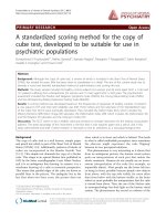
Fig. 7 UV–visible absorption spectra of aqueous solutions of (i) pure
4-aminophenol, (ii) pure 4-nitrophenol, (iii) 4-nitrophenolate ion and
(iv) silver nanoparticles catalyzed product
Fig. 6 Typical TEM image of silver nanoparticles in stabilized in
chitosan. Inset shows the SAED pattern of the silver nanoparticles
and stays a longer time in water. An aqueous solution of
4-aminophenol shows absorption maximum at 297 nm in
UV–vis spectra. An aqueous solution of 4-nitrophenol
shows a distinct spectral profile with absorption maximum
at 317 nm, which shifts to 400 nm in the presence of
sodium borohydride due to the formation of the 4-nitrophenolate ion (Pradhan et al. 2001). The anionic silver
species remain stable for weeks in the absence of any other
reagent. After adding nanoparticles stabilized in chitosan
(0.1 ml, 0.1 %) solution, the cuvette was shaken vigorously
for mixing and kept in UV–vis spectrophotometer. The
peak at 400 nm disappeared, and a new peak at 297 nm
appeared, which is known to be due to absorption of
4-aminophenol (Panigrahi et al. 2007).
The concentration of the sodium borohydride greatly
exceeds that of 4-nitrophenol and the catalyst nanoparticles. The excess of sodium borohydride used increases the
pH of the reacting system, thereby retarding the degradation of the borohydride ions, and the liberated hydrogen is
purged out, thereby checking the aerial oxidation of the
reduced product of 4-nitrophenol. It is well known that the
metal nanoparticles catalyze this reaction by facilitating
electron relay from the donor BH4- to acceptor 4-nitrophenol to overcome the kinetic barrier. The catalytic
reduction proceeds on the surface of the metal nanoparticles. As soon as the electron donor (BH4-) and electron
acceptor (4-nitrophenolate ion) are adsorbed on the surface
of the silver nanoparticles, catalytic reaction starts by the
transfer of electron from BH4- to 4-nitrophenolate ion.
Thus, silver nanoparticles help in facilitating the reduction
of 4-nitrophenol by lowering the activation energy of the
reaction and play the role of catalyst.
Antibacterial activity studies
The silver nanoparticles synthesized by this method
showed 20 mm and 15 mm inhibition zones, respectively,
against E. coli and M. luteus as shown in Fig. 8, which are
greater than that shown by chitosan alone. Here, the antibacterial effect of silver nanoparticles was more pronounced against Gram-negative (E. coli) than Grampositive (M. luteus) as the Gram-positive bacteria possess a
thick cell wall containing high amount of peptidoglycan. In
contrast, Gram-negative bacteria have two layers of cell
membranes. The outer membrane comprises lipopolysaccharide, which protects it from several antibiotics, detergents and drugs that would normally damage the inner
Fig. 8 Antibacterial test results for E. coli and M. leteus after 24 h of
incubation. 1 chitosan 2 silver nanoparticles stabilized in chitosan
123
118
Appl Nanosci (2014) 4:113–119
membrane or cell wall (peptidoglycan). The outer membrane provides these bacteria with resistance to many
antibacterial drugs.
Conclusions
A simple, one step green approach was developed for synthesis of silver nanoparticles using chitosan. Chitosan acted
as both reducing and stabilizing agent. The silver nanoparticles that are formed are highly stable and have significant
catalytic activity toward reduction of 4-nitrophenol to
4-aminophenol. These green synthesized silver nanoparticles
had significant antibacterial action on E. coli and M. luteus
bacteria. It was also observed that these nanoparticles showed
higher antibacterial property on Gram-negative (E. coli) than
Gram-positive (M. luteus) bacteria.
Open Access This article is distributed under the terms of the
Creative Commons Attribution License which permits any use, distribution, and reproduction in any medium, provided the original
author(s) and the source are credited.
References
Abdul kareem K, Anu kaliani A (2011) Synthesis and thermal study
of octahedral silver nano-plates in polyvinyl alcohol (PVA).
Arab J Chem 4:325–331
Bhui DK, Misra A (2012) Synthesis of worm like silver nanoparticles
in methyl cellulose polymeric matrix and its catalytic activity.
Carbohydr Polym 89:830–835
Bouazza S, Alonzo V, Hauchard D (2009) Synthesis and characterization of Ag nanoparticles–polyaniline composite powder
material. Synth Metals 159:1612–1619
Byeon JH, Kim YW (2012) A novel polyol method to synthesize
colloidal silver nanoparticles by ultrasonic irradiation. Ultrason
Sonochem 19:209–215
Campelo JM, Luna D, Luque R, Marinas JM, Romero AA (2009)
Sustainable preparation of supported metal nanoparticles and
their applications in catalysis. Chem Sus Chem 2:18–45
Chimentao RJ, Kirm I, Medina F, Rodrı´guez X, Cesteros Y, Salagre
P, Sueiras JE (2004) Different morphologies of silver nanoparticles as catalysts for the selective oxidation of styrene in the gas
phase. Chem Commun (7):846–847
Debarre A, Jaffiol R, Julien C, Tchenio P, Mostafavi M (2004) Raman
scattering from single Ag aggregates in presence of EDTA.
Chem Phys Lett 386:244–247
Gong J, Mullins CB (2009) Surface science investigations of
oxidative chemistry on gold. Acc Chem Res 42:1063–1073
Hettiarachchi MA, Wickramarachchi PASR (2011) Synthesis of
chitosan stabilized silver nanoparticles using gamma ray irradiation and characterization. J Sci Univ Kelaniya 6:65–75
Hosseini M, Momeni MM (2010) Silver nanoparticles dispersed in
polyaniline matrixes coated on titanium substrate as a novel
electrode for electro-oxidation of hydrazine. J Mater Sci 45:
3304–3310
Hu B, Wang SB, Wang K, Zhang M, Yu SH (2008) Microwaveassisted rapid facile ‘‘green’’ synthesis of uniform silver
123
nanoparticles: self-assembly into multilayered films and their
optical properties. J Phys Chem C 112:11169–11174
Huang NM, Radiman S, Lim HN, Khiew PS, Chiu WS, Lee KH,
Syahida A, Hashim R, Chia CH (2009) Gamma-ray assisted
synthesis of silver nanoparticles in chitosan solution and the
antibacterial properties. Chem Eng J 155:499–507
Jigar MJ, Sinha VK (2007) Ceric ammonium nitrate induced grafting
of polyacrylamide onto carboxymethyl chitosan. Carbohydr
Polym 67:427–435
Krishna Rao KSV, Ramasubba Reddy P, Lee YI, Kim C (2012)
Synthesis and characterization of chitosan–PEG–Ag nano composites for antimicrobial application. Carbohydr Polym 87:
920–925
Kutsenko AS, Granchak VM (2009) Photochemical synthesis of silver
nanoparticles in polyvinyl alcohol matrices. Theor Exp Chem
45:313–318
Li X, Wang J, Zhang Y, Li M, Liu J (2010) Surfactant less synthesis
and the surface-enhanced Raman spectra and catalytic activity of
differently shaped silver nanomaterials. Eur J Inorg Chem
2010:1806–1812
Link S, Wang ZL, El-Sayed MA (1999) Alloy formation of gold–
silver nanoparticles and the dependence of the plasmon absorption on their composition. J Phys Chem B 103:3529–3533
Liu I, Sonshine DA, Shervani S, Hurt RH (2010) Controlled release of
biologically active silver from nanosilver surfaces. ACS Nano
4:6903–6913
Ma GP, Yang DZ, Zhou YS, Xiao M, Kennedy JF, Nie J (2008)
Preparation and characterization of water-soluble N-alkylated
chitosan. Carbohydr Polym 74:121–126
Nadagouda MN, Speth TF, Varma RS (2011) Microwave-assisted green
synthesis of silver nanostructures. Acc Chem Res 44:469–478
Onishi H, Machida Y (1999) Biodegradation and distribution of
water-soluble chitosan in mice. Biomaterials 20:175–182
Panigrahi S, Basu S, Praharaj S, Pande S, Jana S, Pal A, Ghosh KS,
Pal T (2007) Synthesis and size-selective catalysis by supported
gold nanoparticles: study on heterogeneous and homogeneous
catalytic process. J Phys Chem C 111:4596–4605
Pocurull E, Marce RM, Borrull F (1996) Determination of phenolic
compounds in natural waters by liquid chromatography with
ultraviolet and electrochemical detection after on-line trace
enrichment. J Chromatogr A 738:1–9
Pradhan N, Pal A, Pal T (2001) Catalytic reduction of aromatic nitro
compounds by coinage metal nanoparticles. Langmuir 17:1800–1802
Rao YN, Banerjee D, Datta A, Das SK, Guin R, Saha A (2010)
Gamma irradiation route to synthesis of highly re-dispersible
natural polymer capped silver nanoparticles. Radiat Phys Chem
79:1240–1246
Shimizu K, Miyamoto Y, Satsuma A (2010) Silica-supported silver
nanoparticles with surface oxygen species as a reusable catalyst
for alkylation of arenes. ChemCatChem 2:84–91
Spadaro D, Barletta E, Barreca F, CurrO G, Neri F (2010) Synthesis
of PMA stabilized silver nanoparticles by chemical reduction
process under a two-step UV irradiation. Appl Surf Sci 256:
3812–3816
Starowicz M, Stypula B, Banas J (2006) Electrochemical synthesis of
silver nanoparticles. Electrochem Commun 8:227–230
Tan Y, Dai X, Li Y, Zhu D (2003) Preparation of gold, platinum,
palladium and silver nanoparticles by the reduction of their salts
with a weak reductant-potassium bitartrate. J Mater Chem
13:1069–1075
Terekhov SN, Mojzes P, Kachan SM, Mukhurov NI, Zhvavyi SP,
Panarin AY, Khodasevich IA, Orlovich VA, Thorel A, Grillon F,
Turpin PY (2011) A comparative study of surface-enhanced
Appl Nanosci (2014) 4:113–119
Raman scattering from silver-coated anodic aluminum oxide and
porous silicon. J Raman Spectrosc 42:12–20
Tsujino K, Matsumura M (2005) Boring deep cylindrical nanoholes in
silicon using silver nanoparticles as a catalyst. Adv Mater
17:1045–1047
Xu X, Yang Q, Wang Y, Yu H, Chen X, Jing X (2006) Biodegradable
electrospun poly(L-lactide) fibers containing antibacterial silver
nanoparticles. Eur Polymer J 42:2081–2087
119
Zhang D, Liu X, Wang X, Yang X, Lu L (2011) Optical properties of
monodispersed silver nanoparticles produced via reverse micelle
microemulsion. Phys B Condens Matter 406:1389–1394
123