báo cáo hóa học:" A rapid method for the generation of uniform acellular bone explants: a technical note" pot
Bạn đang xem bản rút gọn của tài liệu. Xem và tải ngay bản đầy đủ của tài liệu tại đây (519.22 KB, 4 trang )
Jähn et al. Journal of Orthopaedic Surgery and Research 2010, 5:32
/>Open Access
TECHNICAL NOTE
BioMed Central
© 2010 Jähn et al; licensee BioMed Central Ltd. This is an Open Access article distributed under the terms of the Creative Commons At-
tribution License ( which permits unrestricted use, distribution, and reproduction in any
medium, provided the original work is properly cited.
Technical Note
A rapid method for the generation of uniform
acellular bone explants: a technical note
Katharina Jähn
1,2
, Volker Braunstein
3
, Pamela I Furlong
1
, Angharad E Simpson
1
, R Geoff Richards
1,2
and
Martin J Stoddart*
1
Abstract
Background: Bone graft studies lack standardized controls. We aim to present a quick and reliable method for the
intra-operative generation of acellular bone explants.
Methods: Therefore, ovine cancellous bone explants from the iliac crest were prepared and used to test several
methods for the induction of cell death. Over night heat inactivation was used as positive treatment control, methods
to be investigated included UV light, or X- ray exposure, incubation in a hypotonic solution (salt-free water) and a short
cycle of repeated freezing and thawing.
Results: Viability of treated and 2 days cultured bone explants was investigated by lactate dehydrogenase assay. Non-
treated cultured control explants maintained around 50% osteocyte viability, while osteocyte survival after the positive
treatment control was abolished. The most dramatic loss in cell viability, together with a low standard deviation, was a
repeated cycle of freezing and thawing.
Conclusions: To summarize, we present a freeze-thaw method for the creation of acellular bone explants, which is
easy to perform, not time-consuming and provides consistent results.
Background
Large bone defects remain a clinical challenge and the
development of novel therapies requires adequate con-
trols. The use of acellular bone explants within bone graft
studies is of crucial importance [1]. Results of trans-
planted bone material can not be interpreted correctly
without the presence of a bone matrix control which does
not contain viable cells. It is well known that the success
of bone grafts is determined by three factors - osteoin-
duction, osteoconductivity and osteogenesis [1]. The
concept of osteoinduction involves mitogenesis of undif-
ferentiated host mesenchymal stem cells towards the for-
mation of osteoprogenitors by i.e. molecules of the TGFβ
family which are stored in huge amounts within bone
matrix [2]. Osteoconductivity is achieved when the
implanted bone is used by the host cells as scaffold for the
formation of new bone. An acellular bone explant control
aids to evaluate the effects of active bone cells (osteogen-
esis) versus the effect of the bone matrix alone (osteoin-
duction and osteoconductivity) during bone
transplantation studies. Unfortunately, a reliable control
with a uniformly diminished cell survival is not standard-
ized yet.
Acellular bone grafts are often prepared using γ-irradi-
ation or ethylene oxide sterilization of allogenic bone
explant material [3]. Yet, both methods have been shown
to reduce the mechanical properties of bone matrix and
negatively affect osteogenesis by the host cells due to
either residual ethylene oxide or radiation effects. The
use of demineralized bone grafts, available from tissue
banks, is very popular due to the ease of purchasing. The
acid extracted (demineralized) bone allograft is of acellu-
lar type. However, the demineralization process is
impractically long, if this method would be intended to
be used for the generation of acellular autologous bone
material intraoperative.
Therefore, the aim of this study is to present a reliable,
quick and easy method for the generation of an acellular
bone explant control, which could be performed during
* Correspondence:
1
AO Research Institute, Clavadelerstrasse 8, 7270 Davos, Switzerland
Full list of author information is available at the end of the article
Jähn et al. Journal of Orthopaedic Surgery and Research 2010, 5:32
/>Page 2 of 4
transplantation operation on possibly autologous bone.
The method must routinely result in a totally acellular
sample. We demonstrate that a rapid cycle of freeze-
thawing is more effective over the use of radiation expo-
sure (X-ray, or UV-C light) or incubation in salt-free
water, and less time consuming as an over night incuba-
tion at high temperatures (>50°C) to generate uniformly
acellular cancellous bone.
Methods
Cancellous Bone Harvest and Treatment
Cancellous bone was obtained from the iliac crest of 3
Swiss Alpine sheep, which were euthanized due to
involvement in separate studies (approved by cantonal
ethics committee). Cylindrical bone explants were drilled
from the iliac crest using a 9.5 mm Synthes drill bit (Ref:
387.661, Synthes, Bettlach, Switzerland). Bone explants
were randomly assigned into 6 different treatment groups
with 3 explants per group. The first treatment group was
exposed to 5 freeze-thaw cycles. Therefore cores were put
in a metal beaker and placed into liquid nitrogen for 1
min. After each freezing cycle, explants were thawed at
56°C for 5 min. The second and third treatment groups
were exposed to radiation. While explants of one group
were treated with 80 kV X-rays for 30 min, group 3 was
placed 20 cm from an UV-C light source (Osram HNS, 30
W) for 60 min. The last group was placed in salt-free
water (purified with Mili-Q Synthesis) for 1 h treatment.
The positive treatment control group was incubated at
56°C for 18 h.
Culture and Viability Analysis
All treated bone explants, together with a non-treatment
control group were cultured for 2 d in DMEM high glu-
cose (Invitrogen) supplemented with 4 mM sodium
hydrogen carbonate (Merck), 10 mM HEPES, 5 mM glyc-
erol-2-phoshate salt hydrate (Sigma), 10 mg/l L-ascorbic
acid phosphate magnesium salt n-hydrate (Wako Chemi-
cals), 50,000 U/l Penicillin, 50 mg/l Streptomycin (Invit-
rogen), and 10% fetal calf serum (FCS; South American
origin, biowest). Viability was analyzed using a lactate
dehydrogenase (LDH) assay [4], with a day 0 non-culture
group to provide baseline viability levels. Harvested
explants were cut with a Leica annular saw (Leica AG,
Glattbrugg, Switzerland) into 300 μm sections. LDH
staining was performed using a 5% Polypep base solution
containing 0.75% sodium chloride and 2 mM Glycyl-Gly-
cine buffer (adjust to pH 8). Lactic acid (60 mM), 1.75
mg/ml NAD
+
(pH 8), and 3 mg/ml nitroblue tetrazolium
(all Sigma) were added freshly to the solution on the day
of the assay. Visualization and quantification of viable
osteocytes per mm
2
bone area was performed as previ-
ously described [5]. In brief, fluorescence micrographs
(excitation BP450-490 nm, beam splitter FT510 nm,
emission BP515-565 nm) were taken from the central
regions of the bone explants using a 20× objective. Bone
matrix area per micrograph was determined. Due to the
achieved depth of field of 4.12-4.52 μm, dark violet
stained osteocytes in focus could be accounted as viable
osteocytes per bone area [Fig. 1D].
Statistical Analysis
The Mann-Whitney Rank Sum test and Bonferroni cor-
rection was used to compare the different methods,
which were analyzed to generate acellular cancellous
bone samples. A p-value ≤ 0.05 was considered to be sig-
nificant.
Results
Due to the fact that there is so much cell death at the
periphery induced during the cutting process, we did not
analyze this region to remove any effect of preparation
artifact. Macroscopical analysis of LDH viability staining
on non-cultured bone explants, showed overall uniform
cell viability. Whereas viability of non-treated 2 days cul-
tured bone demonstrated a central area of decreased
marrow survival. Macroscopically, all treatment groups,
cultured on for 2 days, showed almost no detectable mar-
row LDH viability staining (data not shown).
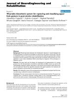
Osteocyte survival was determined and quantified at
higher magnification. Presence of formazan crystals
within osteocytes demonstrating cellular LDH activity
and therefore cell viability was demonstrated in non-cul-
tured and non-treated bone explants. The positive 56°C
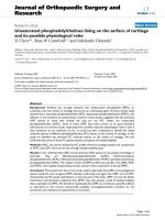
Figure 1 Four representative micrographs from LDH labeled
ovine cancellous bone sections visualized with Axioplan (Fluo-
rescence 515-565 emission filter). A: Non-treatment control cul-
tured for 2 days with presence of viable osteocytes; B: Positive
treatment control incubated at 56°C over night prior to 2 days culture;
C: Freeze-thawing treatment after 2 days culture showing no viable os-
teocytes; D: Representative image to show the method of analysis -
bone matrix area is marked in red, viable osteocytes are marked in blue.
Jähn et al. Journal of Orthopaedic Surgery and Research 2010, 5:32
/>Page 3 of 4
heat incubation treatment group, as well as other treat-
ment groups, presented no or decreased osteocyte LDH
activity [for representative images see Fig. 1].
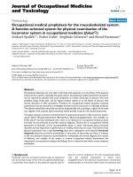
During static culture of 2 days the average cell viability
of bone explants dropped over 50% from 279 (+/- 58) to
119 (+/- 25) viable osteocytes per bone matrix area [Fig.
2]. Not all treatment groups significantly decreased
osteocyte survival compared to the non-treated culture
control sample. The radiation-induced cell death groups
(X-ray and UV light) showed no significant decrease in
cell viability. Thirty minutes X-ray exposure declined the
number of viable osteocytes per mm
2
bone matrix area
from 119 (average of non-treatment control) to an aver-
age of 97 (+/- 46) surviving cells. Treatment with UV light
demonstrated a similar cell survival decrease to 93 (+/-
44) average viable osteocytes per mm
2
. The number of
surviving osteocytes was however significantly reduced
by exposure to salt-free water (p ≤ 0.05), with an average
of 45 (+/- 74) surviving osteocytes per bone matrix area
[Fig. 2]. Moreover, in all of treatments - X-ray radiation,
UV light exposure and salt-free water - a great standard
deviation of cell survival was detected. The lowest num-
ber of surviving osteocytes after treatment, in combina-
tion with a low standard deviation, was detected after
repeated freeze thawing cycles. An average of only 6 (+/-
6) surviving osteocytes per mm
2
bone area was detected
after this treatment.
The positive treatment control, which experienced
56°C over night heat incubation, showed an average of 5
(+/- 10) viable osteocytes per bone matrix area. A signifi-
cant difference in osteocyte survival after treatment with
either radiation treatments (X-ray and UV light) to the
positive treatment control was demonstrated. However,
exposure to salt-free water or repeated freeze thawing
cycles did show a decrease in osteocyte survival, which
was not significantly different to the 56°C heat incubation
control group.
Discussion
Within this study ovine cancellous bone explants were
successfully treated to induce osteocyte death. Moderate
heat incubation is one of the most commonly used meth-
ods for the treatment of allogenic bone explants prior to
transplantation. While the conventionally used steriliza-
tion methods with vapor temperatures over 100°C (121°C
for 20 min, or 134°C for 5 min) lead to extreme loss of
bone strength and osteoconductivity [6]. Different
authors report that a temperature of 50°C is efficient to
kill bone cells via thermal necrosis [7]. We chose to use an
18 h over night incubation at 56°C as positive treatment
control, as it was previously shown to induce cell death in
5 × 10 mm cancellous bone explants [5]. However, this
method is time-consuming and seems most impractical
to be used as a cell-free control group.
The exposure of ovine cancellous bone explants to
either UV-C or X-ray radiation showed no significant
decrease in osteocyte survival over the 2 days culture
control. Cell viability was significantly greater than the
56°C over night heat incubation control. X-rays are a
form of electromagnetic radiation with a wavelength
range of 10-0.1 nm. In medicine X-rays are mainly used
for diagnostic imaging - radiography, or computer
tomography of skeletal fractures [8]. Due to their high
energy, X-rays are able to ionize (remove electrons)
atoms, and destroy chemical compounds by the forma-
tion of radicals i.e. reactive oxygen species. Radiation
induced DNA damage caused by X-ray exposure ranges
from double-stand breaks, to ionization of the desoxyri-
boses [9]. If the changes in the genetic material of a cell
are too dramatic - as it is intended during therapeutically
radiotherapy of cancer treatment - cells can undergo
apoptotic cell death [10]. UV radiation in its shorter
wavelengths - below 200 nm - is also known permit ion-
ization. Additionally, UV-C radiation (100-290 nm) can
be the cause of thymine dimers in the DNA which than
stops the DNA polymerase during replication. Cell death
can be induced [10]. For this purpose, UV-C radiation is
commonly as sterilization method.
The exposure of cells to hypotonic solutions, such as
salt-free water which was used within this study, is caus-
ing cell swelling that terminates in cell death. This occur-
rence has been used occasionally in clinics, where
surgeons washed the abdominal cavity with distilled
water with the intention of lysing isolated cancer cells
which are left after surgery [11]. In an in vitro study
Selzner et al. could show that human colon cancer cells
undergo apoptotic cell death triggered by short-term
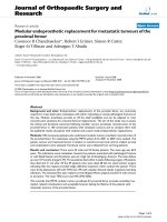
Figure 2 Graph shows boxplots of the datasets for viable osteo-
cytes per bone matrix area under the different treatment condi-
tions. Osteocyte survival was greatly diminished during 56°C over
night heat incubation, freeze-thawing and salt-free water treatment
compared to non-treated 2 days culture control (* p ≤ 0.05). Moreover,
UV light and X-ray exposure were significantly different to the positive
treatment control of 56°C incubation (# p ≤ 0.05). Boxplots show the
median line, the 25% and the 75% quartiles which define the box.
Jähn et al. Journal of Orthopaedic Surgery and Research 2010, 5:32
/>Page 4 of 4
exposure (1-5 min) to a hypotonic solution [12]. The
amount of surviving osteocytes after salt-free water treat-
ment was significantly smaller than the non-treated 2
days cultured control and non-significantly different
from the 56°C over night heat incubated control group.
However, within this group a high standard deviation of
surviving osteocytes was determined, resulting in a rela-
tively questionable prediction of a uniform overall cell
death. Probably this result was due to the presence of
remaining salts within the bone marrow, protecting the
cells from the osmotic shock.
The most promising treatment for the generation of an
acellular bone explant control to be used within an oper-
ating theatre is to our knowledge the repeated use of
freeze-thawing cycles. This method is one of the classical
procedures for experimental in vitro cell lysis [13]. The
rapid freezing procedure causes cells to swell and ulti-
mately break due to massive ice-crystal formation, which
is normally avoided using cryo-preservatives and con-
trolled 1°C/min freezing during cryo preservation of cells
[14]. The method could be time consuming if thawing
would be permitted at room temperature. Therefore we
recommend a 5 min thawing period at 56°C to be per-
formed.
We did not perform additional tests to evaluate the
mechanical properties of the cancellous bone biopsies
after treatment. From previous studies within our group,
we know that the Young's modulus of cancellous bone
pieces does not significantly alter due to over night treat-
ment at 56°C (unpublished observations). The mechani-
cal properties of cancellous bone cylinders (5 mm × 10
mm) taken from bovine humeri were investigated in a
study by Borchers et al. (1995) after long-term, repeated
freezing thawing cycles and compared to 'non-treated'
control cylinders [15]. No significant differences in com-
pressive strength, elastic modulus, or mineral density
could be determined. Therefore, we are likely to expect
that the material properties of the rapid freeze-thaw
treated bone explants in our study were not affected by
treatment.
Conclusion
Taken together, we demonstrate that a rapid cycle of
freeze-thawing is an efficient, reliable, quick and easy
method for the generation of acellular bone explants to be
used as controls in bone graft studies.
Competing interests
The authors declare that they have no competing interests.
Authors' contributions
KJ performed most of the practical work, planned the experiments, analyzed
the data and prepared the manuscript. VB participated in the preparation of
the bone cores and exposure of the samples to the different treatments, per-
formed the microscopic analysis and helped with the planning and prepara-
tion of the manuscript. PIF and AES participated in the cutting and staining of
the bone tissue. RGR participated in the planning of the experiments, data
analysis and the preparation of the manuscript. MJS supervised the study plan-
ning, data analysis and preparation of the manuscript. All authors read and
approved the final manuscript.
Acknowledgements
The authors would like to thank the surgical veterinary team of the AO
Research Institute Davos (CH). The project was partially funded by the ESA MAP
grant #AO99-122 and the AO Foundation. The funding bodies had no influ-
ence on the study or on the decision to publish.
Author Details
1
AO Research Institute, Clavadelerstrasse 8, 7270 Davos, Switzerland,
2
Cardiff
University, School of Biosciences, Cardiff University, Cardiff, (UK), CF103AX and
3
Chirurgische Klinik und Poliklinik Innenstadt, Nußbaumstr. 20, 80336,
München, Germany
References
1. De Long WG Jr, Einhorn TA, Koval K, McKee M, Smith W, Sanders R, Watson
T: Bone grafts and bone graft substitutes in orthopaedic trauma
surgery. A critical analysis. J Bone Joint Surg Am 2007, 89:649-658.
2. Aubin JE, Triffitt JT: Mesenchymal Stem Cells and Osteoblast
Differentiation. In Principles of Bone Biology Edited by: Bilezikian JP, Raisz
LG, Rodan GA. Academic Press; 2002:59-82.
3. Nguyen H, Morgan DA, Forwood MR: Sterilization of allograft bone:
effects of gamma irradiation on allograft biology and biomechanics.
Cell Tissue Bank 2007, 8:93-105.
4. Noble BS, Stevens HY: Techniques for the Study of Apoptosis in Bone. In
Bone Research Protocols Edited by: Helfrich MH, Ralston SH. Totowa:
Humana Press; 2003:225-236.
5. Stoddart MJ, Furlong PI, Simpson A, Davies CM, Richards RG: A
comparison of non-radioactive methods for assessing viability in ex
vivo cultured cancellous bone: technical note. Eur Cell Mater 2006,
12:16-25.
6. Köhler P, Kreiebergs ASL: Physical properties of autoclaved bone. Acta
Orthop Scand 1986, 58:141-145.
7. Karmani S: The thermal properties of bone and the effects of surgical
intervention. Current Orthopaedics 2006, 20:52-58.
8. Ron E: Cancer risks from medical radiation. Health Phy 2003, 85:47-59.
9. Watson JD, Baker TA, Bell SP, Gann A, Levine M, Losick R: The Mutability
and Repair of DNA. In Molecular Biology of the Gene Edited by: Benjamin
Cummings. Cold Spring Harbour Laboratory Press; 2004:235-258.
10. Belka C, Marini P, Lepple-Wienhues A, Budach W, Jekle A, Los M, Lang F,
Schulze-Osthoff K, Gulbins E, Bamberg M: The tyrosine kinase lck is
required for CD95-independent caspase-8 activation and apoptosis in
response to ionizing radiation. Oncogene 1999, 18:4983-4992.
11. Collins S: Decreasing the risk of implantation of cancer cells
intraoperatively. Laryngoscope 1993, 103:825-827.
12. Selzner N, Selzner M, Graf R, Ungethuem U, Fitz JG, Clavien PA: Water
induces autocrine stimulation of tumor cell killing through ATP release
and P2 receptor binding. Cell Death Differ 2004, 11(Suppl 2):S172-S180.
13. DiSilvio L, Jameson J, Gamie Z, Giannoudis PV, Tsiridis E: In vitro
evaluation of the direct effect of estradiol on human osteoblasts (HOB)
and human mesenchymal stem cells (h-MSCs). Injury 2006, 37(Suppl
3):S33-S42.
14. Freshney RI: Culture of Animal Cells New York: John Wiley & Sons, Inc; 2000.
15. Borchers RE, Gibson LJ, Burchardt H, Hayes WC: Effects of selected
thermal variables on the mechanical properties of trabecular bone.
Biomaterials 1995, 16:545-551.
doi: 10.1186/1749-799X-5-32
Cite this article as: Jähn et al., A rapid method for the generation of uniform
acellular bone explants: a technical note Journal of Orthopaedic Surgery and
Research 2010, 5:32
Received: 20 November 2009 Accepted: 10 May 2010
Published: 10 May 2010
This article is available from: 2010 Jähn et al; licensee BioMed Central Ltd. This is an Open Access article distributed under the terms of the Creative Commons Attribution License ( which permits unrestricted use, distribution, and reproduction in any medium, provided the original work is properly cited.Journal of Orthopaedic Surgery and Research 2010, 5:32