Tài liệu PDF The Stomach
Bạn đang xem bản rút gọn của tài liệu. Xem và tải ngay bản đầy đủ của tài liệu tại đây (1.62 MB, 12 trang )
The Stomach
The Stomach
Bởi:
OpenStaxCollege
Although a minimal amount of carbohydrate digestion occurs in the mouth, chemical
digestion really gets underway in the stomach. An expansion of the alimentary canal
that lies immediately inferior to the esophagus, the stomach links the esophagus to
the first part of the small intestine (the duodenum) and is relatively fixed in place
at its esophageal and duodenal ends. In between, however, it can be a highly active
structure, contracting and continually changing position and size. These contractions
provide mechanical assistance to digestion. The empty stomach is only about the size
of your fist, but can stretch to hold as much as 4 liters of food and fluid, or more than
75 times its empty volume, and then return to its resting size when empty. Although
you might think that the size of a person’s stomach is related to how much food that
individual consumes, body weight does not correlate with stomach size. Rather, when
you eat greater quantities of food—such as at holiday dinner—you stretch the stomach
more than when you eat less.
Popular culture tends to refer to the stomach as the location where all digestion takes
place. Of course, this is not true. An important function of the stomach is to serve as
a temporary holding chamber. You can ingest a meal far more quickly than it can be
digested and absorbed by the small intestine. Thus, the stomach holds food and parses
only small amounts into the small intestine at a time. Foods are not processed in the
order they are eaten; rather, they are mixed together with digestive juices in the stomach
until they are converted into chyme, which is released into the small intestine.
As you will see in the sections that follow, the stomach plays several important roles
in chemical digestion, including the continued digestion of carbohydrates and the initial
digestion of proteins and triglycerides. Little if any nutrient absorption occurs in the
stomach, with the exception of the negligible amount of nutrients in alcohol.
Structure
There are four main regions in the stomach: the cardia, fundus, body, and pylorus
([link]). The cardia (or cardiac region) is the point where the esophagus connects to
the stomach and through which food passes into the stomach. Located inferior to the
diaphragm, above and to the left of the cardia, is the dome-shaped fundus. Below the
fundus is the body, the main part of the stomach. The funnel-shaped pylorus connects
1/12
The Stomach
the stomach to the duodenum. The wider end of the funnel, the pyloric antrum, connects
to the body of the stomach. The narrower end is called the pyloric canal, which connects
to the duodenum. The smooth muscle pyloric sphincter is located at this latter point of
connection and controls stomach emptying. In the absence of food, the stomach deflates
inward, and its mucosa and submucosa fall into a large fold called a ruga.
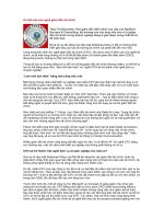
Stomach
The stomach has four major regions: the cardia, fundus, body, and pylorus. The addition of an
inner oblique smooth muscle layer gives the muscularis the ability to vigorously churn and mix
food.
The convex lateral surface of the stomach is called the greater curvature; the concave
medial border is the lesser curvature. The stomach is held in place by the lesser
omentum, which extends from the liver to the lesser curvature, and the greater omentum,
which runs from the greater curvature to the posterior abdominal wall.
Histology
The wall of the stomach is made of the same four layers as most of the rest of the
alimentary canal, but with adaptations to the mucosa and muscularis for the unique
functions of this organ. In addition to the typical circular and longitudinal smooth
muscle layers, the muscularis has an inner oblique smooth muscle layer ([link]). As a
result, in addition to moving food through the canal, the stomach can vigorously churn
food, mechanically breaking it down into smaller particles.
2/12
The Stomach
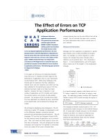
Histology of the Stomach
The stomach wall is adapted for the functions of the stomach. In the epithelium, gastric pits lead
to gastric glands that secrete gastric juice. The gastric glands (one gland is shown enlarged on
the right) contain different types of cells that secrete a variety of enzymes, including
hydrochloride acid, which activates the protein-digesting enzyme pepsin.
The stomach mucosa’s epithelial lining consists only of surface mucus cells, which
secrete a protective coat of alkaline mucus. A vast number of gastric pits dot the surface
of the epithelium, giving it the appearance of a well-used pincushion, and mark the entry
to each gastric gland, which secretes a complex digestive fluid referred to as gastric
juice.
Although the walls of the gastric pits are made up primarily of mucus cells, the gastric
glands are made up of different types of cells. The glands of the cardia and pylorus
are composed primarily of mucus-secreting cells. Cells that make up the pyloric antrum
secrete mucus and a number of hormones, including the majority of the stimulatory
hormone, gastrin. The much larger glands of the fundus and body of the stomach, the
site of most chemical digestion, produce most of the gastric secretions. These glands are
made up of a variety of secretory cells. These include parietal cells, chief cells, mucous
neck cells, and enteroendocrine cells.
Parietal cells—Located primarily in the middle region of the gastric glands are parietal
cells, which are among the most highly differentiated of the body’s epithelial cells.
These relatively large cells produce both hydrochloric acid (HCl) and intrinsic factor.
HCl is responsible for the high acidity (pH 1.5 to 3.5) of the stomach contents and is
needed to activate the protein-digesting enzyme, pepsin. The acidity also kills much of
the bacteria you ingest with food and helps to denature proteins, making them more
available for enzymatic digestion. Intrinsic factor is a glycoprotein necessary for the
absorption of vitamin B12 in the small intestine.
3/12
The Stomach
Chief cells—Located primarily in the basal regions of gastric glands are chief cells,
which secrete pepsinogen, the inactive proenzyme form of pepsin. HCl is necessary for
the conversion of pepsinogen to pepsin.
Mucous neck cells—Gastric glands in the upper part of the stomach contain mucous
neck cells that secrete thin, acidic mucus that is much different from the mucus secreted
by the goblet cells of the surface epithelium. The role of this mucus is not currently
known.
Enteroendocrine cells—Finally, enteroendocrine cells found in the gastric glands
secrete various hormones into the interstitial fluid of the lamina propria. These include
gastrin, which is released mainly by enteroendocrine G cells.
[link] describes the digestive functions of important hormones secreted by the stomach.
Watch this animation that depicts the structure of the stomach and how this structure
functions in the initiation of protein digestion. This view of the stomach shows the
characteristic rugae. What is the function of these rugae?
Hormones
Secreted by
the Stomach
Hormone
Production site
Production
stimulus
Gastrin
Stomach
mucosa, mainly
G cells of the
pyloric antrum
Gastrin
Stomach
mucosa, mainly
G cells of the
pyloric antrum
Target organ
Action
Presence of
peptides and
amino acids in
stomach
Stomach
Increases
secretion by
gastric glands;
promotes gastric
emptying
Presence of
peptides and
amino acids in
stomach
Small
intestine
Promotes
intestinal muscle
contraction
4/12
The Stomach
Hormones
Secreted by
the Stomach
Hormone
Production site
Production
stimulus
Target organ
Action
Gastrin
Stomach
mucosa, mainly
G cells of the
pyloric antrum
Presence of
peptides and
amino acids in
stomach
Ileocecal
valve
Relaxes valve
Gastrin
Stomach
mucosa, mainly
G cells of the
pyloric antrum
Presence of
peptides and
amino acids in
stomach
Large
intestine
Triggers mass
movements
Ghrelin
Stomach
mucosa, mainly
fundus
Fasting state
(levels increase
just prior to
meals)
Regulates food
intake, primarily
Hypothalamus by stimulating
hunger and
satiety
Histamine
Stomach
mucosa
Presence of food
Stomach
in the stomach
Stimulates
parietal cells to
release HCl
Serotonin
Stomach
mucosa
Presence of food
Stomach
in the stomach
Contracts
stomach muscle
Mucosa of
stomach,
Somatostatin especially
pyloric antrum;
also duodenum
Presence of food
in the stomach;
Stomach
sympathetic
axon stimulation
Restricts all
gastric secretions,
gastric motility,
and emptying
Mucosa of
stomach,
Somatostatin especially
pyloric antrum;
also duodenum
Presence of food
in the stomach;
Pancreas
sympathetic
axon stimulation
Restricts
pancreatic
secretions
Mucosa of
Somatostatin stomach,
especially
Presence of food
in the stomach; Small
sympathetic
intestine
axon stimulation
Reduces
intestinal
absorption by
5/12
The Stomach
Hormones
Secreted by
the Stomach
Hormone
Production site
pyloric antrum;
also duodenum
Production
stimulus
Target organ
Action
reducing blood
flow
Gastric Secretion
The secretion of gastric juice is controlled by both nerves and hormones. Stimuli in the
brain, stomach, and small intestine activate or inhibit gastric juice production. This is
why the three phases of gastric secretion are called the cephalic, gastric, and intestinal
phases ([link]). However, once gastric secretion begins, all three phases can occur
simultaneously.
6/12
The Stomach
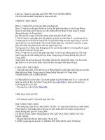
The Three Phases of Gastric Secretion
Gastric secretion occurs in three phases: cephalic, gastric, and intestinal. During each phase,
the secretion of gastric juice can be stimulated or inhibited.
The cephalic phase (reflex phase) of gastric secretion, which is relatively brief, takes
place before food enters the stomach. The smell, taste, sight, or thought of food triggers
this phase. For example, when you bring a piece of sushi to your lips, impulses from
receptors in your taste buds or the nose are relayed to your brain, which returns signals
that increase gastric secretion to prepare your stomach for digestion. This enhanced
secretion is a conditioned reflex, meaning it occurs only if you like or want a particular
food. Depression and loss of appetite can suppress the cephalic reflex.
7/12
The Stomach
The gastric phase of secretion lasts 3 to 4 hours, and is set in motion by local neural
and hormonal mechanisms triggered by the entry of food into the stomach. For example,
when your sushi reaches the stomach, it creates distention that activates the stretch
receptors. This stimulates parasympathetic neurons to release acetylcholine, which then
provokes increased secretion of gastric juice. Partially digested proteins, caffeine, and
rising pH stimulate the release of gastrin from enteroendocrine G cells, which in turn
induces parietal cells to increase their production of HCl, which is needed to create an
acidic environment for the conversion of pepsinogen to pepsin, and protein digestion.
Additionally, the release of gastrin activates vigorous smooth muscle contractions.
However, it should be noted that the stomach does have a natural means of avoiding
excessive acid secretion and potential heartburn. Whenever pH levels drop too low, cells
in the stomach react by suspending HCl secretion and increasing mucous secretions.
The intestinal phase of gastric secretion has both excitatory and inhibitory elements.
The duodenum has a major role in regulating the stomach and its emptying. When
partially digested food fills the duodenum, intestinal mucosal cells release a hormone
called intestinal (enteric) gastrin, which further excites gastric juice secretion. This
stimulatory activity is brief, however, because when the intestine distends with chyme,
the enterogastric reflex inhibits secretion. One of the effects of this reflex is to close the
pyloric sphincter, which blocks additional chyme from entering the duodenum.
The Mucosal Barrier
The mucosa of the stomach is exposed to the highly corrosive acidity of gastric juice.
Gastric enzymes that can digest protein can also digest the stomach itself. The stomach
is protected from self-digestion by the mucosal barrier. This barrier has several
components. First, the stomach wall is covered by a thick coating of bicarbonate-rich
mucus. This mucus forms a physical barrier, and its bicarbonate ions neutralize acid.
Second, the epithelial cells of the stomach's mucosa meet at tight junctions, which block
gastric juice from penetrating the underlying tissue layers. Finally, stem cells located
where gastric glands join the gastric pits quickly replace damaged epithelial mucosal
cells, when the epithelial cells are shed. In fact, the surface epithelium of the stomach is
completely replaced every 3 to 6 days.
Homeostatic Imbalances
Ulcers: When the Mucosal Barrier Breaks Down As effective as the mucosal barrier is,
it is not a “fail-safe” mechanism. Sometimes, gastric juice eats away at the superficial
lining of the stomach mucosa, creating erosions, which mostly heal on their own. Deeper
and larger erosions are called ulcers.
Why does the mucosal barrier break down? A number of factors can interfere with its
ability to protect the stomach lining. The majority of all ulcers are caused by either
8/12
The Stomach
excessive intake of non-steroidal anti-inflammatory drugs (NSAIDs), including aspirin,
or Helicobacter pylori infection.
Antacids help relieve symptoms of ulcers such as “burning” pain and indigestion. When
ulcers are caused by NSAID use, switching to other classes of pain relievers allows
healing. When caused by H. pylori infection, antibiotics are effective.
A potential complication of ulcers is perforation: Perforated ulcers create a hole in the
stomach wall, resulting in peritonitis (inflammation of the peritoneum). These ulcers
must be repaired surgically.
Digestive Functions of the Stomach
The stomach participates in virtually all the digestive activities with the exception
of ingestion and defecation. Although almost all absorption takes place in the small
intestine, the stomach does absorb some nonpolar substances, such as alcohol and
aspirin.
Mechanical Digestion
Within a few moments after food after enters your stomach, mixing waves begin to
occur at intervals of approximately 20 seconds. A mixing wave is a unique type of
peristalsis that mixes and softens the food with gastric juices to create chyme. The
initial mixing waves are relatively gentle, but these are followed by more intense waves,
starting at the body of the stomach and increasing in force as they reach the pylorus. It
is fair to say that long before your sushi exits through the pyloric sphincter, it bears little
resemblance to the sushi you ate.
The pylorus, which holds around 30 mL (1 fluid ounce) of chyme, acts as a filter,
permitting only liquids and small food particles to pass through the mostly, but not
fully, closed pyloric sphincter. In a process called gastric emptying, rhythmic mixing
waves force about 3 mL of chyme at a time through the pyloric sphincter and into the
duodenum. Release of a greater amount of chyme at one time would overwhelm the
capacity of the small intestine to handle it. The rest of the chyme is pushed back into the
body of the stomach, where it continues mixing. This process is repeated when the next
mixing waves force more chyme into the duodenum.
Gastric emptying is regulated by both the stomach and the duodenum. The presence of
chyme in the duodenum activates receptors that inhibit gastric secretion. This prevents
additional chyme from being released by the stomach before the duodenum is ready to
process it.
9/12
The Stomach
Chemical Digestion
The fundus plays an important role, because it stores both undigested food and gases
that are released during the process of chemical digestion. Food may sit in the fundus
of the stomach for a while before being mixed with the chyme. While the food is in
the fundus, the digestive activities of salivary amylase continue until the food begins
mixing with the acidic chyme. Ultimately, mixing waves incorporate this food with the
chyme, the acidity of which inactivates salivary amylase and activates lingual lipase.
Lingual lipase then begins breaking down triglycerides into free fatty acids, and monoand diglycerides.
The breakdown of protein begins in the stomach through the actions of HCl and the
enzyme pepsin. During infancy, gastric glands also produce rennin, an enzyme that
helps digest milk protein.
Its numerous digestive functions notwithstanding, there is only one stomach function
necessary to life: the production of intrinsic factor. The intestinal absorption of vitamin
B12, which is necessary for both the production of mature red blood cells and normal
neurological functioning, cannot occur without intrinsic factor. People who undergo
total gastrectomy (stomach removal)—for life-threatening stomach cancer, for
example—can survive with minimal digestive dysfunction if they receive vitamin B12
injections.
The contents of the stomach are completely emptied into the duodenum within 2 to 4
hours after you eat a meal. Different types of food take different amounts of time to
process. Foods heavy in carbohydrates empty fastest, followed by high-protein foods.
Meals with a high triglyceride content remain in the stomach the longest. Since enzymes
in the small intestine digest fats slowly, food can stay in the stomach for 6 hours or
longer when the duodenum is processing fatty chyme. However, note that this is still a
fraction of the 24 to 72 hours that full digestion typically takes from start to finish.
Chapter Review
The stomach participates in all digestive activities except ingestion and defecation. It
vigorously churns food. It secretes gastric juices that break down food and absorbs
certain drugs, including aspirin and some alcohol. The stomach begins the digestion of
protein and continues the digestion of carbohydrates and fats. It stores food as an acidic
liquid called chyme, and releases it gradually into the small intestine through the pyloric
sphincter.
10/12
The Stomach
Interactive Link Questions
Watch this animation that depicts the structure of the stomach and how this structure
functions in the initiation of protein digestion. This view of the stomach shows the
characteristic rugae. What is the function of these rugae?
Answers may vary.
Review Questions
Which of these cells secrete hormones?
1.
2.
3.
4.
parietal cells
mucous neck cells
enteroendocrine cells
chief cells
C
Where does the majority of chemical digestion in the stomach occur?
1.
2.
3.
4.
fundus and body
cardia and fundus
body and pylorus
body
A
During gastric emptying, chyme is released into the duodenum through the ________.
1.
2.
3.
4.
esophageal hiatus
pyloric antrum
pyloric canal
pyloric sphincter
D
Parietal cells secrete ________.
1.
2.
3.
4.
gastrin
hydrochloric acid
pepsin
pepsinogen
11/12
The Stomach
B
Critical Thinking Questions
Explain how the stomach is protected from self-digestion and why this is necessary.
The mucosal barrier protects the stomach from self-digestion. It includes a thick coating
of bicarbonate-rich mucus; the mucus is physically protective, and bicarbonate
neutralizes gastric acid. Epithelial cells meet at tight junctions, which block gastric juice
from penetrating the underlying tissue layers, and stem cells quickly replace sloughed
off epithelial mucosal cells.
Describe unique anatomical features that enable the stomach to perform digestive
functions.
The stomach has an additional inner oblique smooth muscle layer that helps the
muscularis churn and mix food. The epithelium includes gastric glands that secrete
gastric fluid. The gastric fluid consists mainly of mucous, HCl, and the enzyme pepsin
released as pepsinogen.
12/12