DSpace at VNU: Synthesis, magnetic properties and enhanced photoluminescence of Fe3O4-ZnO heterostructure multifunctional nanoparticles
Bạn đang xem bản rút gọn của tài liệu. Xem và tải ngay bản đầy đủ của tài liệu tại đây (721.65 KB, 8 trang )
VNU Journal of Science: Mathematics – Physics, Vol. 33, No. 1 (2017) 14-21
Synthesis, Magnetic Properties and Enhanced
Photoluminescence of Fe3O4-ZnO Heterostructure
Multifunctional Nanoparticles
Chu Tien Dung1,2, Luu Manh Quynh1, Tran Thi Hong3, Nguyen Hoang Nam1,*
1
Faculty of Physics, VNU University of Science, 334 Nguyen Trai, Thanh Xuan, Hanoi, Vietnam
Department of Physics, University of Transport and Communications, 3 Cau Giay, Hanoi, Vietnam
3
VNU University of Science, 334 Nguyen Trai, Thanh Xuan, Hanoi, Vietnam
2
Received 10 January 2017
Revised 25 February 2017; Accepted 20 March 2017
Abstract: In this paper, Fe3O4-ZnO heterostructure multifunctional nanoparticles (NPs) were
successfully prepared in aqueous solution by ultrasound assisted thermolysis. First, iron oxide
(Fe3O4) magnetic NPs were prepared by co-precipitation method. The Fe3O4 NPs were modified
by 3-aminopropyltriethoxysilane (APTES) to have free amine (-NH2) groups on their surface,
then, Zn2+ ions were added and stirred to adsorb onto the surface of Fe3O4-NH2 NPs in alkaline
solution at a pH of 11. This solution was decomposed through thermolysis in ultrasound bath. The
results of measurements show that photoluminescence of Fe3O4-ZnO heterostructure
multifunctional NPs was enhanced in visible light at 565 nm wavelength to allow detection,
labeling, diagnosis, and therapy in biomedicine. Moreover, they exhibit superparamagnetic
properties of Fe3O4 with high saturation magnetization (MS), which can be used for seperation
application in biomedicine under an external magnetic field.
Keywords:
Fe3O4-ZnO,
multifunctional
nanoparticles,
heterostructure,
enhanced
photoluminescence.
1. Introduction
In recent years, multifunctional nanoparticles (MNPs) based on fluorescent, magnetic, and
plamonic nano-materials are becoming very important materials, which have been a hot research field
in material sciences. Among these MNPs, the NPs combining luminescent and magnetic are the most
popular because of their wide variety of applications in magnetic resonance imaging (MRI), targeted
drug delivery, separation, biodetection, and photodynamic therapy [1-4].
For the magnetic obligation, Fe3O4 NPs are one of the most studied popular materials, are due to
not only their novel properties such as superparamagnetism but also for their potential applications
such as MRI constrast agent [1], separation [2], and hyperthermia therapy to targeted delivery [3],.
_______
Corresponding author. Tel.: 84-913020286
Email:
14
C.T. Dung et al. / VNU Journal of Science: Mathematics – Physics, Vol. 33, No. 1 (2017) 14-21
15
Furthermore, quantum dots (QDs) are particularly attractive for the luminescent properties because of
their tunable emission peak, high photoluminescence quantum yield, and remarkable photostability [57]. The QDs are ideal materials for using as luminescent probes for bioapplications [2-4,8-10]. Among
these QDs, zinc oxide (ZnO) NPs emerge with unique physical and chemical properties such as high
chemical stability, high photoluminescence, bio-compatibility, and nontoxicity [5,8,9]. Therefore, it
would be significant to combine the Fe3O4 NPs with the QDs to form the MNPs with potential
applications for detecting, labeling, and separating biological entities by using luminescence imaging
method under an external magnetic field [4]. Nowadays, the MNPs such as Fe2O3-CdS, Fe3O4-CdS
have synthesized extensively [11, 12]. Although these magnetic-optical materials have disadvantage
due to unbiocompability, toxicity of the QDs (CdSe, CdS), and complicated synthesis protocol [6,13].
Herein, we have synthesized Fe3O4-ZnO heterostructure MNPs in aqueous solution by ultrasound
assisted thermolysis and investigated their characterizations. It is promised that the MNPs are an ideal
system for bioapplications because of their superparamagnetic feature and enhancing luminescent
properties.
2. Experimental
The Fe3O4 was synthesized by co-precipitation method using Fe2+/Fe3+ with 1:2 molar ratios from
the two chloride salts of FeCl2 and FeCl3, was evenly dispersed in ethanol according to our
research [14].
10.5 mg Fe3O4 was equally dispersed in 20 ml ethanol by ultrasound waves, before 5.5 ml
ammonia solution 28% and 4.5 ml aminopropyltriethoxysilane H2N(CH2)3Si(OC2H5)3 (APTES) were
added and vibrated in ultrasound bath with keeping stable temperature at 45oC to form –O–Si–(CH2)3–
NH2 group onto the surface of the Fe3O4 NPs [15]. The solution is then washed several times with
ethanol to collect Fe3O4–NH2.
The Fe3O4–NH2 were dispersed in 20 ml ethanol by ultrasound waves before ammonia solution
28% was added to have solution at a pH of 11. Then, 1ml ion Zinc (Zn2+) 0.2 M was injected and
stirred to adsorb onto the surface of the Fe3O4-NH2. Finally, this solution was decomposed through
thermolysis in ultrasound bath for 2h. The Fe3O4-ZnO NPs were collected after washing several times
with ethanol and distilled water.
ZnO NPs was synthesized at the same condition with the Fe3O4-ZnO sample. As, 1ml ion Zinc
(Zn2+) 0.2 M was injected, stirred in 20 ml Ethanol at a pH of 11, was then decomposed through
thermolysis in ultrasound bath for 2h. The ZnO NPs were collected after washing several times with
ethanol and distilled water.
The crystalline structure was observed by x-ray diffraction (XRD), recorded by using a SIEMENS
D5005 (Bruker-Germany) diffractometer. To study the formation of chemical bonds in the Fe3O4,
Fe3O4–NH2, ZnO and Fe3O4-ZnO NPs, Fourier transform infrared (FTIR) spectroscopy measurement
was carried out on 8400S Shimadzu-Japan. Sample qualitative and quantitative compositions were
investigated by using energy dispersive x-ray spectroscopy (EDS), obtained with ISIS 300 system
(Oxford - UK). The size and morphology of the Fe3O4 and Fe3O4-ZnO were investigated by a JEOL
JEM-1010 transmission electron microscope (TEM), which was operated at 80 kV. The magnetic
hysteresis loops of samples were characterised by vibrating sample magnetometer (VSM) on Digital
Measurement Systems 880 (from USA). Photoluminescence (PL) spectra were recorded by using an
Fluorolog FL3-22 (Jobin-Yvon-Spex, USA) spectrophotometer.
16
C.T. Dung et al. / VNU Journal of Science: Mathematics – Physics, Vol. 33, No. 1 (2017) 14-21
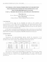
Figure 1. XRD of Fe3O4 (a); ZnO (b); Fe3O4-ZnO (c); insets show zoom-in overlap spectra of all samples at
34.6o-37.5o (d) and 55o-59o (e).
3. Results and discussion
The phase and crystallinity of the as-prepared samples were shown in Figure 1. In the Figure 1(a)
shows the XRD patterns of the Fe3O4. Six diffraction peaks observed at 30.03o, 35.49o, 43.35o, 53.55o,
57.39o, 62.87o, marked by their Miller indices (220), (311), (400), (422), (511), and (440) respectively.
They can be well indexed to the face centered cubic spinel structure of the Fe3O4 in JCPDS cards No.
19-0629. Average lattice parameter of the Fe3O4 crystal was calculated as
, which is in
agreement with published results [16]. Average crystalline size of the Fe3O4 was calculated as
from the full width at half maximum (FWHM) of the most intense diffraction
peaks by using the Debye-Scherrer equation [16]. In the Figure 1(b), the peaks located at
,
,
,
,
,
,
,
, and
were
respectively indexed to the (100), (002), (101), (102), (110), (103), (200), (112), and (201) diffraction
planes of ZnO crystals with a hexagonal wurtzite structure when compared to the standard diffraction
in JCPDS cards No. 36-1451. The average lattice parameter of the ZnO is calculated as
and
, which are consistent with reported papers [5,9,10]. The XRD patterns of the Fe3O4ZnO MNPs in the Figure 1(c) shows that it has discernible diffraction peaks locating at
,
,
,
,
,
,
, and
, were indexed to
diffraction planes of the ZnO. In the Figure 1(c) exposes very low diffraction peaks locating at
and
, were indexed to diffraction planes of the Fe3O4. Moreover, the diffraction peaks of the
Fe3O4-ZnO were broaden to cover simultaneously diffraction peaks of the Fe3O4 and the ZnO. In the
Figure 1(d-e), insets show zoom-in overlap spectra of the Fe3O4, ZnO, and the Fe3O4-ZnO at
, and
, respectively, indicating that the MNPs compose of crystallite the Fe3O4 and the
ZnO. Herein, the ZnO NPs is deposited onto the surface of the Fe3O4-NH2 to form shell layer
covering the Fe3O4 core, which hinders x-rays from penetrating deeper into the Fe3O4-ZnO NPs.
Hence, a direct observation of the Fe3O4 phases cannot be clearly seen [17].
C.T. Dung et al. / VNU Journal of Science: Mathematics – Physics, Vol. 33, No. 1 (2017) 14-21
a
17
b
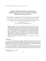
Figure 2. TEM image of Fe3O4 (a); Fe3O4-ZnO (b)
Figure 2(a) is a TEM image of the as-prepared Fe3O4 NPs. It reveals almost homogeneous,
relatively spherical shape and a diameter of 6 nm to 14 nm, which is consistent with calculation from
XRD patterns for the diameter of the Fe3O4 NPs. The TEM image of the Fe3O4-ZnO MNPs in Figure
2(b) shows that in red circles, it can be seen two parts: interior part is high contrast, which may be the
Fe3O4 modified by APTES; exterior part is lower contrast, which may be the ZnO nanocrystals.
Agglomeration of the Fe3O4-ZnO as presented in Figure 2(b) can be due to modification of the Fe 3O4
NPs with APTES and the existence of small remanence magnetization of the Fe3O4 [15].
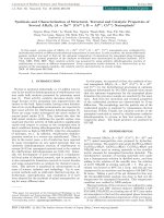
Figure 3. Energy dispersive x-ray spectro-scopy of Fe3O4-ZnO and insert table of percent
of elements composition.
Figure 3 displays energy dispersive X-ray spectroscopy of the Fe3O4-ZnO MNPs and insert a table
of percent of elements composition. The presence of Fe and Zn determines that the MNPs are
simultaneously composed of the Fe3O4, ZnO nanocrystals. Furthermore, the existence of C and Si
provide direct proof for the successful modification of APTES on the surface of the Fe3O4 NPs.
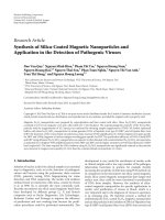
Figure 4(left) shows FTIR spectra of the Fe3O4, Fe3O4-NH2, ZnO and Fe3O4-ZnO samples. It can
be seen that in the Fe3O4, Fe3O4-NH2, and Fe3O4-ZnO samples have absorption peaks aroud 425 cm-1,
628 cm-1 corresponding to the Fe-O vibration of the Fe3O4 phase [18]. Peak at 628 cm-1 has shifted to
650 cm-1 in the Fe3O4-NH2 samples, which is clear evidence to prove the successful modification of
APTES on the surface of the Fe3O4, and agrees well with the result from the EDS [19].
18
C.T. Dung et al. / VNU Journal of Science: Mathematics – Physics, Vol. 33, No. 1 (2017) 14-21
Figure 4. FTIR (left) and Raman shift (right) spectra of Fe3O4; Fe3O4-NH2; ZnO and Fe3O4-ZnO.
Figure 5. Photoluminesences spectra of ZnO (a); Fe 3O4-ZnO (b) and inset shows
luminescence image using excited laser beam 325 nm wavelength.
Further, the band at 1012 cm-1 and 1223 cm-1 only observed in the Fe3O4-NH2, are assigned to
stretching vibrations of Si-O-Si [15, 19] and vibration of –CH2 [18]. And shoulder at 1072 cm-1 is
corresponding to stretching vibrations of C-N, which is relating to the –(CH2)3–NH2 group onto the
C.T. Dung et al. / VNU Journal of Science: Mathematics – Physics, Vol. 33, No. 1 (2017) 14-21
19
surface of the Fe3O4. The peaks at 1384 cm-1 are because of the existence of CO2 molecule in air [20].
The peaks, observed between 1450 cm-1 and 1700 cm-1, are allotted the crucial stretching mode of OH
[18]. These vibrations indicate the presence of H2O molecules on the surface of the all samples. On the
other hand, the band at 556 cm-1 and 712 cm-1 is only observed in the ZnO and Fe3O4-ZnO samples,
can be attributed to the Zn-O stretching mode in the ZnO lattice [20,21]. Interestingly, the peak of the
Fe-O vibration in the Fe3O4-ZnO MNPs has lower absorption intensity than in the Fe3O4 and the
Fe3O4-NH2, which indicates that ZnO nanocrystals are assembled onto the surface of the Fe3O4-NH2.
Combining with XRD results, it is concluded that the as-prepared Fe3O4-ZnO MNPs have core-shell
structure.
Raman spectroscopy is useful to probe the local structure. Figure 4(right) presents the Raman
spectra of all samples. The sharp peak at 204 cm-1, 287 cm-1, and 611 cm-1, only observed in the Fe3O4
and the Fe3O4-NH2 samples, are characterization of the T2g, Eg, and A1g modes of the Fe3O4 [22]. The
intense Raman shift peaks at 998 cm-1 and 1601 cm-1 in the Fe3O4-NH2 sample are corresponding to
vibration of Si-C [23] and C-C [24], allowing to determine the existence of –O–Si–(CH2)3–NH2 group
onto the surface of the Fe3O4 NPs, which is suitable to the results of FTIR. Raman shift spectrum of
the Fe3O4-ZnO MNPs shows that the intense and sharp peaks at 332 cm-1 and 438 cm-1 are
characterization of the E2(high) - E2(low) and E2(high) modes of the hexagonal ZnO crystalline
structure, which are agreement with that of the ZnO sample [25, 26]. The high frequency E2(high)
mode involves the vibration of oxygen atoms, which is attributed to low intrinsic defects only
associated with oxygen such as oxygen vacancies (VO). In addition, the stronger intensity of the
E2(high) peak is the better quality crystals [25, 26]. The low peak observed at 207 cm-1, is assigned to
the 2TA mode of the ZnO nanocrystals and is the agreement with T 2g mode in the Fe3O4 [26]. These
results indicate that ZnO nanocrystals coat surrounding of the Fe3O4 NPs, which is consistent with the
results of XRD and FTIR.
Figure 6. The magnetic hysteresis loops of Fe3O4 (a); Fe3O4-NH2 (b) and Fe3O4-ZnO (c) at room temperature.
Figure 6 presents the room temperature PL spectra of the ZnO NPs and the Fe3O4-ZnO MNPs. The
PL spectra shows a broad emission spectrum covering from near ultraviolet to whole of the visible
region. All the samples show a small ultraviolet emission peak at approximately 387 nm (equivalent to
3.2 eV), which can be attributed to the near band edge transitions in the ZnO nanocrystals, originating
from the recombination of free exciton through an exciton-exciton collision process [27]. Whereas the
visible band emission is known to be due to an electronic transition from the conduction band edge to
a defect of associated trap state such as an oxygen vacancy, which is consistent with the appearance of
E2(high) mode in Raman shift spectra [28, 29]. Nevertheless, intensity of visible band emission in the
20
C.T. Dung et al. / VNU Journal of Science: Mathematics – Physics, Vol. 33, No. 1 (2017) 14-21
Fe3O4-ZnO MNPs was enhanced and was higher than that of the ZnO many times. It can be explained
that the formation of core-shell structure of the Fe3O4-ZnO MNPs makes trap states, as suggesting
valence band and conduction band of the Fe3O4 core [26]. So, the center of visible band emission at
565 nm (equivalent to 2.21 eV) in the Fe3O4-ZnO MNPs is attributed to can be due to an electronic
transition from the conduction band edge of the ZnO nanocrystal to the trap states such as conduction
band edge of the Fe3O4 core. With high photoluminescence in visible light, the Fe3O4-ZnO MNPs are
promised application for biosensor [10,26], bioimaging [8,9].
The magnetic hysteresis loops of the Fe3O4, Fe3O4-NH2, and the Fe3O4-ZnO were measured at
room temperature, were displayed in Figure 6. The MS of the Fe3O4, Fe3O4-NH2, and the Fe3O4-ZnO
have value as 51.8, 40.9, and 19.6 (emu/g), respectively. The decrease of the M S values can be
explained by the successful modification of the APTES on the surface of the Fe3O4 NPs and the
formation of ZnO nanocrystals surrounding the Fe3O4-NH2. This results can be comfirmed that the
MNPs contain simultaneously the Fe3O4 and the ZnO nanocrystals. In the Figure 6 shows that the
Fe3O4, Fe3O4-NH2, and the Fe3O4-ZnO are superparamagnetic at room temperature with high MS and
small coercivity (approximately 7 Oe). The magnetic properties results in that the MNPs can be tagged
and separated with biomolecules or targeted drug delivery and magnetic resonance imaging (MRI)
under the induction of an external magnetic field [3,16].
4. Conclusion
In this paper, the Fe3O4-ZnO MNPs were successfully synthesized in aqueous solution by
ultrasound assisted thermolysis. The MNPs compose of the magnetic Fe3O4 NPs and the
photoluminescent ZnO nanocrystals. Moreover, the MNPs not only enhance PL in visible light but
also exhibit superparamagnetism with high saturation magnetization at room temperature. These
MNPs can make wide variety of applications for biomedical.
Acknowledgements
This study was accomplished at the Center for Material Sciences, VNU University of Sciences.
The first author thanks the project 911 - Vietnam education fellowship for financial support.
References
[1] Y.D. Jin, C.X Jia, S.W Huang, M.O. Donnell, and X.H. Gao, “Multifunctional nanoparticles as coupled contrast
agents”. Nature Communications 1, (2010) 41.
[2] G G. Liu, D. Hu, M. Chen, C.C. Wang, and L.M. Wu, “Multifunctional PNIPAM/Fe 3O4–ZnS hybrid hollow
spheres: Synthesis, characterization, and properties”. Journal of Colloid and Interface Science 397 (2013) 73.
[3] H.H. Yiu, “Engineering the multifunctional surface on the magnetic nanoparticles for targeted biomedical
applications”, Nanomedicine. 6(8) (2011) 1429.
[4] L.W. Chan, Y.N. Wang, L.Y. Lin, M.P. Upton, J.H. Hwang, and S.H. Pun, “Synthesis and Characterization of
Anti-EGFR Fluorescent Nanoparticles for Optical Molecular Imaging”, Bioconjugate Chem. 24 (2013) 167.
[5] S.T. Dutta, S. Chattopadhyay, A. Sarkar, M. Chakrabarti, D. Sanyal, and D. Jana, “Role of defects in tailoring
structural, electrical and optical properties of ZnO”, Progress in Materials Science, 54 (2009) 89.
[6] K. Yu, B. Zaman, S. Romanova, D.S.Wang, and J.A. Ripmeester “Sequential Synthesis of Type II Colloidal
CdTe/CdSe Core–Shell Nanocrystals”, small, 1, No. 3 (2005), 332.
C.T. Dung et al. / VNU Journal of Science: Mathematics – Physics, Vol. 33, No. 1 (2017) 14-21
21
[7] D.A. Bussian, S.A. Crooker, M. Yin, M.Brynda, A.L. Efros, and V.I. Klimov, “Tunable magnetic exchange
interactions in manganese-doped inverted core–shell ZnSe–CdSe nanocrystals". Nature materials, 8 (2009) 35
[8] K. Senthilkumar, O. Senthilkumar, K. Yamauchi, M. Sato, S. Morito, T. Ohba, M. Nakamura, and Y. Fujita,
“Preparation of ZnO nanoparticles for bio-imaging applications”, Phys. Status Solid b, 246 (2009) 885.
[9] J.W. Rasmussen, E. Martinez, P. Louka, and D.G. Wingett, “Zinc Oxide Nanoparticles for Selective Destruction
of Tumor cells and Potential for Drug delivery Applications”, Expert Opin Drug Deliv, 7 (2010) 1063.
[10] Z.W.Zhao, W. Lei, X.B. Zhang, B.P. Wang, and H.L. Jiang, “ZnO-Based Amperometric Enzyme Biosensors”,
Sensors. 10 (2010) 1216.
[11] Z. Wu, H. Yu, L. Kuai, H. Wang, T.H. Pei, and B.Y. Geng, “CdS urchin-like microspheres/a-Fe2O3 and
CdS/Fe3O4 nanoparticles heterostructures with improved photocatalytic recycled activities. Journal of Colloid and
Interface Science 426 (2014) 83.
[12] A. Roychowdhury, S.P. Pati, S. Kumar, and D. Das, “Tunable properties of magneto-optical Fe3O4/CdS
nanocomposites on size variation of the magnetic component”, Materials Chemistry and Physics, 151 (2015) 105.
[13] S.A.O. Gomes, C.S. Vieira, D.B. Almeida, J.R. Santos-Mallet, R.F. S. Menna-Barreto, C.L. Cesar, and D. Feder,
“CdTe and CdSe Quantum Dots Cytotoxicity: A Comparative Study on Microorganisms”. Sensors, 11 (2011),
11664.
[14] C.T. Dung, L.M. Quynh, N.P. Linh, N.H. Nam, N.H. Luong, “Synthesis of ZnS:Mn-Fe3O4 bifunctional
nanoparticles by inverse microemulsion method”, Journal of Science: Advanced Materials and Devices 1 (2016)
200.
[15] M. Mahdavi, M.B. Ahmad, M.J. Haron, Y. Gharayebi, K. Shameli, and B. Nadi, “Fabrication and
Characterization of SiO2/(3-Aminopropyl) triethoxysilane-Coated Magnetite Nanoparticles for Lead(II) Removal
from Aqueous Solution”, J Inorg Organomet Polym, 23 (2013) 599.
[16] H.E. Ghandoor, H.M. Zidan, M.M.H. Khalil, and M.I.M. Ismai, “Synthesis and Some Physical Properties of
Magnetite (Fe3O4) Nanoparticles”. Int. J. Electrochem. Sci., 7 (2012) 5734.
[17] M. Stefan, O. Pana, C. Leostean, C. Bele, D. Silipas, M. Senila, and E. Gautron, “Synthesis and characterization
of Fe3O4–TiO2 core-shell nanoparticles”. Journal of Applied Physics 116 (2014) 114312.
[18] Y.S. Li, J.S. Church, A.L. Woodhead, and F. Moussa. “Preparation and characterization of silica coated iron
oxide magnetic nano-particles”. Spectrochimica Acta Part A 76 (2010) 484.
[19] Z. Hassannejad, M.E. Khosroshahi, and M. Firouzi, “Fabrication and Characterization of Magnetoplasmonic
Liposome Carriers”. Journal of Inorganic and Organometallic Polymers and Materials, 23, 3, (2013) 599.
[20] S. Senthilkumaar, K. Rajendran, S. Banerje, T.K. Chini, and V. Sengodan. “Influence of Mn doping on the
microstructure and optical property of ZnO”. Mater. Sci. Semicon. Process. 11 (2008) 6.
[21] P. Patra, S. Mitra, N. Debnath, P. Pramanik, and A. Goswami, “Ciprofloxacin conjugated zinc oxide nanoparticle:
A camouflage towards multidrug resistant bacteria”. Bull. Mater. Sci., 37 (2014) 199.
[22] O.N. Shebanova and P. Lazor. “Raman spectroscopic study of magnetite (FeFe 2O4): a new assignment for the
vibrational spectrum”. Journal of Solid State Chemistry 174 (2003) 424.
[23] X.X. Qi, G.M. Zhai, J. Liang, S.F. Ma, X.G. Liu, and B.S. Xu. “Preparation and characterization of SiC@CNT
coaxial nanocables using CNT as template”. CrystEngComm, 16 (2014), 9697.
[24] E.B. Santos, F.A. Sigoli, and I.O. Mazali. “Facile synthesis of the dendritic structure of silver nanoparticleschitosan and its application as an effective SERS substrate”. New J. Chem. 38 (2014) 5369.
[25] Y.Q. Huang, M.D. Liu, Z. Li, Y. Zeng, and S.B. Liu, “Raman spectroscopy study of ZnO-based ceramic films
fabricated by novel sol/gel process”. Materials Science and Engineering B 97 (2003) 111.
[26] C.Karunakaran and P. Vinayagamoorthy. “Magnetically recoverable Fe3O4-implanted Ag-loaded ZnO
nanoflakes for bacteria-inactivation and photocatalytic degradation of organic pollutants”. New J. Chem., 40
(2016) 1845.
[27] Z.K. Tang, M. Kawasaki, A. Ohtomo, H. Koinuma, and Y. Segawa, “Self-assembled ZnO nano-crystals and
exciton lasing at room temperature”. Journal of Crystal Growth, 287 (2006) 169.
[28] N.S. Norberg, and D.R. Gamelin. “Influence of Surface Modification on the Luminescence of Colloidal ZnO
Nanocrystals”. J.Phys.Chem B, 109 (2005) 20810.
[29] K. Vanheusden, W.L. Warren, C.H. Seager, D.R. Tallant, and J.A. Voigt, “Mechanisms behind green
photoluminescence in ZnO phosphor powders”. Journal of Applied Physics, 79 (1996) 7983.