DSpace at VNU: Kineosporia babensis sp. nov., isolated from plant litter in Vietnam
Bạn đang xem bản rút gọn của tài liệu. Xem và tải ngay bản đầy đủ của tài liệu tại đây (146.18 KB, 5 trang )
International Journal of Systematic and Evolutionary Microbiology (2009), 59, 550–554
DOI 10.1099/ijs.0.002907-0
Kineosporia babensis sp. nov., isolated from plant
litter in Vietnam
Yayoi Sakiyama,1 Nguyen K. N. Thao,2 Nguyen M. Giang,2 Shinji Miyadoh,1
Duong V. Hop2 and Katsuhiko Ando1
Correspondence
1
NITE Biological Resource Center (NBRC), National Institute of Technology and Evaluation (NITE),
Chiba 292-0818, Japan
Yayoi Sakiyama
2
Institute of Microbiology and Biotechnology (IMBT), Vietnam National University, Hanoi (VNUH),
Hanoi, Vietnam
Three actinomycetes, designated strains VN05A0342, VN05A0351 and VN05A0415T, were
isolated from plant-litter samples collected in the north of Vietnam and examined in a polyphasic
taxonomic study. Phylogenetic analysis based on the 16S rRNA gene sequences showed that
these isolates were most closely related to the type strain of Kineosporia mikuniensis (98.5 %
sequence similarity). Morphological properties (the formation of spore domes and motile spores)
and chemotaxonomic data supported the assignment of the three isolates to the genus
Kineosporia. The isolates all contained the following: meso-diaminopimelic acid in the
peptidoglycan (with small amounts of the LL isomer); ribose, mannose, galactose and glucose as
the whole-cell sugars; MK-9(H4) as the predominant isoprenoid quinone; C18 : 1 and C16 : 0 as the
major cellular fatty acids; and phosphatidylcholine, phosphatidylglycerol, diphosphatidylglycerol
and phosphatidylinositol as the phospholipids. The high DNA–DNA relatedness (.71 %) among
the three isolates showed that they represented a single species. On the other hand, the
DNA–DNA relatedness between the novel isolates and all type strains of Kineosporia species
was less than 46 %. The physiological properties of our isolates were distinct from those of all of
the Kineosporia species with validly published names, e.g. decomposition of L-tyrosine and
aesculin and the utilization of raffinose and D-arabitol. Therefore, strains VN05A0342,
VN05A0351 and VN05A0415T represent a novel species of the genus Kineosporia, for which
the name Kineosporia babensis sp. nov. is proposed. The type strain is VN05A0415T
(5VTCC-A-0961T 5NBRC 104154T).
The genus Kineosporia was first reported by Pagani &
Parenti (1978). The genus was described as comprising
organisms that form sporangia (each containing a single
zoospore) at the edge of the substrate mycelium and
contain only LL-diaminopimelic acid (LL-A2pm) in the
peptidoglycan. Subsequently, Itoh et al. (1989) and Kudo
et al. (1998) emended the description of the genus on the
basis of the presence of both LL-A2pm and meso-A2pm and
the similarity of the colonial morphology to ‘spore-dome
actinomycetes’ (Willoughby, 1969). At the time of writing,
the genus Kineosporia comprises five species with validly
Abbreviation: A2pm, diaminopimelic acid.
The GenBank/EMBL/DDBJ accession numbers for 16S rRNA gene
sequences of strains VN05A0415T, VN05A0342 and VN05A0351 are
AB377116, AB377118 and AB377119, respectively.
Cultural characteristics and fatty acid compositions of strains
VN05A0342, VN05A0351 and VN05A0415T and all of the type
strains of Kineosporia species are available as supplementary material
with the online version of this paper.
550
published names. In this study, we used a polyphasic
approach to classify three novel actinomycete isolates. On
the basis of the data from this study, these three isolates
represent a novel species of the genus Kineosporia.
The samples of plant litter were collected in 2005 from the
mountainside at Ba Be National Park, Bac Kan Province, in
northern Vietnam. The samples were dried at room
temperature for 5–7 days and then inoculated using the
rehydration–centrifugation method (Hayakawa et al., 2000)
on humic acid-vitamin agar (Hayakawa & Nonomura,
1987) containing nalidixic acid (20 mg l21) and kabicidin
(0.75 mg l21). Our isolates, designated VN05A0342,
VN05A0351 and VN05A0415T, were isolated after incubation for more than 10 days at room temperature.
Strains VN05A0342, VN05A0351 and VN05A0415T were
incubated on yeast extract-soluble starch medium (YS
medium; 2 g yeast extract, 10 g soluble starch and 15 g
agar l21; pH 7.3) at 28 uC for 10–14 days. The orange
colonies that formed appeared moist and were raised, like
002907 G 2009 IUMS Printed in Great Britain
Kineosporia babensis sp. nov.
phosphatidylethanolamine and phosphatidyl-N-methylethanolamine were absent. The major cellular fatty acids
were C18 : 1 and C16 : 0, but iso- and/or anteiso-branched
fatty acids were not detected (Supplementary Table S2).
The chemotaxonomic data for our isolates were consistent
with the characteristics described for the genus Kineosporia.
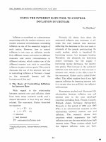
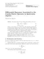
Fig. 1. Scanning electron micrograph of strain VN05A0415T
grown on water agar for 10 days at 28 6C. Bar, 5 mm.
‘spore-dome actinomycetes’ (Willoughby, 1969), from the
surface of the YS medium. Aerial mycelium was absent.
The spore-domes were formed by bunches of single spores
borne on sporophores similar to those described by Itoh et
al. (1989). Scanning electron microscopy showed that the
spores were globular and/or ovoid (1.0–2.0 mm in
diameter) with a smooth surface. The spores seemed to
be enveloped in a club-shaped sporangium (Fig. 1), as
reported by Pagani & Parenti (1978). Light-microscopic
observation of cells suspended in phosphate buffer
(pH 7.0, 1 mM) showed the spores to be motile. The
cultural characteristics of our isolates and all type strains of
Kineosporia species were observed on ISP media 2–7
(Shirling & Gottlieb, 1966) and YS medium after
incubation at 28 uC for 3 weeks (see Supplementary
Table S1, available in IJSEM Online). Our isolates and all
type strains of Kineosporia species showed good growth on
YS medium, ISP 2 and ISP 3. Only our isolates and the type
strain of Kineosporia aurantiaca grew on ISP 6.
For the chemotaxonomic analysis, biomass from each
strain was obtained by centrifugation and lyophilization
after incubation in yeast extract-glucose broth (10 g yeast
extract and 10 g glucose l21; pH 7.3) for 7–10 days at
28 uC. The whole-cell sugars, isoprenoid quinones, phospholipids and cellular fatty acids were analysed as described
by Staneck & Roberts (1974), Minnikin et al. (1984) and
Tamura et al. (1994). The A2pm isomer in the peptidoglycan was analysed as described by Nozawa et al. (2007).
Kudo et al. (1998) reported that Kineosporia strains exhibit
heterogeneity of the A2pm isomer because of the presence
of different isomers in mycelium and spores. Our isolates,
strains VN05A0342, VN05A0351 and VN05A0415T, also
contained both of the A2pm isomers: a small amount of LLA2pm was present, but the main isomer was meso-A2pm.
The whole-cell sugars were ribose, mannose, galactose and
glucose. The isoprenoid quinone was MK-9(H4).
Phosphatidylcholine, phosphatidylglycerol, diphosphatidylglycerol and phosphatidylinositol were detected, but
The DNA was extracted as described by Marmur (1961)
and Saito & Miura (1963), but with a slight modification:
after lysis, we used 20 % SDS and protease K to denature
proteins,
and
phenol/chloroform/isoamyl
alcohol
(25 : 24 : 1, by vol.) to remove denatured proteins. 16S
rRNA gene sequences were analysed as described by
Tamura & Hatano (2001). Sequence analysis was performed with an ABI Prism BigDye Terminator cycle
sequencing kit (PE Applied Biosystems) and an automatic
DNA sequencer (model 3130 Genetic Analyzer; PE Applied
Biosystems). The CLUSTAL_X program (Thompson et al.,
1997) was used to align the 16S rRNA gene sequences with
corresponding sequences (available in the GenBank/EMBL/
DDBJ databases) from all of the type strains of Kineosporia
species and some related actinomycetes of the suborder
Frankineae. Phylogenetic trees were constructed using the
neighbour-joining (Saitou & Nei, 1987) and maximumparsimony (Kluge & Farris, 1969) methods. The topology
of the trees was evaluated by means of bootstrap analysis
based on 1000 replicates (Felsenstein, 1985). DNA–DNA
hybridization was carried out using the method of Ezaki et
al. (1989). The G+C content of the DNA was determined
using the method of Mesbah et al. (1989).
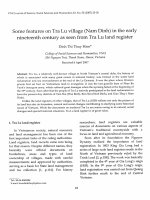
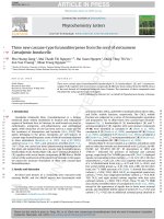
Phylogenetic analysis based on 16S rRNA gene sequences
revealed that our isolates and all type strains of the genus
Kineosporia formed a monophyletic cluster (Fig. 2). The
cluster had bootstrap support in both neighbour-joining
Fig. 2. Neighbour-joining phylogenetic tree, based on 16S rRNA
gene sequences, for strains VN05A0342, VN05A0351 and
VN05A0415T, all type strains of Kineosporia species and some
actinomycetes in suborder Frankineae. Numbers at branch points
are confidence limits estimated by means of bootstrap analysis
based on 1000 replicates; only values .500 are presented. Bar,
0.01 Knuc in nucleotide sequences.
551
Y. Sakiyama and others
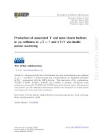
Table 1. DNA–DNA hybridization among strains VN05A0342, VN05A0351 and VN05A0415T
and all type strains of Kineosporia species
Source of unlabelled DNA
1.
2.
3.
4.
5.
6.
7.
8.
VN05A0342
VN05A0351
VN05A415T
K. aurantiaca NBRC 14067T
K. rhamnosa NBRC 16231T
K. succinea NBRC 16232T
K. rhizophila NBRC 16233T
K. mikuniensis NBRC 16234T
DNA–DNA relatedness (%) with labelled DNA from:
1
2
3
4
5
6
7
8
100
82
76
20
5
28
34
28
92
100
71
26
7
40
46
33
108
98
100
17
6
21
27
19
13
13
13
100
9
16
17
15
19
21
21
19
100
22
22
19
23
21
26
20
8
100
25
19
23
23
25
14
7
19
100
22
30
30
32
28
11
23
33
100
and maximum-parsimony phylogenetic trees. Although
our isolates and the type strain of Kineosporia rhizophila
formed a clade in the phylogenetic tree, the tree topology
was not supported by bootstrapping analysis (52.2 %). Our
isolates shared 16S rRNA gene sequence similarity of 99.8–
100 %. The nucleotide sequence similarity between our
isolates and all type strains of the genus Kineosporia ranged
from 96.1 to 98.5 %. Our isolates showed the greatest
similarity with respect to the type strain of Kineosporia
mikuniensis.
DNA–DNA hybridization among our isolates and all type
strains of Kineosporia species was determined (Table 1).
The DNA relatedness among strains VN05A0342,
VN05A0351 and VN05A0415T ranged from 71 to 108 %.
Consequently, our isolates were identified as representing a
single species. The DNA relatedness between our isolates
and all Kineosporia type strains was less than 46 %, being
below the 70 % cut-off point recommended for the
delineation of genomic species (Wayne et al., 1987).
Therefore, strains VN05A0342, VN05A0351 and
VN05A0415T were different from all type strains of
Kineosporia species. The G+C contents of their DNAs
were in the range 69–70 mol%.
supplemented with 1 % organic salts or 0.2 % benzoic acid.
An acid-production test was performed on basal medium
composed of (l21) 10 g peptone and 5 g NaCl (pH 7.2)
plus the test compound (1 %).
The features that served to differentiate strains
VN05A0342, VN05A0351 and VN05A0415T from known
species of the genus Kineosporia were the decomposition of
L-tyrosine and aesculin and the utilization of raffinose and
D-arabitol (Table 2).
On the basis of the results of the polyphasic taxonomic
study presented here, strains VN05A0342, VN05A0351
and VN05A0415T represent a novel species of the genus
Kineosporia, for which the name Kineosporia babensis
sp. nov. is proposed.
Description of Kineosporia babensis sp. nov.
Kineosporia babensis (ba.ben9sis. N.L. fem. adj. babensis
referring to Ba Be National Park, Vietnam, from which the
first strains were isolated).
Physiological and biochemical characteristics of our
isolates were tested after incubation at 28 uC for 3 weeks.
NaCl tolerance was examined on YS medium prepared
with 0, 1, 2, 3, 4, 5 and 6 % NaCl (w/v). ISP 8 (Gordon &
Mihm, 1957) was used to test for nitrate reduction.
Decomposition of urea was determined on Christensen
urea agar containing 2 % urea (Gordon et al., 1974).
Degradation of casein and other compounds (final
concentration 0.5 %) was determined using nutrient agar
as the basal medium (Gordon et al., 1974). Aesculin
hydrolysis and utilization of citrate were examined
according to the methods of Gordon et al. (1974). The
utilization of other carbohydrates was tested on yeast
nitrogen base without amino acids (Bacto), as described by
Goodfellow (1971). The utilization of organic acids was
determined on a medium composed of (l21) 1.0 g
NH4NO3, 1.0 g KH2PO4, 0.5 g MgSO4 . 7H2O and 0.2 g
KCl (pH 7.2), containing 20 ml 0.04 % phenol red and
The orange-coloured colonies grow prolifically on YS
medium and appear moist and raised. Each raised colony
produces clusters of single spores. The spore surface is
smooth and the spores are globular and/or ovoid (1.0–
2.0 mm in diameter). Grows at 10–28 uC, but not at 5 or
37 uC. Grows in the presence of 3 % NaCl (w/v). Melanin is
not produced on ISP 6 or ISP 7. Negative for nitrate
reduction. Decomposes aesculin, arbutin, casein, testosterone, L-tyrosine and urea, but not adenine, hypoxanthine or
xanthine. Utilizes carbon sources such as L-arabinose,
cellobiose, D-fructose, D-galactose, D-glucose, myo-inositol,
D-lactose, maltose, D-mannitol, melezitose, melibiose,
raffinose, L-rhamnose, D-ribose, salicin, D-sorbitol, starch,
sucrose, trehalose and D-xylose, but not adonitol, Darabitol, dulcitol, methyl a-D-glucoside, L-sorbose or
xylitol. Utilizes organic acids such as fumarate, malate
and succinate, but not benzoate, mucate, oxalate or Ltartrate. Produces acid from L-arabinose, cellobiose, Dfructose, D-galactose, D-glucose, L-rhamnose, sucrose and
D-xylose, but not from adonitol, i-erythritol, myo-inositol,
melezitose and D-sorbitol. The cell wall contains major
552
International Journal of Systematic and Evolutionary Microbiology 59
Kineosporia babensis sp. nov.
Table 2. Differential physiological characteristics among
strains VN05A0342, VN05A0351 and VN05A0415T and type
strains of all Kineosporia species
Strains: 1, VN05A415T (strains VN05A0342 and VN05A0351 showed
identical results unless indicated); 2, K. aurantiaca NBRC 14067T; 3,
K. rhamnosa NBRC 16231T; 4, K. succinea NBRC 16232T; 5, K.
rhizophila NBRC 16233T; 6, K. mikuniensis NBRC 16234T. Data for
reference type strains were taken from Kudo et al. (1998).
Strain
Decomposition of:
L-Tyrosine
Aesculin
NaCl tolerance (%, v/v)
Utilization of:
Raffinose
D-Arabitol
1
+ (brown
pigment)
+
4*
+
2
2
3
4
5
6
Gordon, R. E., Barnett, D. A., Handerhan, J. E. & Pang, C. H.-N.
(1974). Nocardia coeliaca, Nocardia autotrophica, and the nocardin
strain. Int J Syst Bacteriol 24, 54–63.
Hayakawa, M. & Nonomura, H. (1987). Humic acid-vitamin agar, a
new medium for selective isolation of soil actinomycetes. J Ferment
Technol 65, 501–509.
Hayakawa, M., Otoguro, M., Takeuchi, T., Yamazaki, T. & Iinuma, Y.
(2000). Application of a method incorporating differential centrifu-
gation for selective isolation of motile actinomycetes in soil and plant
litter. Antonie Van Leeuwenhoek 78, 171–185.
Itoh, T., Kudo, T., Parenti, F. & Seino, A. (1989). Amended description
of the genus Kineosporia, based on chemotaxonomic and morphological studies. Int J Syst Bacteriol 39, 168–173.
2
2
2
2
2
+ 2 2 2 2
,3 ,2 ,5 ,5 ,1
2 ± 2
+ 2 +
+ 2
+ +
*Strain VN05A0351 tolerated 3 % NaCl but not 4 %.
Kluge, A. G. & Farris, J. S. (1969). Quantitative phyletics and the
evolution of anurans. Syst Zool 18, 1–32.
Kudo, T., Matsushima, K., Itoh, T., Sasaki, J. & Suzuki, K. (1998).
Description of four new species of the genus Kineosporia: Kineosporia
succinea sp. nov., Kineosporia rhizophila sp. nov., Kineosporia
mikuniensis sp. nov. and Kineosporia rhamnosa sp. nov., isolated
from plant samples, and amended description of the genus
Kineosporia. Int J Syst Bacteriol 48, 1245–1255.
Marmur, J. (1961). A procedure for the isolation of deoxyribonucleic
acid from microorganisms. J Mol Biol 3, 208–218.
Mesbah, M., Premachandran, U. & Whitman, W. B. (1989).
amounts of meso-A2pm and small amounts of LL-A2pm.
The whole-cell sugars are ribose, mannose, galactose and
glucose. The predominant menaquinone is MK-9(H4). The
phospholipids are phosphatidylcholine, phosphatidylglycerol, phosphatidylglycerol and phosphatidylinositol. The
major cellular fatty acids are C18 : 1 and C16 : 0. The DNA
G+C content is 69–70 mol%.
T
T
The type strain, VN05A0415 (5VTCC-A-0961 5NBRC
104154T), was isolated from plant litter.
Precise measurement of the G+C content of deoxyribonucleic acid
by high-performance liquid chromatography. Int J Syst Bacteriol 39,
159–167.
Minnikin, D. E., O’Donnell, A. G., Goodfellow, M., Alderson, G.,
Athalye, M., Schaal, A. & Parlett, J. H. (1984). An integrated
procedure for the extraction of bacterial isoprenoid quinones and
polar lipids. J Microbiol Methods 2, 233–241.
Nozawa, Y., Sakai, N., Arai, K., Kawasaki, Y. & Harada, K. (2007).
Reliable and sensitive analysis of amino acids in the peptidoglycan of
actinomycetes using the advanced Marfey’s method. J Microbiol
Methods 70, 306–311.
Pagani, H. & Parenti, F. (1978). Kineosporia, a new genus of the order
Acknowledgements
This work was conducted as a joint research project between the
Department of Biotechnology, NITE (NITE-DOB), Japan, and the
IMBT, VNUH, Vietnam. The authors are grateful to Dr Tomohiko
Tamura, Ms Kozue Anzai, Mr Nobuyuki Goto, Dr Misa Otoguro, Dr
Hideki Yamamura, Ms Kayo Tsuruya, Mr Shinpei Ino, Ms Ayako
Hashimoto, Dr Takuji Nakashima (NITE), Dr Dinh Thuy Hang, Dr
Dao Thi Luong (IMBT, VNUH) and all members at IMBT for their
kind help and advice. We also thank Dr Yuriko Nozawa (Taisho
Pharmaceutical Co., Ltd) for analysis of the A2pm isomer by LC-MS.
Actinomycetales. Int J Syst Bacteriol 28, 401–406.
Saito, H. & Miura, K. (1963). Preparation of transforming deoxy-
ribonucleic acid by phenol treatment. Biochim Biophys Acta 72,
619–629.
Saitou, N. & Nei, M. (1987). The neighbor-joining method: a
new method for reconstructing phylogenetic trees. Mol Biol Evol 4,
406–425.
Shirling, E. B. & Gottlieb, D. (1966). Methods for characterization of
Streptomyces species. Int J Syst Bacteriol 16, 313–340.
Staneck, J. L. & Roberts, G. D. (1974). Simplified approach to
identification of aerobic actinomycetes by thin-layer chromatography.
Appl Microbiol 28, 226–231.
References
Ezaki, T., Hashimoto, Y. & Yabuuchi, E. (1989). Fluorometric
deoxyribonucleic acid-deoxyribonucleic acid hybridization in microdilution wells as an alternative to membrane filter hybridization in
which radioisotopes are used to determine genetic relatedness among
bacterial strains. Int J Syst Bacteriol 39, 224–229.
Felsenstein, J. (1985). Confidence limits on phylogenies: an approach
using the bootstrap. Evolution 39, 783–791.
Goodfellow, M. (1971). Numerical taxonomy of some nocardioform
bacteria. J Gen Microbiol 69, 33–80.
Gordon, R. E. & Mihm, J. M. (1957). A comparative study of some
strains received as nocardiae. J Bacteriol 73, 15–27.
Tamura, T. & Hatano, K. (2001). Phylogenetic analysis of the genus
Actinoplanes and transfer of Actinoplanes minutisporangius Ruan et al.
1986 and ‘Actinoplanes aurantiacus’ to Cryptosporangium minutisporangium comb. nov. and Cryptosporangium aurantiacum sp. nov. Int J
Syst Evol Microbiol 51, 2119–2125.
Tamura, T., Nakagaito, Y., Nishii, T., Hasegawa, T., Stackebrandt, E.
& Yokota, A. (1994). A new genus of the order Actinomy-
cetales, Couchioplanes gen. nov., with descriptions of Couchioplanes
caeruleus (Horan and Brodsky 1986) comb. nov. and Couchioplanes caeruleus subsp. azureus subsp. nov. Int J Syst Bacteriol 44,
193–203.
Thompson, J. D., Gibson, T. J., Plewniak, F., Jeanmougin, F. &
Higgins, D. G. (1997). The CLUSTAL_X Windows interface: flexible
553
Y. Sakiyama and others
strategies for multiple sequence alignment aided by quality analysis
tools. Nucleic Acids Res 25, 4876–4882.
Wayne, L. G., Brenner, D. J., Colwell, R. R., Grimont, P. A. D.,
Kandler, O., Krichevsky, M. I., Moore, L. H., Moore, W. E. C., Murray,
R. G. E. & other authors (1987). International Committee on
554
Systematic Bacteriology. Report of the ad hoc committee on
reconciliation of approaches to bacterial systematics. Int J Syst
Bacteriol 37, 463–464.
Willoughby, L. G. (1969). A study on aquatic actinomycetes, the
allochthonous leaf component. Nova Hedwigia 18, 45–113.
International Journal of Systematic and Evolutionary Microbiology 59