DSpace at VNU: Preparation and properties of silver nanoparticles loaded in activated carbon for biological and environmental applications
Bạn đang xem bản rút gọn của tài liệu. Xem và tải ngay bản đầy đủ của tài liệu tại đây (696.57 KB, 6 trang )
Talanta 85 (2011) 1560–1565
Contents lists available at ScienceDirect
Talanta
journal homepage: www.elsevier.com/locate/talanta
Development of interdigitated arrays coated with functional polyaniline/MWCNT
for electrochemical biodetection: Application for human papilloma virus
Lam Dai Tran a,∗ , Dzung Tuan Nguyen b , Binh Hai Nguyen a , Quan Phuc Do c , Huy Le Nguyen d
a
Institute of Materials Science, Vietnam Academy of Science and Technology, 18 Hoang Quoc Viet Road, Ha Noi, Viet Nam
Institute for Tropical Technology, Vietnam Academy of Science and Technology, 18 Hoang Quoc Viet Road, Ha Noi, Viet Nam
c
Research Center for Environmental Technology and Sustainable Development, Hanoi University of Science, 354 Nguyen Trai Road, Ha Noi, Viet Nam
d
School of Chemical Engineering, Hanoi University of Science and Technology, 1 Dai Co Viet Road, Ha Noi, Viet Nam
b
a r t i c l e
i n f o
Article history:
Received 20 April 2011
Received in revised form 16 June 2011
Accepted 16 June 2011
Available online 23 June 2011
Keywords:
Interdigitated arrays (IDA)
Polyaniline-multiwalled carbon nanotube
film (PANi–MWCNT)
Peptide aptamer-antigen affinity
Electrochemical detection
Human papilloma virus (HPV)
a b s t r a c t
In this study, polyaniline-multiwalled carbon nanotube film (PANi–MWCNT) has been polymerized on
interdigitated platinum electrode arrays (IDA), fabricated by MEMS technology for the detection of human
papillomavirus (HPV) infection, using immobilized peptide aptamers as affinity capture reagent. Labelfree, electrochemical detection of the specific immune reaction between antigen peptide aptamer HPV16-L1 (with a molecular weight of 1825 Da), the most common genotype in cytological normal women
worldwide, and its specific antibody of HPV-16 (which is much bigger with molecular weight of ca.
150 kDa) on multifunctional PANi–MWCNT based arrays was reported. The most significant advantage
of this technique consists of reagentless and multiple detection of antigen–antibody complex formation
on well conducting IDA interface of PANi–MWCNT, without intermediate steps or any labeling reagents,
as normally required in the previous works.
© 2011 Elsevier B.V. All rights reserved.
1. Introduction
Cancer of the cervix is the third most common cancer in women
worldwide with an estimated 529,000 new cases in 2008 [1]. The
role of human papilloma virus (HPV) in the etiology of cervical
cancer precursor lesions and invasive carcinoma development has
been well established. It is a member of the papilloma virus family
of viruses that is capable of infecting humans. Like all papilloma
viruses, HPVs establish productive infections only in the stratified
epithelium of the skin or mucous membranes. While the majority
of the nearly 200 known types of HPV cause no symptoms in most
people, some types can cause warts (verrucae), while others can –
in a minority of cases – lead to cancers of the cervix, vulva, vagina,
and anus in women or cancers of the anus and penis in men. Infection with high-risk HPV types is associated with the development of
cervical cancer, currently the second most common cancer among
women worldwide. The most common high-risk or oncogenic HPV
types are HPV-16 and HPV18 [2–8]. These facts showed the importance in detecting the presence of anti-HPV antibody response in
sexual active young people. Until now, most of serological analyses, either in case of natural infection or in prophylactic vaccines
∗ Corresponding author. Tel.: +84 4 37564129; fax: +84 4 38360705.
E-mail address: (L.D. Tran).
0039-9140/$ – see front matter © 2011 Elsevier B.V. All rights reserved.
doi:10.1016/j.talanta.2011.06.048
have relied on enzyme-linked immunosorbent assays (ELISAs) [9].
Owing to the difficulties to perform serological assays and HPV cultures efficiently, some tools based on molecular recognition have
been developed for the diagnosis of HPV infections. At the basis of
molecular recognition, the detection of HPV DNA are in use, based
on the extraction of genomic DNA from clinical samples with posterior PCR amplification and detection. However, due to the high
mutation rates of viruses, detection by PCR is complicated [10].
Electrochemical biosensors have received considerable attention regarding the detection of DNA hybridization due to the
advantages of low cost, simplicity, high sensitivity, compatibility with mass manufacturing, possibility of microfabrication
technologies and portability, making them excellent candidates
for point-of-care DNA diagnostics. Electrochemical detection of
HPV related sequences has been reported in the past by using
methylene-blue as hybridization indicator or secondary probes
labeled with ferrocene [11]. In the first case, a 20-mer probe
sequence was adsorbed on the surface of a graphite electrode
and used for the detection of a 20-mer target related to L1 gene
of identical length by recording the variations in methylene-blue
response before and after target recognition, achieving a limit of
detection of 1.2 ng/L (0.5 nM). The other example involved the
use of a bioelectronic DNA detection platform formerly commercialized as eSensorTM , for the detection of HPV sequences based
on thiolated probes immobilized on the chip surface. After target
L.D. Tran et al. / Talanta 85 (2011) 1560–1565
immobilization, a ferrocene-labeled probe was hybridized and the
current response was measured. These chips were able to detect
86% of the HPV targets contained in clinical samples using a positive/negative type response. In a more recent report, detection of
HPV was carried out by treating a captured dsDNA duplex with acid
and directly measuring the released purine bases by square wave
voltammetry [12]. An electrochemical sensor microarray based on
DNA detection for the individual and simultaneous detection of
specific high-risk HPV sequences, more specifically HPV-16 and
45 and analytical parameters such as sensitivity, specificity and
reproducibility have been studied [13].
The primary objective of this work is to design a sensitive interface for electrochemically multiplexed analyses of biomolecules.
Several advantageous features of this platform will be developed.
First, IDA is attractive for their possibility to eliminate the main
drawbacks of the electrochemical sensors such as the phenomenon
of “electrode fouling”, the “memory effect” from one sample to
another as well as the possibility to be produced inexpensively at
large scale. Second, designed hybrid organic–inorganic electrode
interface is expected to express a synergic effect to the overall
system and thus improve sensing characteristics. Actually, some
metal oxide nanoparticles such as iron oxide (Fe3 O4 ), zinc oxide
(ZnO) and especially carbon nanotube (CNT) and graphene having
a large surface-to-volume ratio, high surface reaction activity, high
catalytic efficiency and strong adsorption ability were proved to be
useful for improving sensor stability and sensitivity [14–16]. In this
study, a specific peptide aptamer as probe was used to target HPV.
Peptide aptamers belong to a promising class of affinity reagents
that can be used to bind target proteins and dissect biological processes. These reagents generally comprise proteins that have been
engineered to mimic antibodies by displaying loops or surfaces that
specifically bind a target protein. Effectively, it has been shown that
the 15 amino acid HPV-16-L1 peptide aptamer (311–325 sequence,
Asn–Leu–Ala–Ser–Ser–Asn–Tyr–Phe–Pro–Thr–Pro–Ser–Gly–Ser–
Met), being a part of the HPV-16 virus capside, is specifically
recognized by antibodies directed against the HPV-16 virus itself.
This approach was first proposed by Piro et al. [17].
For IDA platform, multifunctional PANi–MWCNT composite
film was elaborated. Then, HPV-16-L1 (with a molecular weight
of 1825 Da) was grafted as probe to detect the HPV-16 antibody (Ab) (which is much bigger (ca. 150 kDa) than the peptide
aptamer probe). It is therefore expected that the presence of
the peptide aptamer/Ab complex in the vicinity of the polymer/solution interface strongly influences the electroactivity and
switching rate of the conducting polymer, so that a current change
could be detected after recognition of antigen–antibody, in a
direct and label-free detection format. The significant advantage
of this technique consists of reagentless and multiple detection of
antigen–antibody complex formation on well conducting IDA interface of PANi–MWCNT, without intermediate steps or any labeling
reagents, as normally required in the previous works.
2. Experimental
1561
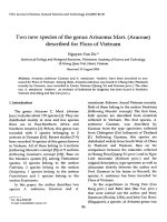
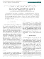
Fig. 1. Schematic representation of IDA electrodes.
Jolla, CA, USA. HPV-16-L1 peptide (311–325 sequence, i.e.
Asn–Leu–Ala–Ser–Ser–Asn–Tyr–Phe–Pro–Thr–Pro–Ser–Gly–Ser–
Met) was purchased from Genscript, USA. Secondary goat F(ab )2
anti-mouse Ig conjugated to horseradish peroxidase was purchased
from Tebu (Le Perray-en-Yvelines, France). Antibodies against HPV
(anti HPV, mouse anti-papillomavirus 16 L1 late protein, monoclonal antibody) and against Ovalbumin (anti-Ovalbumin,
anti-OVA) were obtained from AbD serotec (Morphosys, UK).
2.2. Interdigitated arrays fabrication
The interdigitated arrays (IDA) as shown in Fig. 1 were fabricated
on silicon substrate via lithography technique. Silicon wafers were
covered with a layer of SiO2 1 m thick by means of dry thermal
oxidation. The wafer was spin-coated with a layer of photoresist
AZ5214E (1 m thickness) and the shape of the electrodes was
defined by UV-photolithography. Then, chromium and platinum
were sputtered on the top of the wafer with the thickness of 50
and 500 nm, respectively. The working and counter electrodes were
patterned by a lift-off process (30 s in acetone solution with ultrasonic vibration). A second photolithographic step is carried out to
deposit the silver layer. Next, a 50 mM solution of FeCl3 (Merck)
was applied to the silver surface for 50 s at room temperature, followed by rinsing with DI water to define the reference electrodes.
The final diameter of the working electrodes was 500 m.
2.3. Electropolymerization of PANi–MWCNT film
The PANi–MWCNT film was electropolymerized on IDA using
cyclic voltammetry within the potential range from −0.2 to +1.0 V
(vs. SCE) with sweep rate of 50 mV/s, in a fresh solution containing
0.1 M ANi in 0.5 M H2 SO4 and 0.8 wt% MWCNTc (weight percent
with respect to ANi).
2.1. Chemical and biochemical reagents
N -(3-dimethylaminopropyl)-N-ethylcarbodiimide hydrochloride (EDC), N-hydroxysuccinimide (NHS) were provided by
Sigma. Aqueous solutions were made with DI water (18 M ).
Carboxylic mutilwall carbon nanotube (MWCNTc) was purchased from Shenzhen Nanotech Port Co., Ltd., China (purity
CNTs > 98%, out diameter: 10–20 nm, length: 5–15 m, carboxyl ratio: 2.31 wt%). Aniline (Merck, 99.5%) was distilled
under vacuum prior to polymerization. OVA (egg albumin, 2× crystallized) was purchased from Calbiochem, La
2.4. Peptide aptamer grafting and peptide–antibody reaction
conditions
For HPV-16-L1 grafting, 1.5 × 10⁻⁴ M EDC + 3 × 10⁻⁴ M NHS
were prepared with ultra-pure water. Then 50 nM HPV-16-L1 was
added. PANi–MWCNT electrodes were put into this solution during
2 h under stirring at 37 ◦ C. Afterwards, the electrode was rinsed in
water during 30 min under stirring at 37 ◦ C.
For immune reaction between the peptide aptamer and the antibody, a concentration range from 10 to 50 nM (in DI water) of
1562
L.D. Tran et al. / Talanta 85 (2011) 1560–1565
anti-HPV was used in our tests. The electrodes pre-modified with
HPV-16-L1 aptamer were left to react with anti-HPV for 15 min,
under stirring at 37 ◦ C, and then thoroughly washed in water under
stirring at the same temperature. As for blank experiment with
irrelevant antibodies (anti-OVA and/or anti- Keyhole Limpet Hemocyanin, anti-KLH) the same conditions were applied.
Voltammetric measurements were performed on AUTOLAB
PGSTAT 30 Electrochemical Analyser (EcoChemie, the Netherlands)
under the control of GPES software (ver. 4.9). The parameters for
CV: scan rate: 50 mV/s; potential range of −0.5 to 0.6 V vs. SCE. The
parameters for SWV were optimized as follows: frequency: 12.5 Hz;
start potential: −0.5 V; end potential: +0.5 V; step: 8 mV; amplitude: 25 mV. Prior to SWV measurements, the electrodes were held
for 120 s at the starting potential for conditioning. The SWV scans
were repeated until complete stabilization of the electrochemical signal (i.e., no difference observed between two successive
responses). All electrochemical experiments were conducted at
room temperature.
a
nd
th
2 => 20 cycle
200
150
Oxidation
100
I (μA)
2.5. Electrochemical measurements
250
50
0
Reduction
-50
-100
-150
-0,2
0,0
0,2
0,4
0,6
0,8
1,0
E (V)
250
b
PANi/CNT
PANi
200
3. Results and discussion
150
3.1. Electrochemical synthesis of PANi–MWCNT composite
3.2. PANi–MWCNT composite characterization
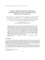
FE-SEM image revealed that PANi–MWCNT composite consists
of porous networks formed by MWCNT and PANi (Fig. 3a). Being
uniform and porous, this structure is well suitable for biocomponent immobilization.
The roughness of the surface of PANi–MWCNT composite was
characterized by using AFM techniques (Fig. 3b). AFM image
showed that the surface of composite was porous with roughness
about 0.33 m; this means the active area of composite was larger
than PANi films and absorption ability was increased.
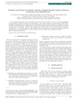
The FTIR spectrum of PANi–MWCNT composite (Fig. 4, solid
curve) presents benzenoid (B) and quinoid (Q) ring stretching bands
(C C) appeared at 1460 and 1612 cm−1 . The peaks at 1110 and
3415 cm−1 can be attributed to B N+ = Q stretching and –N–H
stretching vibrations respectively of PANi in the composite film. The
peak at 1702 cm−1 is unambiguously attributed to –COO− stretching vibration, clearly indicating the presence of carboxyl group
(–COOH) [22]. This fingerprint vibration of –COOH group is very
important for successful immobilization of HPV-16-L1 aptamer via
amine coupling, using the most common approach with aqueous
mixture of EDC/NHS groups to yield amine reactive esters. While
in the pure PANi the intensity of the quinonoid band is obviously
I /μA
100
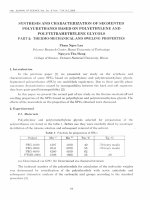
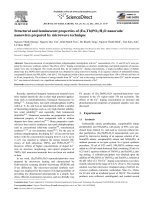
The cyclic voltammograms (CVs) recorded during 20-cycle synthesis of PANi–MWCNT are shown in Fig. 2a. The oxidation peaks
at about 0.16 V are related to the transformation of PANi from leucoemeraldine form (fully reduced state) to emeraldine salt (neutral
state). The small oxidation peaks at about 0.4 V are due to the
branched structure of PANi–MWCNT layers. The oxidation peaks
at about 0.62 V refer to the state transformation from emeraldine
to pernigraniline (fully oxidized state). The peak current increase
of the two main oxidation peaks at about 0.16 and 0.62 V indicates
that well conducting PANi film has been formed. It can be seen from
Fig. 2b that under the same experimental conditions, the current
peak of PANi–MWCNT was almost 4 times larger than that of pure
PANi after 20 cycle formation, which confirms well the role of CNT
in increasing composite conductivity as well as its surface area,
two main parameters that can significantly improve the overall
biosensor performance.
50
0
-50
-100
-150
-0.2
0.0
0.2
0.4
0.6
0.8
1.0
E (V)
Fig. 2. Electropolymerization of PANi–MWCNT (a) and CVs comparison of PANi film
with PANi–MWCNT composite during electropolymerization (b).
higher than that of benzenoid band (meaning that PANi is richer in
quinonoid unit (dotted line), i.e. Q/B > 1), in the PANi–MWCNT the
ratio of Q/B decreases (intensity of the quinonoid band is reduced),
which can be explained by the fact that MWCNT interacts strongly
with the conjugated structure of PANi, especially via quinonoid
unit.
3.3. HPV-16-L1 aptamer grafting on PANi–MWCNT composite
As discussed above, peptide aptamer, made of a few amino acids,
can bind antibodies with high affinity (Kd 106 –109 M) [18–23]. They
are commonly used in ELISA and biosensors as they are more stable,
safer to handle, more available than viral proteins or cells and moreover can be designed and synthesized on purpose. In our study,
HPV-16-L1 was grafted onto PANi–MWCNT coated IDA as described
in Section 2. The immobilization was a determinant step in the
electrochemical biosensor fabrication. HPV-16-L1 is a small peptide
with a molecular weight of 1825 Da therefore can be immobilized
without a significant hinderance of the electrode surface (i.e. does
not produce a complete surface blockage for ion transport into the
polymer film), providing that it is grafted at relatively low surface
densities.
Next, the antibody molecule (ca. 150 kDa for anti-HPV, as it is
an IgG one) is much bigger and more voluminous than HPV-16-L1
L.D. Tran et al. / Talanta 85 (2011) 1560–1565
1563
Fig. 3. FE-SEM image (a) and AFM image (b) of PANi–MWCNT film.
probe. The average surface occupied by one anti-HPV-16 antibody
molecule could be estimated as ca. 104 A˚ 2 [17]. Therefore, the surface of anti-HPV-16 molecules available to form a uniform blocking
layer on the polymer surface would be as low as ca. 15 pmol cm−2 .
On the basis of this estimation, HPV-16-L1 was grafted at low
surface density (i.e. at 50 nmol L−1 or lower, 50 nmol L−1 is the concentration than was normally used in spectrophotometric assays
[17]). This HPV-16-L1 density would warrant an efficient complete
immune reaction between HPV-16-L1 and anti-HPV-16. Effectively,
0.1 nmol L−1 HPV-16-L1 will induce negligible SWV signal after
grafting, whereas for 1 mol L−1 HPV-16-L1 and higher, a full surface blockage is achieved and subsequent complexation cannot be
detected by SWV (results not shown).
As shown in Fig. 5, the cyclic voltammogram shows two wave
pairs at −0.25 V/−0.28 V and +0.1 V/−0.01 V. As it is the faradic
component, which is relevant to characterize diffusion hindering,
square wave voltammetry has been advantageously used in the
following experiments, instead of classical cyclic voltammetry. The
SWV choice instead of CV is rationalized on its ability to reduce
capacitive current as well as the parasite current due to reduction
of dissolved oxygen (in SWV, the currents are sampled in both positive and negative pulses successively, furthermore, the registered
current is the subtraction between oxidation and reduction currents, thus current density in SWV’s are higher that those in CVs,
recorded for the same electrode [24]).
3.4. Electrochemical detection of HPV-16-L1 aptamer:anti HPV
complex
SWV obtained before and after grafting of HPV-16-L1 were
clearly shown in Fig. 6 (curve 1 and curve 2, respectively). Further,
with the use of SWV we could demonstrate the presence of complex
formation between HPV-16-L1 aptamer and its specific (relevant)
anti-HPV. As expected, formation of the HPV-16-L:anti-HPV-16
complex induces significant current drops (Fig. 6, curves 3–7,
depending on added anti-HPV-16 concentration). Furthermore, it is
possible to perform decomplexation followed by re-complexation
1564
L.D. Tran et al. / Talanta 85 (2011) 1560–1565
65
2.6x10
-4
2.4x10
-4
2.2x10
-4
2.0x10
-4
1.8x10
-4
1.6x10
-4
1.4x10
-4
60
1280
I /A
1110
1460
50
1702
1612
Transmittance (%)
55
45
40
35
30
3415
Pure PANi
PANi-MWCNT
25
0
4000
3500
2000
1500
1000
500
-1
Wavenumber (cm )
20
40
60
80
Anti-HPV-16 concentration /nM
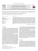
Fig. 7. The response curves of immunosensor with anti-HPV-16 concentration range
from 0 to 80 nM.
Fig. 4. FTIR spectra of PANi and PANi–MWCNT composite.
5.0x10
I /A
0.0
-5.0x10
-4
3.0x10
-4
2.5x10
-4
2.0x10
-4
1.5x10
-4
1.0x10
-4
5.0x10
-5
(1)
(2)
-5
I /A
1.0x10
3.5x10
-4
-5
(3)
0.0
-1.0x10
-1.5x10
-4
-5.0x10
-5
-1.0x10
-4
-0.5
-4
-0.6
-0.4
-0.2
0.0
0.2
0.4
0.6
0.8
(1) + EDC/NHS
(2)+ HPV-16-L1 aptamer
(3)+ Anti-OVA
-0.4
-0.3
-0.2
-0.1
0.0
0.1
0.2
0.3
0.4
0.5
E /V vs. Ag/AgCl
E /V vs. Ag/AgCl
Fig. 8. SWV of PANi–MWCNT IDA recorded in HCl 0.1 M after treatment with
EDC/NHS (curve 1), after grafting of 5 × 10⁻⁸ M HPV-16-L1 (curve 2) and after complexation with 5 × 10⁻⁸ M anti-OVA (curve 3).
Fig. 5. Electroactive CV of PANi–MWCNT composite in 0.1 M HCl. v = 50 mV/s.
and so on for at least 5 times, thus indicating the reversibility of
Ag–Ab interaction as well as the robustness of this IDA based arrays.
To evaluate the analytical performance of above IDA arrays, a
calibration curve was done for a series of anti-HPV-16 concentra3.5x10-4
(1)
3.0x10⁻⁴
2.5x10⁻⁴
I /A
2.0x10⁻⁴
1.5x10⁻⁴
(7)
-4
(1) + EDC/NHS
(2) + HPV-16-L1 aptamer
(3) + 10nM anti-HPV-16
(4) + 20nM anti-HPV-16
(5) + 30nM anti-HPV-16
(6) + 40nM anti-HPV-16
(7) + 50nM anti-HPV-16
1.0x10
-5
5.0x10
0.0
-5.0x10⁻⁵
-1.0x10⁻⁴
-0.5
-0.4
-0.3
-0.2
-0.1
0.0
0.1
0.2
0.3
0.4
tion ranging from 10 to 80 nM. As it can be seen, the signal tends to
saturation for concentrations above 80 nM of target, as expected
according to above estimation for antigen and antibody densities. Assuming a linear behavior at low target concentrations the
electrochemical assays showed a sensitivity of 1.75 ± 0.2 A nM−1
(r2 = 0.997) in the range of 10–50 nM with LOD of 490 pM, respectively (Fig. 7).
Control experiments were also performed to confirm whether
above signal decrease was really come from true complexation but
not any other interfering phenomena like non-specific adsorption
or signal instability. Thus, blank experiments were carried out with
an irrelevant antibody directed against OVA (anti-OVA), whose
molecular weight was 400 kDa, i.e. bigger than that of anti-HPV16. As shown in Fig. 8, no complex formation (no signal drop) was
observed for anti-OVA and HPV-16-L1. Another unspecific antibody
(anti-KLH) has also shown the same results (figure not shown). In
summary these experiments demonstrated clearly that the signal
change was due to specific recognition by anti-HPV-16 antibody
when complexing with HPV-16-L1 peptide aptamer.
0.5
E /V vs. Ag/AgCl
Fig. 6. SWV of PANi–MWCNT IDA recorded in HCl 0.1 M after treatment with
EDC/NHS (curve 1), after grafting of 5 × 10⁻⁸ M HPV-16-L1 (curve 2) and after complexation with 10–50 nM of anti-HPV-16 (curves 3–7).
4. Conclusion
Analytical performance of PANi–MWCNT based IDA arrays was
evaluated. The assays showed a sensitivity of 1.75 ± 0.2 A nM−1
L.D. Tran et al. / Talanta 85 (2011) 1560–1565
(r2 = 0.997) in the range of 10–50 nM of anti-HPV with LOD of
490 pM. With concentration of or above 80 nM the SWV signal
tends to saturation, as expected according to the theoretical estimation for antigen peptide aptamer (Ag) and Ab densities on the
electrode surface. Control experiments with irrelevant antibodies
(anti-OVA and anti-KLH) also confirmed that the signal decrease
was really come from true complexation between Ag and Ab but
not any other interfering phenomena like non-specific adsorption
or signal instability.
One powerfully advantageous aspect of our arrays is the ability
to array multiple copies of the same probes (to control for technical variability) as well as to array multiple probes against the same
target on each electrode of IDA (to control for biological variability). With the functional conducting PANi–MWCNT immobilized
Ag peptide aptamers as affinity capture reagent the concept of
the reagentless electrochemical immunoarrays was proposed. As
for the transduction scheme, it can proposed that the specific formation of Ag/Ab complex could be detected via change in signal
transduction due to a steric hindrance, intervening in ion transport rate at the polymer–solution interface and therefore affects
the redox behavior of PANi–MWCNT composite. Further work will
be required to determine whether above described assays offer
sufficient sensitivity and specificity for clinical use.
Acknowledgements
Funding of this work was mainly provided by Vietnam National
Foundation for Science and Technology Development NAFOSTED
grant (code 104.03-2010.60). Additional logistic support was also
provided from MOST grant (code 59/2615/2010/HÐ-NÐT). We also
acknowledge Prof. M.C. Pham and B. Piro (ITODYS, University of
Paris Diderot, France) for initial suggestion and invaluable discussion regarding HPV choice as a model for Ab-Ag interaction study.
The technical support of IMS-VAST key laboratory was critical for
development and characterization of IDA.
References
[1] J. Ferlay, H.R. Shin, F. Bray, D. Forman, C. Mathers, D.M. Parkin, Estimates of
worldwide burden of cancer in 2008: GLOBOCAN 2008, International Jounal of
Cancer 127 (2010) 2893–2917.
[2] H.H. Handsfield, Clinical presentation and natural course of anogenital warts,
The American Journal of Medicine 102 (1997) 16–20.
˜
[3] F.X. Bosch, M.M. Manos, N. Munoz,
M. Sherman, A.M. Jansen, J. Peto, M.H. Schiffman, V. Moreno, R. Kurman, K.V. Shan, Prevalence of human papillomavirus
in cervical cancer: a worldwide perspective, Journal of the National Cancer
Institute 87 (1995) 796–802.
[4] C.E. Greer, C.M. Wheeler, M.B. Ladner, K. Beutner, M.Y. Coyne, H. Liang, A. Langenberg, T.S. Yen, R. Ralston, Human papillomavirus (HPV) type distribution
and serological response to HPV type 6 virus-like particles in patients with
genital warts, Journal of Clinical Microbiology 33 (8) (1995) 2058–2063.
1565
[5] S. Laurensona, M.R. Petta, K. Hoppe-Seylerb, C. Denkb, F. Hoppe-Seylerb, N.
Colemana, P. Ko Ferrigno, Development of peptide aptamer microarrays for
detection of HPV-16 oncoproteins in cell extracts, Analytical Biochemistry 410
(2011) 161–170.
[6] M. Steben, E.D. Franco, Human papillomavirus infection: epidemiology and
pathophysiology, Gynecologic Oncology 107 (2007) S2–S5.
[7] E.F. Dunne, E.R. Unger, M. Sternberg, G. McQuillan, D.C. Swan, S.S. Patel,
L.E. Markowitz, Prevalence of hpv infection among females in the United
States, The Journal of the American Medical Association 297 (2007)
813–819.
[8] A.R. Kreimer, G.M. Clifford, P. Boyle, et al., Human papillomavirus types in head
and neck squamous cell carcinomas worldwide: a systematic review, Cancer
Epidemiology, Biomarkers & Prevention 14 (2005) 467–475.
[9] J. Dillner, The serological response to papillomaviruses, Seminars in Cancer
Biology 9 (1999) 423–430.
[10] S. Nagao, M. Yoshinouchi, Y. Miyagi, A. Hongo, J. Kodama, S. Itoh, T. Kudo, Rapid
and sensitive detection of physical status of human papillomavirus type 16
DNA by quantitative real-time PCR, Journal of Clinical Microbiology 40 (2002)
863–867.
[11] S.D. Vernon, D.H. Farkas, E.R. Unger, V. Chan, D.L. Miller, Y.P. Chen, G.F. Blackburn, W.C. Reeves, Bioelectronic DNA detection of human papillomaviruses
using eSensorTM : a model system for detection of multiple pathogens, BMC
Infectious Diseases 3 (2003) 12.
[12] N. Zari, A. Amine, M.M. Ennaji, Label-free DNA biosensor for electrochemical
detection of short dna sequences related to human papilloma virus, Analytical
Letters 42 (2009) 519–535.
[13] L. Civit, A. Fragoso, C.K. O’Sullivan, Electrochemical biosensor for the multiplexed detection of human papillomavirus genes, Biosensors & Bioelectronics
26 (2010) 1684–1687.
[14] L. Yang, X. Ren, F. Tang, L. Zhang, A practical glucose biosensor based on Fe3 O4
nanoparticles and chitosan/nafion composite film, Biosensors and Bioelectronics 25 (2009) 889–895.
[15] J. Huang, G. Yanga, W. Meng, L. Wu, A. Zhu, X. Jiao, An electrochemical impedimetric immunosensor for label-free detection of Campylobacter
jejuni in diarrhea patients’ stool based on O-carboxymethylchitosan surface modified Fe3O4 nanoparticles, Biosensors and Bioelectronics 25 (2010)
1204–1211.
˜
˜ J.M. Pingarrón, J. Riu, F. Xavier Rius, Electrochemical sens[16] P. Yánez-Sede
no,
ing based on carbon nanotubes, Trends in Analytical Chemistry 29 (2010)
939–953.
[17] B. Piro, A. Kapella, V.H. Le, G. Anquetin, Q.D. Zhang, S. Reisberg, V. Noel, L.D.
Tran, H.T. Duc, M.C. Pham, Detection of human papilloma virus (HPV) infection
by reagentless electrochemical peptide biosensor, Electrochimica Acta, (2011),
doi:10.1016/j.electacta.2011.04.094, in press.
[18] P.J. Conroy, S. Hearty, P. Leonard, R. O’Kennedy, Antibody production, design
and use for biosensor-based applications, Seminars in Cell & Developmental
Biology 20 (2009) 10–26.
[19] E. Katz, I. Willner, Probing biomolecular interactions at conductive and
semiconductive surfaces by impedance spectroscopy: routes to impedimetric immunosensors, DNA-sensors, and enzyme biosensors, Electroanalysis 15
(2003) 913–947.
[20] P. Holliger, P.J. Hudson, Engineered antibody fragments and the rise of single
domains, Nature Biotechnology 23 (2005) 1126–1136.
[21] S. Wang, E.S. Humphreys, S.Y. Chung, D.F. Delduco, S.R. Lustig, H. Wang, K.N.
Parker, N.W. Rizzo, S. Subramoney, Y.M. Chiang, A. Jagota, Peptides with selective affinity for carbon nanotubes, Nature Materials 2 (2003) 196–200.
[22] M. Yemini, M. Reches, Ehud Gazit, Judith Rishpon, Peptide nanotube-modified
electrodes for enzyme-biosensor applications, Analytical Chemistry 77 (2005)
5155–5159.
[23] P. Samuelson, E. Gunneriusson, P.A. Nygren, S. Ståhl, Display of proteins on
bacteria, Journal of Biotechnology 96 (2002) 129–154.
[24] A.J. Bard, L.R. Faulkner, Electrochemical Methods: Fundamentals and Applications, 2001.