DSpace at VNU: Molecular Cloning, Expression of minD Gene from Lactobacillus acidophilus VTCC-B-871 and Analyses to Identify Lactobacillus rhamnosus PN04 from Vietnam Hottuynia cordata Thunb.
Bạn đang xem bản rút gọn của tài liệu. Xem và tải ngay bản đầy đủ của tài liệu tại đây (433.88 KB, 6 trang )
Indian J Microbiol
DOI 10.1007/s12088-013-0384-1
ORIGINAL ARTICLE
Molecular Cloning, Expression of minD Gene from Lactobacillus
acidophilus VTCC-B-871 and Analyses to Identify Lactobacillus
rhamnosus PN04 from Vietnam Hottuynia cordata Thunb.
Tu Hoang Khue Nguyen • Vinh Thi Thanh Doan
Ly Dieu Ha • Huu Ngoc Nguyen
•
Received: 31 July 2012 / Accepted: 28 February 2013
Ó Association of Microbiologists of India 2013
Abstract The minD gene encoding an inhibitor cell
division MinD homolog from Lactobacillus acidophilus
VTCC-B-871 was cloned. We showed that there were
97 % homology between minD genes of L. acidophilus
VTCC-B-871 and Lactobacillus rhamnosus GG and Lactobacillus rhamnosus Lc705. Based on the analysis of the
DNA sequence data from the L. rhamnosus genome project
and sequenced minD gene of L. acidophilus VTCC-B-871,
a pair of primers was designed to identified the different
minD genes from L. acidophilus ATCC 4356, L. rhamnosus ATCC 11443. Besides, the polymerase chain reaction
product of minD gene was also obtained in L. rhamnosus
PN04, a strain was isolated from Vietnamese Hottuynia
cordata Thunb. In addition, we performed a phylogenetic
analysis of the deduced amino acid sequence of MinD
homologs from L. acidophilus VTCC-B-871 with the other
strains and compared the predicted three-dimension structure of L. acidophilus VTCC-B-871 MinD with Escherichia coli MinD, there are similarity that showed evolution
of these strains. The overexpression of L. acidophilus
VTCC-B-871 MinD in E. coli led to cell filamentation in
IPTG and morphology changes in different sugar stresses,
interestingly. The present study is the first report characterizing the Lactobacilus MinD homolog that will be useful
in probiotic field.
T. H. K. Nguyen (&) Á V. T. T. Doan Á H. N. Nguyen
School of Biotechnology, International University, Hochiminh
City National University, Quarter 6, Linh Trung Ward, Thu Duc
District, Hochiminh City, Vietnam
e-mail:
L. D. Ha
Department of Reference Substances, Institute for Drug Quality
Control, Hochiminh City, Vietnam
Keywords Cell division inhibitor Á Morphology change Á
Lactobacillus Á Comparative analyses Á Hottuynia cordata
Thunb.
Introduction
In Escherichia coli, the proper placement of the cell division site generates two equally sized daughter cells which
are maintained by the MinC, MinD and MinE proteins
encoded by the min locus. In this system, the MinC and
MinD protein acts as the inhibitors of cell division by
blocking septum formation at all potential division sites
(polar and mid sites) [1]. The MinE protein gives topological specificity to the MinCD division inhibitors by
restricting its activity to polar division sites, thus ensuring
that separation is limited to the proper division site at
midcell. MinE binds to the trailing edge of MinD and
stimulating its ATP hydrolysis, which results in the realease of MinD, and thus MinC and MinE, from the membrane [2–4]. In the other hand, Bacillus subtilis contains
MinCD homologues and DivIVA acts topologically, but
not MinE [5]. It was also noticed that the entire nucleotide
sequences of the Streptomyces genomes of Streptomyces
coelicolor and Streptomyces avermitilis have recently
reported, and this strain carried the MinD homolog, but not
MinC or MinE [6, 7]. The MinD homolog harbored by
Streptomyces lavendulae ATCC25233 has also been characterized and this strain did not carry MinC and MinE [8].
Since the genus Streptomyces consists of filamentous
bacteria, minD in S. lavendulae ATCC 25233 may have a
role other than cell division.
Lactic acid bacteria are one of the most commonly used
probiotics. The role of prebiotics in improving human
health has attracted global attention and the research is
123
Indian J Microbiol
mostly focused on the strains belonging to Lactobacillus
[9]. The survival of Lactobacillus probiotics was usually
lower than the amounts noted in probiotic label in their
products. Therefore, to find out the roles of Min system in
Lactobacillus that made a wide genus with the different
survival rates that relate to the cell division are necessary.
From the stated reasons, we cloned and tested whether the
Lactobacillus acidophilus MinD protein is functional in
E. coli cells by overexpression. By analysis the minD gene
from L. acidophilus VTCC-B-871, the minD genes from
L. acidophilus ATCC 4356 and L. acidophilus ATCC
11443 Lactobacillus rhamnosus PN04, a strain isolated and
identified from Vietnamese Hottuynia cordata Thunb. were
identified from which a method for determination of
L. acidophilus and L. rhamnosus will be applied so far.
Materials and Methods
Plasmids, Bacterial Strains, Growth Conditions
The pUC19 and pGEM-T vectors used for molecular
cloning and E. coli JM109, BL21(DE3)pLysS were purchased by Promega. The pET28 (a?) used for overexpression was purchased by Novagen. L. rhamnosus GG,
L. rhamnosus ATCC 11443, L. acidophilus ATCC 4356,
L. acidophilus VTCC-B-871 purchased by Vietnam type
culture collection (VTCC). E. coli JM109 was used as a
host to clone Lactobacillus minD genes. E. coli
BL21(DE3)pLysS was used as an expression strain. Lactobacillus strains were grown on MRS for 72–96 h at
30 °C. E. coli strains were grown in Luria–Bertani for
18–24 h at 37 °C with shaking at 200 rpm. When required,
antibiotics were added to media in the following concentrations: 100 lg of ampicillin/ml, 10 lg of chloramphenicol/ml, 50 lg of kanamycin/ml for E. coli.
DNA and RNA Isolation
Genomic DNA was isolated from Lactobacillus strains that
had been grown for 72–96 h in MRS. The samples were
incubated in MRS according to standard protocols. Total
RNA was purified according to manufacturer’s instructions
(Takara).
Isolation of the Homologous DNA minD Probe
from Lactobacillus rhamnosus GG
Genomic DNA from L. rhamnosus GG was amplified by
PCR using a sense primer OMR1(50 -GAATGCGACCGGG
GCGGCTGACGGTGCGA-30 ) and an anti-sense primer
OMR2 (50 -TCAACGGCACGCTATCACCTAGTAACCG
GC-30 ) which was homologous to sequences between 391
123
and 739 nt of the minD gene (Gene ID: 8422477). The PCR
was done under the following conditions: an initial 2 min
at 95 °C; then, 29 cycles of 1 min at 95 °C followed by
30 s at 55 °C; and 30 s at 72 °C, finally, an extension
period of 30 s at 72 °C. A PCR product of 349 bp corresponding to minD fragment was ligated into the pGEM-T
vector and introduced into E. coli JM109 from the TA
Cloning kit (Promega).
Cloning, Sequencing and DNA Analysis
The genomic DNA from L. acidophilus VTCC-B-871 strain was
digested with restriction enzymes supplied by Takara (Japan).
The digestion was followed as instructions of the company.
Southern hybridization was performed by using a Hybond-N?
(Amersham Biosciences) membrane. Probe labeling, hybridization and detection were performed with AlkPhos Direct
Labeling and Detection System (Amersham Biosciences)
according to the protocol supplied by the manufacturer.
The cloning minD from L. acidophilus was performed
[10]. DNA sequencing was performed with the ABI
PRIZM 310 genetic analyzer using the BigDye terminator
cycle sequencing ready reaction kit according to the
manufacturer’s protocols. The Lactobacillus minD genes
was determined and analyzed using Fasta. The protein
molecular mass, pI were calculated on an ExPASy Proteomics Server. The sequence data obtained in this study
has been submitted to the DDBJ.
Overexpression Studies of MinD and Light Microscopy
The L. acidophilus minD was amplified by PCR with a
sense primer BHE1 (50 -CATATGGGGACAGCGTTAGT
AGTGACTTC-30 ) (the NdeI site is underlined) and an
antisense BHE2 (50 -CTCGAGGATGGCGATGGAACAA
TTTTGAC-30 ) (the XhoI site is underlined). The amplified
minD was subcloned into pGEM-T vector and then was
checked by DNA sequencing. The minD fragment was cut
out from pGEM-T vector by NdeI and XhoI doubledigestion and inserted into the same sites of pET-28(a?) to
produce pET-28(a?)/minD. E. coli BL21(DE3)pLysS
transformed with pET-28(a?)/minD was grown in LB
medium supplemented with appropiate antibiotics at 37 °C
to OD600 = 0.5, after which 0.5 mM IPTG or 1 % glucose, 1 % saccharose, 1 % manitol was added to culture to
induce at 28 °C from 5 to 24 h. Light microscopy was used
to observed the morphological changes in E. coli.
Isolation and Identification of Lactobacillus rhamnosus
from Hottuynia Cordata Thunb.
Hottuynia Cordata Thunb. samples were collected in the
Southern of Vietnam. No specific permits were required for
Indian J Microbiol
the described field studies. The leaves were incubated in
MRS for 72–96 h at 30 °C. The culture was used to spread
onto MRS agar that was incubated in MRS for 72–96 h at
30 °C. The purified colonies were tested by microscopic
examination with gram stain and catalase negative [11].
The isolated strains were identified by biochemical characterization based on the ability of the isolates to utilize
different carbon sources, which determined by API CHL
50 system (bioMe´rieux, Lyon, France) and 16S rRNA
sequencing analysis.
Phylogenetic Analyses, Protein Homology Modelling
and Analysis
Phylogenetic analyses were performed on the MinD
deduced amino acid sequences that were previously
reported. Protein sequences were aligned with ClustalW
software and clustered by using the un-weighted pair group
method for the arithmetic mean. The tertiary structures of
the deduced amino acid sequences of MinD were predicted
by homology modelling using the Swiss-Model Server [12,
13] and MinD from E. coli (PDB: 3q9l) was used as
template. The structural parameters and prediction quality
of the modeled structures were evaluated using the program QMEAN4 with respect to score obtained for
high-resolution experimental structures solved by X-ray
crystallography [14].
Results and Discussion
Analysis of Genomic DNA of Lactobacillus
acidophilus VTCC-B-871
L. rhamnosus GG genome was used as a template to prepare a 349 bp-DNA probe for Southern blotting and colony
hybridization. A 4.0 kb HindIII-PvuII fragment was cloned
from L. acidophilus VTCC-B-871 chromosomal DNA.
A minD gene of 798 bp was sequenced and was deposited
in the DDBJ database under accession no. AB725356. The
MinD protein encoded by L. acidophilus VTCC-B-871
consists of 265 amino acids with a calculated pI of 5.07 and
Mw of 28857.17 kDa. Having 100 % identity, the protein
exhibits highest similarity to MinD (EHJ35458) from
L. rhamnosus ATCC 21052 and 99 % identity to MinD
(YP_00317015, gene ID: 8422477) from L. rhamnosus
GG.
By comparison of nucleotide sequences of the minD
gene from L. acidophilus VTCC-B-871 with L. rhamnosus
strains, there are 779/798 (98 %) identity to L. rhamnosus
ATCC 8530 and 777/798 (97 %) identity to Lc 705 and
strain GG. Interestingly, the analyses the nucleotide
sequence showed that there was 100 % similarity of the 40
nt at 50 end and 40 nt at 30 end of minD gene in L. acidophilus VTCC-B-871 and L. rhamnosus ATCC 8530, Lc
705 as well as GG. Clearly, minD gene will be identified
easily and used in the identification of L. acidophilus or
L. rhamnosus.
Conservation of MinD Proteins from Various Bacteria
and Phylogenic Analysis
Figure 1. shows the alignment of amino acid sequences of
MinD proteins from Lactobacillus strains with 15 strains
listed in Fig. 1. The Walker A and B motifs and the two
Asp residues (Asp38, Asp40) located between them are
conserved in all sequences, suggesting that the Lactobacillus MinD protein possess an ATPase activity like that of
E. coli MinD. Although L. acidophilus and L. rhamnosus
strains are gram positive, the consensus nucleotide binding
sequence of the Walker A motif is known as G/A-X-X-GX-G-K-T/S that overlap with Walker A in gram negative
E. coli, and Neiserria gonorrhoea [15] and that is distinguishable with Streptomyces which are gram-positive and
the seventh Lys is replaced by Ala or Thr [8]. The results
elucidated the relationship of minD from gram negative
and gram positive. The protein or deduced amino acid
MinD sequences from 15 strains were used to generate
phylogenetic trees. Clustal alignment used for phylogenetic
analysis allowed to determine the location of amino acids
expected to have a catalytic role in MinD (Fig. 1). The
combination with the tree analysis showed the early separation between amino acid sequences from the aligned
strains can be explained in terms of the evolution of MinD
for different purposes (Fig. 2). On this background, the
results of the phylogenetic analyses based both on amino
acid sequence similarities as well as their structural features would be strengthen the phylogenetic analysis and to
establish a relationship between the genes encoding MinD
with their three-dimensional structures involved in ATP
binding (Fig. 2). By the comparison, the MinC and MinE
homologs might have been eliminated in the process of the
evolution.
Protein Homology Modelling and Comparisons
of Protein Structures
Once the tertiary structure of MinD was predicted, these
results strongly support the notion that there is a close
relationship between the tertiary structure of MinD and the
lifestyle of the microorganisms. Comparative analyses of
three-dimensional structures have been utilized for different purposes in searching for putative biological functions,
drug design, protein–protein interaction studies [14].
However, to our knowledge, the study that uses a comparative analysis of protein structure in combination with a
123
Indian J Microbiol
Fig. 1 Alignment of conserved motifs in MinD from Lactobacillus
strains and other species. Alignment was carried out with the Clustal
W program. Listed proteins are from the following strains: L. wel,
Listeria welshimeri; B. sub, Bacillus subtilis; L. san, Lactobacillus
sanfranciscensis; L. aVT1, Lactobacillus acidophilus VTCC-B-871,
L. rha, Lactobacillus rhamnosus; L. cas, Lactobacillus casei, E. amyl,
Erwinia amylovora; E. pyr, Erwinia pyrifoliae; P. ana, Pantoea
ananatis; E. coli, Escherichia coli; N. gor, Neisseria gonorrhoeae; S.
cel, Sorangium cellulosum; R. le, Rhizobium leguminosarum; W. suc,
Wolinella succinogenes; S. coe, Streptomyces coelicolor. The
conserved motifs (Walker A and B) and the two Asp residues are
indicated by bars and arrow heads, respectively. Asterisks below the
sequences show the conserved residues in all sequences
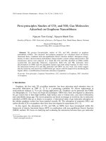
Fig. 2 The phyogenic tree was
used by the un-weighted pair
group method using the
arithmetic mean and clustering
the three-dimensional structures
of MinD. a and c Threedimensional structure of MinD
from Lactobacillus acidophilus
VTCC-B-871 and S. coelicolor
respectively, predicted by
homology modelling using the
Swiss-Model Server. b Threedimensional structure of MinD
from E. coli (PDB: 3q9l)
phylogenetic analysis to explore the evolution of lifestyle.
Using Swiss-model server, the structure of L. acidophilus
was predicted, using the template of E. coli (Fig. 2) with
the final total energy of -8877.295 kJ/mol. The quality of
the structure prediction was estimated by QMEAN4
(Table 1). The structures showed the alpha helix and beta
123
sheet in L. acidophilus MinD and E. coli MinD occuring in
the region of 2–249 amino acids of L. acidophilus (Fig. 2)
with the ligand models of 2 ATP and 1Mg2? molecules
while Streptomyces MinD showed modelling homology in
the region of 149–245 amino acids with no ligand model in
this structure that corresponds to the sequences aligned in
Indian J Microbiol
Table 1 QMEAN4 data for model quality estimation
Scoring function term
Raw score
C-beta interaction energy
-138.89
All-atom pairwise energy
-6408.44
Z-score
0.13
-0.7
Solvation energy
-26.27
-0.06
Torsion angle energy
-55.07
-0.85
QMEAN4 score
0.721
L. rhamnosus NT10. The isolated strain was named
L. rhamnosus PN04 and the 16S rRNA sequence was
deposited in DDBJ (accession number: AB738399). Using
the OMS sense and OMS1 antisense primers, a PCR
product of minD gene was detected, indeed (Fig. 3). The
results also pointed the relationship between L. acidophilus
and Lactobacillus rhamnosus.
-0.90
Overexpression of minD Gene and Light Microscopy
the Fig. 1. Although E. coli MinD exhibited a dimer that
indicated the self-interaction [4] and L. acidophilus MinD
predicted in monomer, the monomers of these structures
are highly similar.
The results meant the comparative analysis can be an
important tool for studying the proteins of microorganisms
but also for the evolution of microorganisms and their
proteins, since structural differences may reflect other
important properties such as substrate specificity and others
that can not be inferred from the analysis of amino acid
sequences only. Therefore, minD gene might also participate in the evolution in microorganisms.
Study the minD Genes in Lactobacillus acidophilus
ATCC 4356 and Lactobacillus rhamnosus ATCC
11443
To find out the relation, the MinD homologs were identified in L. acidophilus ATCC 4356 and L. rhamnosus ATCC
11443. As discussed above, a pair of primers with a sense
primer OMS (50 -ATGGGGACAGCGTTAGTAGTGACT
TC-30 ) and an antisense OMS1 (50 -GATGGCGATGGAAC
AATTTTGAC-30 ) was designed from L. acidophilus VT
CC-B-871 to identify minD gene in L. acidophilus ATCC
4356 and L. rhamnosus ATCC 11443. After isolation, a
minD gene from L. acidophilus ATCC 4356 was sequenced.
A minD gene of 798 bp was sequenced and deposited in the
DDBJ under accession no. AB725355. Similarity, a minD
gene of 798 bp from L. rhamnosus ATCC 11443 was
deposited in the DDBJ under accession no. AB725357. With
the understanding of minD in sequences, the role of the
survival of different strains will be discovered.
To test whether the L. acidophilus MinD protein is functional in E. coli cells, the E. coli BL21(DE3)plysS was
introduces with plasmid pET-28(a?) containing minD.
After expression, cells transformed with the pET-28(a?)
exhibited the normal rod-shaped morphology (Fig. 4a),
while the same strain transformed with pET-28(a?)/minD
exhibited a mixed phenotype of long filaments (Fig. 4b).
The result of filamentous phenotype may have occurred
because Lactobacillus MinD enhanced MinC-mediated
inhibition of cell division at all potential division sites in
E. coli cells. Indeed, it has also been reported that the
overexpression of Neissheria MinD in E. coli cells leads to
filamentation [15]. This result indicated that Lactobacillus
MinD is functional across species. The cells transformed
with the pET-28(a?)/minD were also tested to grow in
glucose, saccharose and manitol. Interestingly, under the
saccharose stress, the cells become long and curled shape
(Fig. 4c). The IPTG inducer was used in pET vector system
because of T7 promoter. However, under the sugar stresses,
the morphology was changeable. The hypothesis was posed
whether the interaction between MinD and sugar. The
Isolation and Identification of Lactobacillus rhamnosus
from Hottuynia Cordata Thunb.
To make a sure of the minD existence and aid for identification, an isolation of L. rhamnosus from H. cordata
Thunb. was done and checked by biochemical tests using
ABI 50CHL and 16S rRNA sequencing. By using the API
50CHL (BioMerieux), a isolated strain showed the result of
L. rhamnosus (Data not shown). By Blast search, the 16S
rRNA sequence of L. rhamnosus shows 99 % identity to
Fig. 3 The PCR product of minD gene from isolated Lactobacillus
rhamnosus. From left to right: 1, k/HindIII marker; 2, PCR product.
The arrow shows the PCR product
123
Indian J Microbiol
Fig. 4 Morphology of Escherichia coli harboring the minD gene
from Lactobacillus acidophilus. Escherichia coli BL21(DE3)plysS
cells harboring pET 28(a?)/minD and pET 28(a?) were analyzed by
light microscopy. a Escherichia coli BL21(DE3)plysS cells harboring
pET 28(a?). b Escherichia coli BL21(DE3)plysS cells harboring pET
28(a?)/minD in IPTG. c Escherichia coli BL21(DE3)plysS cells
harboring pET 28(a?)/minD in saccharose. The scale bar is 5 lm
results were the first reports in the morphological differentiation of E. coli carrying minD gene of Lactobacillus.
Harper D et al (2002) Compete genome sequence of the model
actinomycete Streptomyces coelicolor A3(2). Nature 417:
141–147
Ikeda H, Ishikawa J, Hanomoto A, Shinose M, kikuchi H, Shiba
T, Sakaki Y, Hattori M, Omura S (2003) Complete genome
sequence and comparative analysis of the industrial microorganism Streptomyces avermitilis. Nat Biotechnol 21:526–531
Nguyen HKT, Kumagai T, Matoba Y, Suzaki T, Sugiyama M
(2008) Molecular cloning and functional analysis of minD gene
from Streptomyces lavendulae ATCC 25233. J Biosci Bioeng
106(3):303–305
Chithra M, Muralikrishna G (2012) Prebiotic activity of purified
xylobiose obtained from ragi (Eleusine coracana, Indaf-15) Bran.
Indian J Microbiol 52(2):251–257
Sambrook J, Russell DW (2001) Molecular cloning: a laboratory
manual, 3rd edn. Cold Spring Harbor Laboratories, New York
Seema P, Avishek M, Arun G (2012) Potentials of exopolysaccharides from lactic acid bacteria. Indian J Microbiol 52(1):3–12
Arnold K, Bordoli L, Kopp J, Schwede T (2006) The swiss-model
workspace: a web-based environment for protein structure
homology modeling. Bioinformatics 22:195–201
Schwede T, Kopp J, Guex N, Peitsch MC (2003) Swiss-model: an
automated protein homology-modeling server. Nucleic Acids Res
31:3381–3385
Benkert P, Biasini M, Schwede T (2011) Toward the estimation
of the absolute quality of individual protein structure models.
Bioinformatics 27(3):343–350
Parti RP, Biswas D, Helgeson S, Michael FS, Cox A, Dillon JA
(2011) Attenuated virulence of min operon mutants of Neisseria
gonorrhoeae and their interactions with human urethral epithelial
cells. Microbes Infect 13(6):545–554
Acknowledgments Thanks to the grant supplied by the National
Foundation of Science and Technology Development of Vietnam
(Nafosted) and the support of Hochiminh City International University by which this work has been fullfiled.
7.
8.
References
9.
1. Park KT, Wu W, Lovell S, Lutkenhaus J (2012) Mechanism of
the asymetric activation of the MinD ATPase by MinE. Mol
Microbiol 85(2):271–281
2. Zhou H, Schulze R, Cox S, Saez C, Hu Z, Lutkenhaus J (2005)
Analysis of minD mutations reveals residues required for minE
stimulation of the minD ATPase and residues required for minC
interaction. J Bact 187(2):629–638
3. Ma L, King GF, Rothfield L (2004) Positioning of the MinE
binding site on the MinD surface suggests a plausible mechanism
for activation of the Escherichia coli MinD ATPase during
division site selection. Mol Microbiol 54(1):99–108
4. Lutkenhaus J, Sundaramoorthy M (2003) MinD and role of the
deviant Walker A motif, dimerization and membrane binding in
oscillation. Mol Microbiol 48(2):295–303
5. Stahlber H, Kutejova E, Muchova K, Gregorini M, Lustig A,
Muller SA, Olivieri V, Engel A, Wilkinson AJ, Barak I (2004)
Oligomeric structure of Bacillus subtilis cell division protein
DivIVA determined by transmission electron microscopy. Mol
Microbiol 52(5):1281–1290
6. Bently SD, Chater KF, Cerdeno-Ta´rraga AM, Challis GL,
Thomson NR, James KD, Harris DE, Quail MA, Kieser H,
123
10.
11.
12.
13.
14.
15.