Toxicological effect and histopathological changes in gills and liver of tilapia (oreochromis niloticus) exposed to niclosamide
Bạn đang xem bản rút gọn của tài liệu. Xem và tải ngay bản đầy đủ của tài liệu tại đây (1.09 MB, 40 trang )
THAI NGUYEN UNIVERSITY
UNIVERSITY OF AGRICULTURE AND FORESTRY
DUONG THI HONG NGOC
TOXICOLOGICAL EFFECT AND HISTOPATHOLOGICAL CHANGES
IN GILLS AND LIVER OF TILAPIA EXPOSED TO NICLOSAMIDE
BACHELOR THESIS
Study Mode : Full-Time
Major
: Environmental Science and Management
Faculty
: International Training and Development Center
Batch
: 2012 – 2016
Thai Nguyen, 20/06/2016
DOCUMENTATION PAGE WITH ABSTRACT
Thai Nguyen University Of Agriculture And Forestry
Degree Program:
Bachelor of Environmental Science and Management
Student name:
Duong Thi Hong Ngoc
Student ID:
DTN 1253110106
TOXICOLOGICAL EFFECT AND
HISTOPATHOLOGICAL CHANGES IN GILLS AND
Thesis Title:
LIVER OF TILAPIA (OREOCHROMIS NILOTICUS)
EXPOSED TO NICLOSAMIDE
Dr. Arinafril Naalim
Supervisor (s):
Krisna Murti, MD., M. Biotech. Stud., Ph.D.
Dr. Duong Van Thao
Abstract: Niclosamide commonly used as a pesticide and is able to contaminate the
aquatic ecosystem as a toxic pollutant from agricultural and domestic washouts.
The aim of this study was to investigate the toxic effect of niclosamide on gills and
liver tissues of the tilapia fish Oreochromis Niloticus exposed to sublethal
concentration of 0.035 ppm and 0.05 ppm. This experiment was performed in
period of three months from March 2016 to May 2016 at the Laboratory of
Aquaculture and Pesticide Toxicology of the Integrated Research Laboratory of
Sriwijaya University Graduate School and the Department of Anatomic Pathology,
Faculty of Medicine, University of Sriwijaya/Dr. Mohammad Hoesin General
Hospital Palembang. The most common changes at all doses of niclosamide in
solution were destruction of gill lamellas. While, hepatic lesions were characterized
by blood congestion in central vein. Histological comparison of tissues biopsy
indicated that most damage occurred in the gills rather than in the liver. The
severity of damages on gills and liver of the fish is proportional to the concentration
of the pesticides.
Key-words:
Oreochromis Niloticus, Tilapia, Niclosamide, Toxicity, Gills,
Liver, Histopathology
Number of pages:
40
Date of submission: 20/09/2016
Supervisor’s
signature
ACKNOWLEDGEMENT
I would like to express my deepest appreciation to all those who provided
me the opportunity to complete this research.
Foremost, I would like to express my sincere gratitude and deep regards to
my supervisor: Dr.-phil. Arinafril of Sriwijaya University, Indralaya, Indonesia,
who guided me wholeheartedly when I implemented this research.
My special thanks go to Krisna Murti, Md., M. Biotech. Stud., Ph.D.,
second supervisor - who offered me a warm welcome, assisted me with the
histopathological detection in this dissertation; she was very patient with my
knowledge gaps and gave me the opportunity to use the research facilities in her
department - Department of Anatomic Pathology, Faculty of Medicine, University
of Sriwijaya/Dr. Mohammad Hoesin General Hospital Palembang.
Besides my supervisors, I would like to thank Dr. Duong Van Thao,
Adviser, for his supervision, encouragement, advice, and guidance in writing this
thesis.
In addition, formal thanks should be offered to the Rector of Sriwijaya
University, Prof. Dr. Badia Perizade, MBA., for granting my internship
acceptance.
I also want to express my thanks to the Dean of Faculty of Medicine in
Sriwijaya University, Dr. dr. Mohammad Zulkarnain, M. Med.Sc, PKK., and
Director of Dr. Mohammad Hoesin General Hospital Palembang, Dr. Mohammad
Syahril, Sp.P., MPH., who gave the permission to use all required equipment and
the necessary materials to conduct my research in Laboratory of Department of
Anatomic Pathology, Faculty of Medicine, University of Sriwijaya/Dr. Mohammad
Hoesin General Hospital Palembang.
I gratefully acknowledge Ms. Mirna Fitrani, Mbak. Ana Nyayu and Mr.
Mohammad Zainuri, Laboratory of Aquaculture, Sriwijaya University for helping
and providing me necessary equipment as well as knowledge for fish anatomy and
how to dissected the fish.
I wish to thank the technicians of Department of Anatomic Pathology,
Faculty of Medicine, University of Sriwijaya/Dr. Mohammad Hoesin General
Hospital Palembang for their help in tissue preparation.
My sincere thanks also go to all my classmates – k44 AEP for helping me
finish the study.
Special thanks to Ate Jelly, Van, Dung, Phong, Keraia, Ye, Indonesian
friends and all the people who helped me when I stayed in Palembang – Indonesia.
Finally, I would like to thank my family, for their love and supporting me
throughout my life.
Palembang, April 2016
Student
Duong Thi Hong Ngoc
TABLE OF CONTENTS
LIST OF FIGURES ............................................................................................. 1
LIST OF TABLES ............................................................................................... 2
PART I. INTRODUCTION ............................................................................... 3
1.1
Background and rationale ......................................................................... 3
1.2
Research’s Objectives ............................................................................... 5
1.3
Research questions and hypotheses ........................................................... 5
1.4
Limitations ................................................................................................ 6
PART II. LITERATURE REVIEW ................................................................... 7
2.1
Niclosamide .............................................................................................. 7
2.2
Toxic effects of Niclosamide on organisms ............................................... 9
2.2.1
Toxicity .............................................................................................. 9
2.2.2
Histological Effects .......................................................................... 12
2.3
Test Species ............................................................................................ 14
PART III.MATERIALS AND METHODS ...................................................... 15
3.1
Time and Place ....................................................................................... 15
3.2
Materials ................................................................................................. 15
3.3
Equipment .............................................................................................. 16
3.4
Methods .................................................................................................. 17
3.4.1
Toxicity test ..................................................................................... 17
3.4.2
Histopathological examination ......................................................... 17
PART IV.RESULTS .......................................................................................... 21
4.1
Behavioral changes ................................................................................. 21
4.2
Histopathological changes ...................................................................... 21
4.2.1
Gills ................................................................................................. 21
4.2.2
Liver ................................................................................................ 23
PART V. DISCUSSION AND CONCLUSION ................................................ 26
5.1
Discussion .............................................................................................. 26
5.2
Conclusion .............................................................................................. 28
BIBLIOGRAPHY ...............................................................................................29
LIST OF FIGURES
Figure 3.2: Stock solution of Niclosamide - bayluscide .................................................. 15
Figure 4.2.1: Histopathological changes observed in the gills.. ....................................... 22
Figure 4.2.2a: Histopathological alteration of liver in central vein after treated fish with
niclosamide.. .................................................................................................................. 24
Figure 4.2.2b. Histopathological alteration of liver in Portal Vein after treated fish with
niclosamide.. .................................................................................................................. 25
LIST OF TABLES
Table 2.1: Physical and Chemical Properties of Niclosamide ............................................. 7
Table 2.2.1: Toxicology profile of the niclosamide .......................................................... 10
Table 3.3: Listed of Equipment used for this study ........................................................... 16
PART I.
1.1
INTRODUCTION
Background and rationale
Molluscicides are toxic to non-target animals and cause environmental
pollution (Wang R. & Chen C., 2003; Huang S.S. & Zhu H.G., et al., 2014).
Some plant molluscicides low toxicity or (and) high for the fish or other animals
is not its goal (Wei F.H. & Xu X.J., et al., 2002). In the reports of recent years,
some moluscicides supposedly are less harmful and effectiveness, for example
niclosamide, a potential molluscicide derived from the plant Solanum
xanthocarpum, a tropical plant species (Dai J.R. & Wang W., et al, 2008).
To reduce the harmful effect of plant molluscicides, niclosamide - a new
developing molluscicide have been developed lately with desirable and natural
molluscicide content. Hence, niclosamide becomes more popular due to its
nature origin, moreover it is showed less toxic to environment (Rapado L.N., de
Sá Pinheiro A., et al., 2013).
Since 1960s, niclosamide has been recommending by the WHO (World
Health Organization) for usage as a molluscicide and it is still molluscicide of
choice up to now (WHO, 1992). It is commercially available as a 50% wettable
powder and its content is widely known as niclosamide ethanolamine salt
(WPN) (Yang G.J. & Li W., et al., 2010). However, WPN is expensive and
highly toxic to fish and other aquatic animal, therefore, some economically poor
areas of crabs or fish farming are not used it (Fang Y.M & Huang Y.X, 2007;
Zhu M.D. & Hong L.D., et al., 2005).
3
Niclosamide is a relatively selective molluscicide because it is mainly
used to fight the freshwater snails - intermediate hosts of schistosomiasis and
fascioliasis. It is highly toxic to aquatic vertebrates but less effective for the
mammals
e.g.
fish,
amphibians
and
crustaceans
(Oliveira-Filho
and
Paumgartten, 2000), this agent also has a slight effect on aquatic plants and
zooplankton (WHO, 2007). Molluscicide niclosamide or Bayluscide has been
reported to be effective with all development stages of snails and schistosomiasis
(Tchounwou, et al., 1991 & Lowe, et al., 2005).
Moreover, niclosamide is sometimes used as a lampricide (Nettles, et al.,
2001; WHO, 2003). However, in conditions of certain water quality, dose of
Bayluscide that used to kill sea lampreys can also kill rainbow trout (Nettles, et
al., 2001). Niclosamide has been used with TFM (a lampricide) to supplement
this product and increase its efficacy as a lampricide. It kills a wide variety of
snails, cestodes and cercariae by affecting the respiration and the carbohydrate
metabolism (Nettles, et al., 2001).
Worldwide, however, niclosamide is used primarily as a molluscicide
(WHO, 2003) and is recommended by WHO for control of schistosome-bearing
snails’ (WHO, 2003), because of its high toxicity to aquatic snails (e.g. Helisoma
trivolvis and Biomphalaria havanensis) (Tchounwou, et al., 1991).
The present study is aimed to investigate the effects of niclosamide on the
gills and liver of the tilapia - Oreochromis Niloticus. Histopathological changes
in the organs that are directly exposed to the pesticide such as the gills were
taken as a parameter to assess the impact or toxic effect of niclosamide. In
4
addition to this, histopathological studies of the liver were performed to
understand the effect of absorbed niclosamide on the internal soft tissue.
Furthermore, the lethal concentrations of most of the pesticides cause
various degrees of histopathological alterations of different organs of fish.
Therefore, morphologic studies including histopathological analysis could be
used as bio-monitoring tools or indicators of health condition in scope of toxicity
studies since they provide early signs of disease (Meyers & Hendricks., 1985).
Due to being exposed to pollutants, major structural damages may occur in their
target organs, histological structure may change and physiological stress may
occur. This stress may be caused by some alterations in the metabolic functions.
The changes in the functions are initiated by the changes in the tissue and
cellular level (Van der Oost, et al., 2003).
1.2
Research’s Objectives
The objectives of this study are to evaluate the toxicological effects of
niclosamide on Tilapia as well as to investigate the histopathological alterations
on its gills and liver under different concentrations of niclosamide.
1.3
Research questions and hypotheses
Research questions:
− How does niclosamide affect the gills of Tilapia?
− How does niclosamide affect the liver of Tilapia?
5
Hypotheses:
Hypothesis 1:
HO (Null Hypothesis): Exposure to niclosamide will not result in changes
in gills histology of Oreochromis Niloticus.
HA (Alternative Hypothesis): Exposure to niclosamide will result in
changes in gills histology of Oreochromis Niloticus.
Hypothesis 2:
HO: Exposure to niclosamide will not result in changes in liver histology
of Oreochromis Niloticus.
HA: Exposure to niclosamide will result in changes in liver histology of
Oreochromis Niloticus.
1.4
Limitations
As far as niclosamide is considered, there is a lack of experimental results
about the histopathological effects on gills and liver tissues of fish of
niclosamide in the literature.
6
PART II.
2.1
LITERATURE REVIEW
Niclosamide
Table 2.1: Physical and Chemical Properties of Niclosamide
Chemical Name
5-Chloro-N-(2-chloro-4-nitrophenyl)-2hydroxybenzamide
Niclosamide
Common Name
Structural Formula
Trade name
Niclocide, Fenasal, Phenasal
Chemical Formula
C13H8Cl2N2O4
CAS Registry
Number
Molecular Weight
50-65-7
327.119 g/mol
No data available
Specific Gravity
Yellowish to grey-greenish powder
Physical State
599
CIPAC code numbers
227 to 232°C
Melting point or
Solubility
No data available
9.9 x 10⁻⁹ mm Hg at 25EC.
Vapor Pressure
Octanol/Water
Partition Coefficient
log POW = 10 at 20°C at pH 9.6
9.6
Ph
7
In 1964s, the U.S. Department of Agriculture (USDA) has recognized
niclosamide as a pesticide and it has 5 products niclosamide, which registered in
the Environmental Protection Agency as a 70% WP for sea lamprey control, two
Special Local Needs labels with the 70% WP, one 3.2% granular formulation,
and one 5% granular formulation. The registrant has requested voluntary
cancellation of the 5% granular product (Environmental Protection Agency,
2004).
Although niclosamide is used to remove oxidative phosphorylation, but
we still do not fully understand its mode of operation. In vitro studies
demonstrated that niclosamide inhibited rat liver mitochondrial synthesis of ATP
(WHO, 2003). Bayluscide inhibits succinate oxidation and causes oxaloacetate
accumulation (Ishak, et al., 1972).
Niclosamide is relatively selective; primarily used against aquatic snails,
but it is also used as an anti-parasitic drug in human medicine and veterinary
medicine. Although it may be toxic to aquatic vertebrates (e.g. fishes and
amphibians) and crustaceans, but it has slight effect on mammals. However,
niclosamide is non-persistent in the aquatic environment, it has a very low
toxicity to aquatic plants and plankton (WHO, 2003).
It is relatively harmless to humans. It is also uses as an antihelminthic in
humans, livestock and pets (WHO, 2003). In addition, the side effects of it are
frequent gastrointestinal discomfort. It has been also reported that the workers
8
experienced skin irritation after applying niclosamide, but this is thought to be
caused rather by formulation ingredients other than niclosamide (WHO, 2003).
2.2
Toxic effects of Niclosamide on organisms
2.2.1 Toxicity
Pesticides, including molluscicides, must be applied in accordance with
the full product label as registered by the U.S. Environmental Protection Agency
(USEPA). U.S. Environmental Protection Agency (2004) recommends that:
“Niclosamide has acute oral LD50 values of >1000 mg/kg (Toxicity
Category III). The acute dermal toxicity is minimal, as indicated by a
LD50 > 2000 mg/kg (Toxicity Category III) (EPA, 2004). It produces
slight skin irritation (Toxicity Category IV) and caused eye irritation
(unclassified Toxicity Category based on short time interval of eye
examination). It was a moderate skin sensitizer. The acute inhalation data
are not available. Niclosamide showed no evidence of causing
developmental toxicity, mutagenicity or carcinogenicity (EPA, 2004).”
The toxicological and ecotoxicological data included in the summary
below was derived from niclosamide having impurity (table 2.2.1).
9
Table 2.2.1: Toxicology profile of the niclosamide
Species
Test
Duration
and Result [(isomer/form)]
conditions
or
guideline adopted
rats, male and oral
rabbits, male and LD50dermal
³
female
female
(Hecht
mg/kg
bw
10.000
&
Gloxhuber, 1962)
rats, female
oral
LD50 > 5000 mg/kg bw
(Flucke, 1978)
rats, male and Dermal
not stated
LD50 = >4000 mg/kg bw
female
only determined for EC
250, not TC, Kröthlinger,
1997
rabbits,
male Dermal
not stated
LD50 >2000 mg/kg bw
and female
only determined for WP
70, not TC, Nelson &
Bauman, 1969
rats, male and Inhalation
dust,
female
exposure
Crawford et al, 1970
not stated
irritating,
Rabbits
skin
irritation
1
h LC50 = >20.000 mg/m3 ,
high
especially
doses
repeated
or
at
with
application,
Kimmerle, 1971, Lorke &
Lischka, 1965, Crawford
& Roney, 1971
Rabbits
eye irritation
not stated
strongly irritating to eyes,
locally corrosive to cornea,
Crawford & Roney, 1971,
Nelson, 1969, Kimmerle,
1971
10
guinea pigs
skin
Buehler patch test not
sensitization
sensitizing,
result
obtained from EC 250, not
for TC, Stropp, 1997
guinea pigs
skin
not stated
sensitization
moderate
dermal
sensitizer, Frost, 1988
For environmental applications against snails, the concentration of 0.6 to
1.0 mg / L niclosamide is effective. In humans over the age of six, two oral
doses of 1g each, one hour apart for five successive days are usually effective
against dwarf tapeworm and 500 mg for younger children. From 83–500 mg/kg
are recommended used for animals (WHO, 2003).
Niclosamide can be fatal to sea lamprey larvae (LC100 = 0:06 to 0:15 mg
/ L). Meanwhile it is more toxic to free-swimming sea lamprey juvenile (12-h
LC50 = 0.0625 mg / L) and the larvae burrowed sea lampreys (12-h LC50 =
0110 mg / L) (Nettles, et al., 2001). The 24-h LC50 for teleost fish species
(juveniles) ranges from 0.052 to 0.143 mg/L, with salmonids tending to be more
susceptible than other species (Nettles, et al., 2001). With the concentration of
0.5 mg / L of niclosamide (48h exposure) is toxic to all species of fish, but not to
the zooplankton and aquatic plants, which are need higher concentrations
(WHO, 2003). Overall, the soft-bodied invertebrates such as worms, leeches,
snails are vulnerable to poisoning of niclosamide (24-h LC50 = 0.03–0.4 mg/L)
than hard-bodied invertebrate, such as insect larvae or crustaceans (24-h LC50 =
0.8– > 50.0 mg/L) (Nettles, et al., 2001).
11
Niclosamide is relatively non-toxic to birds and bees if applied as
recommended (WHO, 2003). Concentration range tested the impact of the acute
toxicity of low toxicity to highly toxic of niclosamide to aquatic animals is 0.034
to > 50 mg / L. The most tolerant species tested were crayfish, dragonflies,
snipeflies, and dobsonflies. The most sensitive species were turbellarians, snails,
and aquatic earthworms and appeared to affect organisms inhabiting sediments.
With the potential to absorb sediments of niclosamide, the use of formulations
specifically designed to slowly release the chemical at the water-sediment
interface, and the acute toxicity of niclosamide to aquatic invertebrates is
necessary (EPA, 2004).
2.2.2 Histological Effects
A large number of agricultural runoff flowing into the aquatic
environment, including pesticides, herbicides, fungicides, fertilizer residues,
heavy metals, etc. In addition, the aquatic environment must also receive a large
variety of industrial waste, domestic waste human and oil compounds (El-Sayed,
2006). Eutrophication, environmental degradation, imbalance of biodiversity and
bioaccumulation are impacts likely due to these organic compounds and
inorganic caused. Therefore, wild and farmed fishes, including tilapia, are
predicted to be affected by these pollutions. These effects depend on the
concentration of pollutants, exposure duration, species, size and environmental
conditions. However, little pay attention to the impact of environmental
pollution on tilapia farming, primarily interested in wild tilapia. Despite the fact
that hundreds of chemical compounds might be hazardous to these fish, even at
12
low concentrations, only a few compounds have been considered (El-Sayed,
2006).
One of these organic, phenolic compound, seriously contaminates to
aquatic environment. These chemical is very toxic to aquatic animals and can
cause serious damage to the economic and ecological. (El-Sayed, 2006). For
example, Hart, et al., (1998) discovered that Nile tilapia subjected to the
carcinogenic
polycyclic
aromatic
hydrocarbon
(PAH)
7,
12-
dimethylbenzanthracene (DMBA) suffered from reduced spleen, pronephros and
total white blood cell counts. This fish shows decreased expression of swimming
activities and feeding, but increased mortality and skin pigmentation. In another
research, Mehrim (2001) have assessed the acceptable maximum level of
phonelic to Nile tilapia fingerlings. He noticed that the acceptable maximum
level was 30ppm, and if more than that, fish exhibited a respiratory
manifestation and hyper-irritability, followed by lethargy, increased mucus
secretion, skin darkness, fin erosion, gill and liver congestion and distension of
the gall bladder.
There is a phenolic compound widely used as a molluscicide, it is
Bayluscide - one of the chloronitrophenol derivatives (niclosamide ethanolamine
salt) (5,2-dichloro-4-nitro-salicylic-anilide). It is often used to eradicate the
intermediate host snails of schistosomiasis (bilharzia) and fascioliasis. It is very
toxic to snails and fish (El-Sayed, 2006). Acute toxicity of Nile tilapia after
exposed with Bayluscide included erratic and nervous swimming, continuous
opening of mouth and gill cover, haemorrhage under the scales and at the base of
13
the fins, degenerative and necrotic changes in the liver, kidney, spleen, heart and
gills (Marzouk and baker, 1991). Abdel-Fattah M. El-Sayed discovered that 0.3
mg/l was lethal to Nile tilapia, while 0.1 mg/l was sublethal. Fish exposed to
0.15 mg/l (50% of the median lethal concentration (LC50) suffered from nervous
and respiratory manifestations, corneal opacity, decrease in red and white blood
cell counts, haemoglobin concentration and phagocytic activity, and a high
accumulation of Bayluscide in the gills, liver and muscles (El-Sayed, 2006).
2.3
Test Species
Scientific classification
Kingdom:
Animalia
Phylum:
Chordata
Class:
Actinopterygii
Order:
Perciformes
Family:
Cichlidae
Subfamily:
Pseudocrenilabrinae
Genus:
Tilapia
Tribe:
Tilapiini
Species:
Niloticus
Binomial name: Oreochromis Niloticus (Smith, 1840)
The Mozambique tilapia, Oreochromis Niloticus, is an African tilapia
cichlid fish, used as food and thereby introduced in aquaculture for commercial
products and may be (erroneously) called "Java tilapia" in trade, which was
chosen for the present study (Nagl, S. & Tichy, H., el at., 2001).
14
PART III. MATERIALS AND METHODS
3.1
Time and Place
This experiment was performed in period of 3 months from March 2016
to May 2016 at Laboratory of Aquaculture, and Pesticide Toxicology Laboratory
of the Integrated Research Laboratory of Sriwijaya University Graduate School
and the Department of Anatomic Pathology, Faculty of Medicine, University of
Sriwijaya/Dr. Mohammad Hoesin General Hospital Palembang.
3.2
Materials
Toxicity Experiment
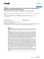
Tilapia (O. Niloticus) were contaminated by a stock solution of
Niclosamide
(C13H8Cl2N2O4)
5-Chloro-N-(2-chloro-4-nitrophenyl)-2-
hydroxybenzamide.
Figure 3.2: Stock solution of Niclosamide - bayluscide
15
Histopathological Examination
The gills and liver tissues of control and treated fish were fixed in 10 %
neutral buffered formalin solution pH 7.0 for 24 hours. Fixation volume should
be 10 to 20 times of the volume of fish tissues.
3.3
Equipment
Table 3.3: Listed of Equipment used for this study
Apparatus and Instruments used in laboratory
- Aprons
- Mask
- Automatic tissue processor
- Microtome
- Cassettes
- Microscope slides
- Chopping board
- Rubber gloves
- Containers
- Scalpel
- Dissecting Knife
- Scissors
- Embedding centers histology
- Thin glass
- Graduated cylinder
- Tissue floating bath
16
3.4
Methods
3.4.1 Toxicity test
A total of 6 juvenile fishes were used with the average weight of the fish
was 101.4±2·9 g. The experiments were conducted in aerated glass aquariums
(120 x 40 x 30 cm) each containing 2 fishes in 10 L of contaminated test solution
and tap water for the control and allowing one hour for acclimation to laboratory
conditions. Stock solution of Niclosamide effective concentration (EC) was
prepared by diluting 1ml pesticide in 100ml of distilled water to different
concentrations of 0.035 and 0.05 ppm, which were used as experimental water
for toxicity study of tilapia. The fishes were exposed to dissolve Niclosamide
until died. The last tank was left untreated as a control group. During acute
toxicity experiment fishes were not fed. All the test exposures were carried out
in duplicate. Dead individuals were dissected for observing the histopathological
alterations in the gills and livers.
3.4.2 Histopathological examination
At the end of exposure period, the 6 fishes were taken from each replicate
tank. Dead fishes were taken out and the gills arches of the fishes were excised
from both sides. Fishes were dissected, the abdominal cavity was operated and
the livers excised quickly.
Procedures required in preparing tissues for light microscopy include
following steps (Raphael, 1976; Bancroft & Gamble, 2002). These steps were
helped by technicians of Laboratory of Aquaculture and Pesticide Toxicology
17
Laboratory of the Integrated Research Laboratory of Sriwijaya University
Graduate School and the Department of Anatomic Pathology, Faculty of
Medicine, University of Sriwijaya/Dr. Mohammad Hoesin General Hospital
Palembang.
Fixations
The gills and livers tissues of the control and the experimental fishes were
dissected and immediately fixed in 10 % neutral buffered formalin solution pH
7.0 for 24 hours. Volume of the formalin is 10 – 20 times of tissues volume.
Tissue Processing
An automatic tissues processor was employed. The selected tissues were
dehydrated, cleared, impregnated and embedded in paraffin wax following standard
procedures. Dehydration involved passing the tissues through increasing strengths of
alcohol, the duration of the procedure can be noted down as:
• 70% alcohol – 1 hour
• 80% alcohol – 1 hour
• 96% alcohol – 1 hour
• 96% alcohol – 1 hour
• Ethanol – 1 hour
• Ethanol – 1 hour
• Xylene – 1 hour
• Xylene – 1 hour
• Paraffin liquid – 1 hour
• Paraffin liquid – 1 hour
18
Dehydration was carried out hence, that the wax i.e Paraffin, which is
used for impregnation, can be easily miscible as it is immiscible with water.
Removal of alcohol with "xylene" that was mixable with the embedding medium
(paraffin) and left overnight.
Embedding
It was conducted by transferring the tissues which has been cleared to a
mold filled with molten wax and allowed to cool and solidified. After
solidification, a paraffin block is obtained which is then sectioned to obtain
ribbons.
Sectioning
The paraffin blocks were sectioned at 3µm thickness by rotary microtome
and the ribbons were floated in hot water bath. The sections from water were
then mounted on clean glass slides. They were then dried on a hot plate at about
50°C for 10 minutes. The sections on the slides were kept ready for staining.
Staining procedure
Initially, the slides were immersed in xylene 3 times for 5 minutes each, a
hydrocarbon solvent, to dissolve paraffin. They were hydrated 3 times by
passing through a series of alcohol 96% for 3 minutes each. Next step, they were
washed in running tap water for 3 minutes and stained using hematoxylin for 5
minutes. The slides then were washed under running tap water for 3 minutes.
Then, they were dipped 2 times in 80% alcohol followed by staining step with
19