Nghiên cứu áp dụng kỹ thuật thông khí áp lực dương liên tục boussignac (CPAP b) trong xử trí trước bệnh viện khó thở cấp cứu luan án tom tat (english)
Bạn đang xem bản rút gọn của tài liệu. Xem và tải ngay bản đầy đủ của tài liệu tại đây (1.37 MB, 27 trang )
MINISTRY OF EDUCATION & TRAINING
MINISTRY OF NATIONAL DEFENCE
108 INSTITUTE OF CLINICAL MEDICAL AND
PHARMACEUTICAL SCIENCES
-----------
NGUYỄN THÀNH
APPLICATION STUDY OF CONTINUOUS
POSITIVE AIRWAY PRESSURE BOUSSIGNAC
(CPAP-B) FOR EMERGENCY DYSPNEA
IN PRE-HOSPITAL SETTING
Specialty: Anesthesia – Critical care
Code: 62720122
DISSERTATION SUMMARY
Name of supervisors:
1. Professor VŨ VĂN ĐÍNH
2. Professor LÊ ANH TUẤN PhD.
Hà Nội – 2018
THIS DISSERTATION WAS FULFILLED
AT 108 INSTITUTE OF CLINICAL MEDICAL AND
PHARMACEUTICAL SCIENCES
Name of supervisors:
1. Professor VŨ VĂN ĐÍNH
2. Professor LÊ ANH TUẤN PhD.
Reviewer 1: ……………………………………………………
Reviewer 2: ……………………………………………………
Reviewer 3: ……………………………………………………
This Dissertation will be defended in front of Dissertation committee
of the Institute at:
date: /
/
This Dissertation can be found at:
1. National Library
2. Library of 108 Institute of clinical medical and pharmaceutical sciences
1
RATIONAL AND JUSTIFICATION
Emergency dyspnea is a common pathologic presentation in prehospital setting, about 25% of total cases on ambulances [18]. This
may be a sign of life threatening condition, especially with warning
signs such as altered mental status, respiratory failure, unstable
hemodynamic [9].
General principles for approaching patient with emergency
dyspnea include: Airway management, breathing support, circulation
support. Non-invasive ventilation, BiPAP, CPAP can be choices for
emergency dyspnea patients with respiratory failure [5].
CPAP Boussignac (CPAP-B) is a non-invasive ventilation device.
it can generate continuous positive airway pressure which increase
alveoli ventilation and oxygenation. This is a simple, light weight,
portable device that is convenient for using on ambulances [4] and it
has been applied in pre-hospital setting in many countries with
positive outcomes. In Vietnam, no study on this device in pre-hospital
care for patients with emergency dyspnea is found.
Objectives
1. Surveillance clinical presentation, arterial blood gas of patients
with emergency dyspnea in pre-hospital setting
2. Evaluation the effectiveness of
Continuous Positive Airway
Pressure Boussignac (CPAP-B) for patients with emergency
dyspnea in pre-hospital setting
3. Evaluation undesired effects during application of CPAP-B
2
CHAPTER 1
LITERATURE REVIEW
1.1. Emergency dyspnea
1.1.1. Concept of emergency dyspnea
Emergency dyspnea is defined as the newly onset or acute on
chronic of uncomfortable breathing arising within 24 to 48 hours and
accompanied by “warning signs” such as:
Airway obstruction: wheezing, foreign body in the airway
Respiratory failure, hypoxia, altered mental status, difficult
speaking, using accessory muscles, respiratory muscles fatigue,
tachypnea, pursed lips, diminishing breath sound one or both sides
Unstable hemodynamic: chest pain, tachycardia, hypotension
Reduced oxygen saturation [16,18]
Those warning signs along with emergency dyspnea required
immediate critical interventions for saving patient’s life, and those
signs are also significant for making diagnosis [26]
1.1.2. Principle of emergency dyspnea management in the prehospital setting
Emergency dyspnea is a life-threatening situation, in the context
of pre-hospital care, Management of cases with emergency dyspnea
focus to airway assessment, breathing and circulation support in order
to secure patient life and safe transport to hospitals.
1.2. CPAP Boussignac (CPAP-B)
1.2.1. Mechanism of action
CPAP-B is a non-invasive ventilation support device. It generates
airway continuous positive pressure from oxygen flow. That helps
improve alveoli ventilation and oxygenation. Mechanism of action of
CPAP-B is based on principle of Bernoulli. The air flow from a larger
3
diameter pipe to a smaller diameter one is accelerated to the speed of
sound. The interference of those air flow within Boussignac valve
creates a turbulent flow which in turn working as a virtual valve
generates positive pressure toward patients.
1.2.2. Indication: Acute respiratory failure because of [24]
- Acute pulmonary edema
- Post-operation patients
- Chest wall trauma
- Support ETT intubation
- Pneumonia
- Support Weaning
- Acute COPD exacerbation
- Asthma attack
- Sleep apnea
1.2.3. Contraindication: [24]
- Cardiac arrest
- Active vomiting, aspiration risk
- Apnea
- Face burn, trauma
- Unconscious
- ENT, face active bleeding
- Systolic pressure < 90 mm Hg
- Pneumothorax
- Serious Chest trauma
- Skull base fracture
- Agitation, un-cooperation
- Dyspnea due to neuro-
- Inability to protect airway
- Profuse secretion or coughing
muscular conditions
inability
1.2.4. Effectiveness of CPAP-B for management respiratory failure
in the prehospital setting
Templier studied 57 patients with acute pulmonary edema those
who were applied CPAP-B in pre-hospital setting. The result shown
that this device helped improving respiratory rate and SpO2
significantly [29]. D.T Wong revealed that CPAP-B helped not only
improving SpO2 and respiratory rate significantly but also reduced
intubation rate down to 20% [31]. Research of Eva Eiske Spijker et. al
gave similar positive results [19].
4
Thomas Luiz (2016) applied CPAP-B for 57 patients with
respiratory failure in the pre-hospital setting, including of 35 patients
with acute pulmonary edema and 22 patients with COPD. The result
revealed that respiratory rate, oxygenation was improved similarly
between two group but the intubation rate in acute pulmonary edema
group was higher than COPD group (17.1% vs. 4.5%) [30]. This result
was similar to a study of Willi Schmidbauer 2010 [27].
In Vietnam, up to date, There is no research on the effectiveness
of CPAP-B applying on ambulances or in the pre-hospital setting.
1.2.5. Undesired effects of CPAP Boussignac
Since 2009, John Bosomworth had found some undesired effects of
CPAP-B such as:
- Pain or ulcer over the nasal bridge
- Mucosal dryness
- Pneumothorax (very rare)
- Fear of closed space
- Aspiration or gastric insufflation (rare)
- Eye irritation
However this author did not mention which side effect was the
most common as well as the rate of each side effect or what kind of
patient with what kind of problem [24]. Eva Eiske Spijker (2013)
conducted a study to assess effectiveness and related complication of
CPAP-B when applying for patients with acute pulmonary edema in
the pre-hospital setting. The result shown that this device was safe and
had no complication [19]. Similarly, Thomas Luiz et al (2016)
announced that “CPAP-B can be used safely and effectively in the prehospital setting for patients who suffering from acute pulmonary
edema and COPD” [30].
5
CHAPTER 2
METHODOLOGY
2.1. Target population
All patients with emergency dyspnea, those who were provided prehospital care and transported to hospital by 115 Hanoi Emergency
Center from January 2015 to December 2015.
2.1.1. Eligible criteria
Patient was recruited to this research when they had newly onset
or acute on chronic of dyspnea arising within 24 to 48 hours and
accompanied by at least one of following symptom:
Unstable hemodynamic: chest pain, tachycardia, hypotension
Reduced oxygen saturation [16,18]
Tachypnea with respiratory rate ≥ 25 breath/min; or accessory
muscle breathing or paradoxical abdominal movements
Cyanosis; or SpO2 < 95 %
Tachycardia: heart rate > 100 beat/min
AND SpO2 < 95 % after 5 minutes on oxygen therapy with 5 liter per
minute via face mask or nasal cannula
2.1.2. Exclusion criteria
Any patient with at least one of following criteria:
age < 18
uncooperative patients
contraindication with non-invasive ventilation
Pneumothorax without chest decompression
Open chest injury
Abnormal or any trauma of facial structure
Foreign body of upper airway is suspected
Systolic Blood pressure < 90 mmHg
Respiratory rate <10 breath/min
Glasgow score < 8 or at U level (on AVPU scale)
6
2.2. Methodology
2.2.1. Research design: prospective interventional study
2.2.2. Sample size
Sample size was estimated according to the following formula :
n
2 C (1 r ) 2 19,84 1 0,6
142,87 150
0,3333 2
ES 2
C :
constant; C = 19,84 with = 0,01; = 0,05
r :
relative risk, estimated equal 0,6
ES:
Effect size: ES = d /s
d was average difference of SpO2 before and after
intervention (estimated 5%), s was standard deviation
(estimated 5%) [10,32].
2.2.3. Interventional process
At the scene, pre-hospital staffs took patient’s medical history,
chief complain and clinical assessment. In case of emergency dyspnea
was detected, patient was put on oxygen therapy via face mask or nasal
cannula. After 5 minutes, all unresponsive cases (SpO2 < 95% with 5
liter/min) were applied CPAP-B for breathing support. Portable
monitor was also attached for SpO2 and heart rate monitoring. CPAP
level was initiated at 5 cm water, increasing every 2.5 cm water by
adjusting oxygen flow in order to maintain SpO2 ≥ 95%. Maximum
CPAP level was 10 cm water. Those patients with CPAP-B whose
signs and symptoms got worse during intervention process such as
more severe respiratory, unstable hemodynamic, SpO2 < 95% with
CPAP of 10 cm water or having any risk of complication such as
vomiting, pneumothorax, aspiration must be terminated the CPAP-B
therapy for other emergency procedures such as: intubation, larynx
mask airway, AMBU. On arrival at the Emergency room, CPAP-B
7
therapy could be ended for other intervention depend on the indication
of physician in charge there. Two samples for arterial blood gas
analysis were taken before and after using CPAP-B.
2.2.4. Research variables
- General characteristics of target population: Age, gender, medical
history, time of service, duration of intervention
- Clinical characteristics of patients with emergency dyspnea: Level of
consciousness, signs and symptoms of respiratory failure, vital signs.
- Characteristics of arterial blood gas of patients with emergency
dyspnea: pH, PaO2, PaCO2, HCO3- Interventional result and related undesired effects:
Success rate
Comparison clinical, vital signs changes before and after
intervention
Comparison arterial blood gas before and after intervention
Proportion of undesired effects related to CPAP-B
2.2.5. Research criteria
Level of consciousness:
In pre-hospital setting, we applied AVPU scale to assess level of
consciousness [25].
Diagnoses in pre-hospital setting:
In this research, we used clinical practice guideline of Queensland
ambulance service Queensland Australia [12,13,14,15] for diagnosis
of respiratory failure and its causes. In the pre-hospital setting, those
diagnoses mainly based on clinical signs and symptoms.
Respiratory failure classification: criteria of Allal 2012 [8]
8
Success criteria (all following:)
SpO2 maintained above 95%
Heart rate reduction > 20%
Stable hemodynamic
Respiratory rate < 25 breath/min
Improving clinical signs and symptoms [3,7]
Failure criteria (one or more of following)
Worsen respiratory failure lead to termination of therapy and
deploying other methods for breathing support such as intubation,
laryngeal mask airway, AMBU
Unstable hemodynamic
SpO2 < 95% with CPAP level up to 10 cm water
Uncooperation or appearance of related complications lead to
termination of therapy[3,7]
2.3. Statistical analysis
Data was analyzed by medical statistic methods. Mean,
standard diviation was performed as X ± SD (standard distribution)
or as median, quartile (non-standard distribution). Percentage was
compared by χ2 test (or Fisher test). Mean of two independent groups
were compaired by t - test (standard distribution) or Mann-Whitney
test (non-standard distribution). Paired-t-test (standard distribution)
or Wilcoxon (non-standard distribution) was used for before – after
comparison. One way ANOVA test (standard distribution) and
Kruskal-Wallis test (non-standard distribution) was for comparison
of multiple means. p value < 0,05 was consider statistical
significance.
9
CHAPTER 3
RESULT
3.1. General characteristic of target population
There were 150 patients enrolled in this study, mean of age was
73.5 ± 14.7 year old, youngest was 22, oldest was 97 year old.
There were more male than female, gender proportion of male over
female was 2.26:1 aproximately.
Time of service was similar between day time (6h-18h) and night
time (18h-6h). Average duration of care from on-scene to hospotal
was 29.5 ± 11 minutes, minimum was 10 minutes, maximum was
66 minutes. 68.7% patients was transported within 11 to 30 minutes
In this research, there were 5 causes of emergency dypsnea. They
were pneumonia (53 patients, 35.3%), acute COPD exacerbation
(44 patients, 29.3%), pulmonary edema (31 patients, 20.7%),
asthma attack (18 patients, 12.0%), 4 patients (2.7%) with prior
diagnosis of lung cancer.
3.2. Characteristics of clinical manifestation and ABG of
emergency dyspnea patients
3.2.1. Characteristics of clinical manifestation
Table 3.5. Level of consciousness
Level of consciousness
n
Percentage
Alert
121
80.7 %
Response to Verbal
24
16.0 %
Response to Pain
5
3.3 %
Unresponsive
0
0.0 %
150
100 %
Total
Comment: 19.3 % patients altered mental status, no patient had U
level on AVPU scale
10
Table 3.6. Cyanosis
Cyanosis
n
Percentage
Yes
150
100 %
No
0
0%
Comment: Before intervention, all patients had cyanosis sign at
different level.
Table 3.7. Difficulty in taking
n
Percentage
Talks in sentence
1
0.7 %
Talks in phases
63
42.0 %
Talks in words
48
32.0 %
Unable to talk
38
25.3 %
Total
150
100 %
Comment: Most of patients (99.3%) could not talks in sentence
Table 3.8. Diaphoresis
Diaphoresis
n
Percentage
Yes
45
30 %
No
105
70 %
Total
150
100 %
Comment: Only 30% patients had diaphoresis
Table 3.9. Paradoxical abdominal movements
Paradoxical abdominal movements
n
Percentage
Yes
33
22 %
No
117
78 %
Total
150
100 %
Comment: 1/5 patients had sign of paradoxical abdominal movements
11
Table 3.10. Accessory muscle breathing
Accessory muscle breathing
n
Percentage
Yes
144
96 %
No
6
4%
150
100 %
Total
Comment: Most of patients had sign of accessory muscle breathing
Table 3.11. Level of clinical respiratory failure
Clinical respiratory failure
n
Percentage
Level I
10
6.7 %
Level II
110
73.3 %
Level III
30
20.0 %
Level IV
0
0.0 %
150
100 %
Total
Comment: All patients with emergency dyspnea had respiratory
failure clinically at different level, mostly level II (73.3%)
3.2.2. Vital signs of patients with emergency dyspnea
Table 3.13. Pre-intervention vital signs
̅ ± SD)
Vital signs
(𝐗
Min
Max
125.2 ± 12.0
90
156
Respiratory rate (breath/min)
32.5 ± 5.3
20
56
SPO2 (%)
71.6 ± 8.1
46
86
Systolic BP (mmHg)
141.9 ± 36.6
90
250
Diastolic BP (mmHg)
81.1 ± 16.1
40
140
Heart rate (beat/min)
Comment: Before intervention, heart rate, respiratory rate, blood
pressure increased, SpO2 decreased
12
3.2.3. Arterial blood gas of patients with emergency dyspnea
Table 3.14. Pre-intervention arterial blood gas
̅ ± SD)
ABG
n
(𝐗
Min
Max
PaO2 (mmHg)
150
60.98 ± 10.14
38.00
79.70
PaCO2 (mmHg)
150
44.51 ± 13.55
20.10
82.20
HCO3 (mmol/L)
150
24.65 ± 4.84
13.20
37.10
Comment: PaO2 decreased, PaCO2 was at upper level of normal range
Table 3.15. Pre-intervention acid-base balance
pH
n
Percentage
<7.35
45
30 %
7.35-7.45
57
38 %
>7.45
48
32 %
Total
150
100 %
Comment: 62% patients had acid-base imbalance prior intervention
3.3. Effectiveness of CPAP Boussignac in pre-hospital emergency
dyspnea management
3.3.1. Clinical changes before and after intervention
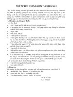
p <0,01
Chart 3.5. Mental status changes before and after intervention
Comment: after intervention, mental status was improved significantly
13
Table 3.18. Cyanosis before and after intervention
Phase
sign
n
Percentage Total
p
Cyanosis
150
100 %
Pre150
intervention Non-Cyanosis
(100%)
0
0%
< 0,01
Cyanosis
3
2%
Post150
intervention Non-Cyanosis
(100%)
147
98 %
Comment: there was no cyanosis in most of patient after intervention,
Table 3.19. Difficulty in taking before and after intervention
Phase
sign
n
Percentage Total
p
Talks in sentence
1
0.7 %
PreTalks in phases
63
42.0%
150
intervention
(100%)
Talks in words
48
32.0%
Unable to talk
Talks in sentence
38
5
25.3%
3.3 %
< 0.01
Talks in phases
123
82.0 %
Post150
intervention Talks in words
(100%)
7
4.7 %
Unable to talk
15
10.0 %
Comment: Taking ability’s improved significantly after intervention
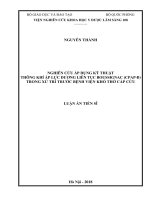
p <0,01
Chart 3.7. Paradoxical abdominal movements before and after
intervention
Comment: after intervention this sign was improved significantly
14
Table 3.20. Accessory muscle breathing before and after
intervention
Phase
Accessory muscle
breathing
n
Percentage Total
Preintervention
Yes
144
96.0%
150
No
6
4.0%
(100%)
Post-
Yes
136
90.7 %
150
intervention
No
14
9.3 %
(100%)
p
> 0,05
Comment: Accessory muscle breathing before and after intervention
had no difference
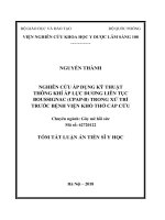
p <0,01
Chart 3.8. Level of clinical respiratory failure before and after
intervention
Comment: Level of clinical respiratory failure was deducted
significantly after intervention
15
Table 3.21. Vital signs before and after intervention
̅ ± SD)
Variable
Phase
(𝐗
Min
Max
p
Pre
71.6 ± 8.1
46
86
SpO2
< 0.01
(%)
Post
98.1 ± 2.4
88
100
Pre 125.2 ± 12.0
90
156
Heat rare
< 0.01
(beat/min)
Post 107.6 ± 10.1
87
134
Pre
32.5 ± 5.3
20
56
Respiratory rate
< 0.01
(beat/min)
Post
23.2 ± 3.0
17
32
Comment: After intervention, SpO2 was increased, heart rate,
respiratory rate were decreased significantly
3.3.2. Arterial blood gas changes before and after intervention
Table 3.25. Arterial blood gas before and after intervention
̅ ± SD)
Variable
Phase
(𝐗
Min
Max
p
Pre
60.98 ± 10.14
38.0
79.7
PaO2
< 0.01
(mmHg)
Post
110.70 ± 19.19 82.5 187.6
Pre
44.51 ± 13.55
20.1
82.2
PaCO2
< 0.01
(mmHg)
Post
41.48 ± 9.76
19.6
69.9
Pre
24.65 ± 4.84
13.2
37.1
HCO30.439
(mmol/L)
Post
24.62 ± 4.31
16.3
35.4
Pre
7.38 ± 0.89
7.15
7.58
pH
< 0.01
Post
7.40 ± 0.74
7.24
7.56
Comment: after intervention, PaO2, PaCO2, pH improved significantly
3.4. Result of CPAP-B therapy and related undesired effects
Table 3.35. Result of CPAP-B therapy in pre-hospital setting
Result
n
Percentage
Success
143
95.3 %
Failure
7
4.7 %
Total
150
100 %
Comment: 95.3% patients with CPAP-B therapy got improvement.
16
Table 3.40. Undesired effects related to CPAP-B therapy
Undesired effects
n
Percentage
Skin redness around mask area
11
7.3 %
Vomiting
2
1.3 %
Abdominal distension
2
1.3 %
Comment: There was no life-threatening complication during
intervention. The most common undesired effect was redness of skin
around mask area.
CHAPTER 4
DISCUSSION
4.1. Characteristics of clinical manifestation and arterial blood gas
of patients with emergency dyspnea
4.1.1. Clinical manifestation
In this research, all patients with emergency dyspnea developed
signs and symptoms of acute respiratory failure. 100% target group
was cyanosis, 99.3% had difficulty in talking (42% talking in phrase,
32 % talking in word, 25.3% unable to talk). 30% patients were with
diaphoresis, 22% were with sign of Paradoxical abdominal
movements, 96% with sign of accessory muscle breathing.
Vital sign prior intervention reflexed status of respiratory failure.
Tachycardia with heart rate was 125.2 ± 12.0 beat/min, tachypnea with
respiratory rate was 32.5 ± 5.3 breath/min; hypoxemia with SpO2 was
71.6 ± 8.1 %.
Foreign and domestic relevant researches did not mention about
classical signs and symptoms of respiratory failure. This can be
understood that clinical manifestations were subjective and nonspecific therefore they were not paid attention in other researches.
However, clinical signs and symptoms are still playing important roles
17
in the context of pre-hospital care in Viet Nam where modern monitors
and equipment are not always available.
4.1.2. Arterial blood gas of target population
Mean PaO2 was 60.98 ± 10.14 mm Hg, tantamount to moderate and
severe respiratory failure, this matched with clinical level of
respiratory failure. Patient’s level of pre-interventional PaO2 in this
research was not as severe as those in research of Nguyễn Thị Thanh
Thủy (55.9 ± 9.4 mmHg) [7] and Lê Đức Nhân ( 50.9 ± 9.5 mmHg)
[3], but similar to result of Phùng Nam Lâm (61.6 ± 18.10 mmHg) [9].
Nguyễn Thị Thanh Thủy and Lê Đức Nhân focused to patients with
acute pulmonary edema only, hence level of PaO2 in their study was
much higher than research of Phùng Nam Lâm and ours since our
target population were patients with respiratory failure from all causes,
not only acute pulmonary edema.
Pre-interventional PaCO2 (44.51 ± 13.55 mmHg) was higher than
patients in researches of Nguyễn Thị Thanh Thủy (39.7 ± 11.4 mmHg)
and Lê Đức Nhân (36.8 ± 12.6 mmHg). Doing deep analysis in each
group of diagnosis, we revealed that patients with high level of PaCO2
prior intervention were those who suffered from acute COPD
exacerbation (59.4 ± 9.97 mmHg), PaCO2 level of patients with acute
pulmonary edema were 36.9 ± 8.3 mmHg, similar to result of two
above studies.
Mean HCO3- level was in physiological range (24.65 ± 4.84
mmHg). This might be because kidneys and buffer systems must take
hours to days to balance acid-base. Therefore we assumed that level
of HCO3- had not much change during acute phase of respiratory
failure, especially in pre-hospital setting. There were 30% patient had
acidosis, 32% had alkalosis. This abnormality was because of different
causes of acute respiratory failure.
18
4.2. Effectiveness of CPAP-B in emergency dyspnea management
4.2.1. Clinical effectiveness of CPAP-B
CPAP-B help improving clinical symptoms: 98% patients had no
cyanosis after intervention, 59.9% improved talking ability,
paradoxical abdominal movements was reduced to 12% in comparison
with pre-intervention.
Clinical level of respiratory failure was improved significantly
(p<0.01). Before intervention, 93.3% patients were at level 2 and 3 of
respiratory failure, after intervention there was only 4% had the same
level, most of patients got deduction in respiratory failure to level 1.
CPAP-B is a breathing support device, help improving ventilation and
oxygenation so that secure patient’s life on rout of transport. However
CPAP-B cannot treat causes of respiratory failure therefore respiratory
status of patients in this research could only be improved, not resolved
completely.
4.2.2. Vital signs changes before and after intervention
4.2.2.1. SpO2
CPAP-B helped improving level of SpO2 rapidly. 5 minutes after
stating therapy, mean SpO2 went from 71.6 ± 8.1% up to 89.36 ±
3.29% and kept increasing in the next point of times. This change was
significant with p value p < 0.001 and similar to research of Eva Eiske
Spijker. Mean level of SpO2 after intervention in Eva Spijker’s
research (98.23 ± 2.64%) was lower than ours [19]. The difference in
target population might explain the gap between two researches. Eva
Spijker’s population were only 16 patients with acute pulmonary
edema, in our research, sample size was significantly larger including
150 patients with respiratory failure from all causes.
19
4.2.2.2. Heart rate
In this research, heart rate was decreased rapidly and significantly,
similar phenomena was announced by other authors. Lê Đức Nhân
found that after intervention by CPAP-B, heart rate reduced from
140.2 ± 15.7 beat/min to 97.0 ± 12.7 beat/min [3], Nguyễn Thanh
Thủy: from 141.7 ± 15.7 beat/min to 95.0 ± 10.6 beat/min [7]. Lưu
Quang Thùy [6], Đào Khắc Hùng were also witnessed heart rate of
post-operative patients were slowed down rapidly when the
intervention was finished [1].
4.2.2.3. Respiratory rate
CPAP-B was effective in reducing respiratory rate of patients
with emergency dyspnea. However, at the point of intervention
cessation, patient’s respiratory rate was 23.2 ± 3.0 breath/min, still
higher than normal value. This can be explained that breathing support
duration by CPAP-B in pre-hospital setting was short (24.53 ± 9.9
minutes on average) therefore dyspnea could only get improvement
and could not be fully recovery
Thomas in 2016 also revealed that respiratory rate of target
population reduced, among those, COPD patients from 27.0 ± 9.0
breath/min to 18.0 ± 4.7 breath/min, acute pulmonary edema patients
from 28.8 ± 11.2 breath/min to 19.0 ± 6.7 breath/min. according to
this author, the difference of respiratory rate before and after
intervention was significant in each group of diagnosis (p<0.01) but
no significant when compared between different group. [30]. Role of
CPAP-B in reducing respiratory rate was also reported similarly in
studies of Templier [29], Fyntanidou [21], Freitas [20], Dieperink [17].
20
4.2.3. Arterial blood gas changes before and after intervention
4.2.3.1. PaO2 level
After intervention, PaO2 was increased significantly with p value
< 0.01. Garutti reported similar result when he applied CPAP-B for
post–operative patients who undergoing lung resection surgery. His
result shown that PaO2 was increased from 75.7 mmHg to 98.3 mmHg
after 15 minutes on CPAP-B [22]. Publication of Đào Khắc Hùng
shown that PaO2 kept improving during process of breathing support
by CPAP. In that study, pre-intervention PaO2 was 60.5 ± 10.5 mmHg,
after120 minutes up to 72.8 ± 12 mmHg and 98.4 ± 10.8 mmHg by the
end of intervention [1]. The author also found that the differences of
PaO2 level in different point of time was significant with p value <
0.05. Similar change of PaO2 level during CPAP-B intervention was
recorded by Nguyễn Thanh Thủy [7], and Lê Đức Nhân [3].
4.2.3.2. PaCO2 level
After intervention, PaCO2 level was decreased significantly
(from 44.51 ± 13.55 mmHg to 41.48 ± 9.76 mmHg) with p value less
than 0.01. The change of PaCO2 in different groups of diagnosis were
different. Level of PaCO2 of COPD patients was at highest before
intervention but they had the most significant change when the therapy
ended. This result was matched with hypothesis of Stefano Nava, he
supposed that CPAP was useful for COPD patients with hypercapnia
since it could resolve intrinsic PEEP in those patients therefore,
reduced the work of breathing, respiratory muscle fatigue, improved
alveoli ventilation [28]. Đào Khắc Hùng had same opinion, he
assumed that CPAP-B could help decreasing PaCO2 level at type 2
respiratory failure patients (with hypercapnia) [1]. In this research we
found similar result. In group with normocapnia, PaCO2 lever before
and after intervention was no difference.
21
4.2.3.3. HCO3- level
Almost no fluctuation of HCO3- level was found during
intervention with CPAP Boussignac. All patients in this research were
emergency dyspnea due to hypoxemia in the acute phase, it was not
long enough for bicarbonate buffer system responded with those acute
clinical change, therefore we could not find significant change of
HCO3- level at different point of time during intervention. That result
was similar to reports of Lưu Quang Thùy [6], Nguyễn Thị Thanh
Thủy [7], Phùng Nam Lâm [2]. Claude Guérin studied on 64 patients
with acute on chronic respiratory diseases also concluded that normal
HCO3- was common among those patients [11].
4.2.3.4. pH
In general, change in pH level of target population was significant
before and after intervention, however, there were 45 patients (30%)
whose acid-base imbalance was still existed after intervention. This
was the result of short intervention time in pre-hospital phase when
interventions such as breathing support, medication (bronchodilator,
steroid…) could not be optimized, therefor CO2 elimination had not
been as effective as in hospital setting.
4.3. Result of CPAP-B therapy in pre-hospital setting
Success rate in our research was 95.3%. There were 7 cases of
failure (4.7%). Our research was the very first one which applied CPAPB for breathing support in pre-hospital setting. As the result, there was
no similar domestic research to compare. Some similar international
studies report different success rate from ours. Thomas Luiz (2016)
announced 7% patients must ended CPAP-B therapy before reaching
hospitals, including 3 cases required endotracheal intubation on
ambulance [30]. In publication of Eva Spijker, 6,3% patients failed to
response to CPAP-B therapy [19]. Failure rate of that two publication
22
were higher than our result. We supposed that unequal severity of target
population made this difference. In Thomas Luiz study, there were 3
over 5 cases with intubation developed signs and symptom of acute
coronary disease. In Eva Spijker study, 31% of patients had ST segment
elevation myocardial infarction. We did not get any case with very
severe diseases such as myocardial infarction, severe respiratory failure,
that may contributed to our higher success rate.
4.4. Undersigned effects of CPAP Boussignac in breathing support
Applying CPAP-B via face mask on ambulances, we found 11 cases
with skin redness around mask area (7.3%). This kind of undesired
effect was common among relevant researches, however in ours,
proportion of this one is much lower (Lê Đức Nhân 13.9% [3], Lưu
Quang Thùy 14.28% [6], Đào Khắc Hùng 20%) [1]. Others authors
reported some cases with conjunctival irritation, but we found none in
our research. Reason of the difference may because duration of applying
CPAP-B in pre-hospital setting was shorter than other researches in
hospital, therefore proportion of undesired effects was less.
There were 2 cases with abdominal distension (1.3%), however, it
caused no impact to the positive effect of CPAP-B on clinical and
arterial blood gas were not impacted. We found no life-threatening
complication related to CPAP Boussignac such as pneumothorax,
hypotension. Some foreign studies on CPAP-B for patients in
emergency rooms as well as on ambulances recommended that CPAPB was safe therapy for breathing support.
23
CONCLUSION
This research was recruited 150 patients with emergency dyspnea
those who were got breathing support by CPAP Boussignac in prehospital setting. According the research result, we came to following
conclusions:
1. Clinical manifestations, arterial blood gas of patients with
emergency dyspnea in pre-hospital setting
In this research, majority of patients with emergency dyspnea were
elderly, mean age was 73.5 ± 14.7, more male than female
Common causes of emergency dyspnea were main causes of
emergency dypsnea were pneumonia, acute COPD exacerbation,
Pulmonary edema, asthma attack, lung cancer.
Clinical manefestations and arterial blood gas revealed status of
acute respiratory failure mostly at moderate level (73,3%)
Clincal manifestations: cyanosis (100%), difficulty in talking
(99.3%), diaphoresis (30%), paradoxical abdominal movements
(32%), accessory musscle brearthing (96%), tachycardia (125.2 ± 12.0
beat/min), tachypnea (32.5 ± 5.3 breath/min), low SpO2 (71.6 ± 8.1%).
Systolic and diastolic blood pressure were 141.9 ± 36.6 mmHg and
81.1 ± 16.1 mmHg respectively.
Arterial blood gas: low PaO2 (60.98 ± 10.14 mmHg). pH, PaCO2,
HCO3- were in normal range. Patients with acute COPD exacerbation
trended to be hypercapnia and acidosis.
2. CPAP-B was effective for management of emergency dyspnea
in pre-hospital setting
CPAP Boussignac helped improving clinical signs and symptoms:
98% of patients had no cyanosis after intervention, sign of difficulty
in talking was improved, proportion of paradoxical abdominal
movements was reduced from 22% to 12 % after intervention.