Medical computer vision algorithms for big data
Bạn đang xem bản rút gọn của tài liệu. Xem và tải ngay bản đầy đủ của tài liệu tại đây (45.98 MB, 187 trang )
LNCS 9601
Bjoern Menze · Georg Langs
Albert Montillo · Michael Kelm
Henning Müller · Shaoting Zhang
Weidong Cai · Dimitris Metaxas (Eds.)
Medical Computer
Vision: Algorithms
for Big Data
International Workshop, MCV 2015
Held in Conjunction with MICCAI 2015
Munich, Germany, October 9, 2015, Revised Selected Papers
123
Lecture Notes in Computer Science
Commenced Publication in 1973
Founding and Former Series Editors:
Gerhard Goos, Juris Hartmanis, and Jan van Leeuwen
Editorial Board
David Hutchison
Lancaster University, Lancaster, UK
Takeo Kanade
Carnegie Mellon University, Pittsburgh, PA, USA
Josef Kittler
University of Surrey, Guildford, UK
Jon M. Kleinberg
Cornell University, Ithaca, NY, USA
Friedemann Mattern
ETH Zurich, Zürich, Switzerland
John C. Mitchell
Stanford University, Stanford, CA, USA
Moni Naor
Weizmann Institute of Science, Rehovot, Israel
C. Pandu Rangan
Indian Institute of Technology, Madras, India
Bernhard Steffen
TU Dortmund University, Dortmund, Germany
Demetri Terzopoulos
University of California, Los Angeles, CA, USA
Doug Tygar
University of California, Berkeley, CA, USA
Gerhard Weikum
Max Planck Institute for Informatics, Saarbrücken, Germany
9601
More information about this series at />
Bjoern Menze Georg Langs
Albert Montillo Michael Kelm
Henning Müller Shaoting Zhang
Weidong Cai Dimitris Metaxas (Eds.)
•
•
•
•
Medical Computer
Vision: Algorithms
for Big Data
International Workshop, MCV 2015
Held in Conjunction with MICCAI 2015
Munich, Germany, October 9, 2015
Revised Selected Papers
123
Editors
Bjoern Menze
TU München
Munich
Germany
Georg Langs
Medical University of Vienna
Wien
Austria
Albert Montillo
University of Texas Southwestern Medical
Center
Dallas, TX
USA
Michael Kelm
Siemens AG
Erlangen
Germany
Henning Müller
University of Applied Sciences Western
Switzerland (HES-SO)
Sierre
Switzerland
Shaoting Zhang
University of North Carolina
Charlotte
USA
Weidong Cai
University of Sydney
Sydney
Australia
Dimitris Metaxas
State University of New Jersey Rutgers
Piscataway, NJ
USA
ISSN 0302-9743
ISSN 1611-3349 (electronic)
Lecture Notes in Computer Science
ISBN 978-3-319-42015-8
ISBN 978-3-319-42016-5 (eBook)
DOI 10.1007/978-3-319-42016-5
Library of Congress Control Number: 2016946962
LNCS Sublibrary: SL6 – Image Processing, Computer Vision, Pattern Recognition, and Graphics
© Springer International Publishing Switzerland 2016
This work is subject to copyright. All rights are reserved by the Publisher, whether the whole or part of the
material is concerned, specifically the rights of translation, reprinting, reuse of illustrations, recitation,
broadcasting, reproduction on microfilms or in any other physical way, and transmission or information
storage and retrieval, electronic adaptation, computer software, or by similar or dissimilar methodology now
known or hereafter developed.
The use of general descriptive names, registered names, trademarks, service marks, etc. in this publication
does not imply, even in the absence of a specific statement, that such names are exempt from the relevant
protective laws and regulations and therefore free for general use.
The publisher, the authors and the editors are safe to assume that the advice and information in this book are
believed to be true and accurate at the date of publication. Neither the publisher nor the authors or the editors
give a warranty, express or implied, with respect to the material contained herein or for any errors or
omissions that may have been made.
Printed on acid-free paper
This Springer imprint is published by Springer Nature
The registered company is Springer International Publishing AG Switzerland
Preface
This book includes articles from the 2015 MICCAI (Medical Image Computing for
Computer Assisted Intervention) workshop on Medical Computer Vision (MCV) that
was held on October 9, 2015, in Munich, Germany. The workshop followed up on
similar events in the past years held in conjunction with MICCAI and CVPR.
The workshop obtained 22 high-quality submissions that were all reviewed by at
least three external reviewers. Borderline papers were further reviewed by the organizers to obtain the most objective decisions for the final paper selection. Ten papers
(45%) were accepted as oral presentations and another five as posters after the authors
responded to all review comments. The review process was double-blind.
In addition to the accepted oral presentations and posters, the workshop had three
invited speakers. Volker Tresp, both at Siemens and Ludwig Maximilians University of
Munich, Germany, presented large-scale learning in medical applications. This covered
aspects of image analysis but also the inclusion of clinical data.
Pascal Fua of EPFL, Switzerland, discussed multi-scale analysis using
machine-learning techniques in the delineation of curvilinear structures. Antonio Criminisi presented a comparison of deep learning approaches with random forests and his
personal experiences in working with and comparing the two approaches.
The workshop resulted in many lively discussions and showed well the current
trends and tendencies in medical computer vision and how the techniques can be used
in clinical work and on large data sets.
These proceedings start with a short overview of the topics that were discussed
during the workshop and the discussions that took place during the sessions, followed
by the one invited and 15 accepted papers of the workshop.
We would like to thank all the reviewers who helped select high-quality papers for
the workshop and the authors for submitting and presenting high-quality research, all of
which made MICCAI-MCV 2015 a great success. We plan to organize a similar
workshop at next year’s MICCAI conference in Athens.
December 2015
Bjoern Menze
Georg Langs
Henning Müller
Albert Montillo
Michael Kelm
Shaoting Zhang
Weidong Cai
Dimitris Metaxas
Organization
General Co-chairs
Bjoern Menze, Switzerland
Georg Langs, Austria
Albert Montillo, USA
Michael Kelm, Germany
Henning Müller, Switzerland
Shaoting Zhang, USA
Weidong Cai, Australia
Dimitris Metaxas, USA
Publication Chair
Henning Müller, Switzerland
International Program Committee
Allison Nobel
Cagatay Demiralp
Christian Barrillot
Daniel Rueckert
Diana Mateus
Dinggang Shen
Ender Konukoglu
Guorong Wu
Hayit Greenspan
Hien Nguyen
Horst Bischof
Jan Margeta
Juan Iglesias
Jurgen Gall
Kayhan Batmanghelich
Kilian Pohl
Le Lu
Lin Yang
Luping Zhou
Marleen de Bruijne
Matthew Blaschko
Matthew Toews
University of Oxford, UK
Stanford University, USA
IRISA Rennes, France
Imperial College London, UK
TU München, Germany
UNC Chapel Hill, USA
Harvard Medical School, USA
UNC Chapel Hill, USA
Tel Aviv University, Israel
Siemens, USA
TU Graz, Austria
Inria, France
Harvard Medical School, USA
Bonn University, Germany
MIT, USA
Stanford University, USA
NIH, USA
University of Florida, USA
University of Wollongong, Australia
EMC Rotterdam, The Netherlands
Ecole Centrale Paris, France
Harvard BWH, USA
VIII
Organization
Matthias Schneider
Michael Wels
Paul Suetens
Ron Kikinis
Ruogu Fang
Tom Vercauteren
Vasileios Zografos
Yang Song
Yiqiang Zhan
Yefeng Zheng
Yong Xia
Yong Fan
Yue Gao
ETH Zurich, Switzerland
Siemens Healthcare, Germany
KU Leuven, Belgium
Harvard Medical School, USA
Florida International University, USA
University College London, UK
TU München, Germany
University of Sydney, Australia
Siemens, USA
Siemens Corporate Research, USA
Northwestern Polytechnical University, China
University of Pennsylvania, USA
UNC Chapel Hill, USA
Sponsors
European Commission 7th Framework Programme, VISCERAL (318068).
Modeling Brain Circuitry
over a Wide Range of Scales
(Invited Paper)
Pascal Fua and Graham Knott
EPFL, 1015 Lausanne, Switzerland
Pascal.Fua@epfl.ch, Graham.Knott@epfl.ch
fl.ch/research
Abstract. We briefly review the Computer Vision techniques we have developed at EPFL to automate the analysis of Correlative Light and Electron
Microscopy data. They include delineating dendritic arbors from LM imagery,
segmenting organelles from EM, and combining the two into a consistent
representation.
Keywords: Brain Connectivity Á Microscopy Á Delineation Á Segmentation Á
Registration
Overview
If we are ever to unravel the mysteries of brain function at its most fundamental level,
we will need a precise understanding of how its component neurons connect to each
other. Electron Microscopes (EM) can now provide the nanometer resolution that is
needed to image synapses, and therefore connections, while Light Microscopes
(LM) see at the micrometer resolution required to model the 3D structure of the
dendritic network. Since both the topology and the connection strength are integral
parts of the brain's wiring diagram, being able to combine these two modalities is
critically important.
In fact, these microscopes now routinely produce high-resolution imagery in such
large quantities that the bottleneck becomes automated processing and interpretation,
which is needed for such data to be exploited to its full potential.
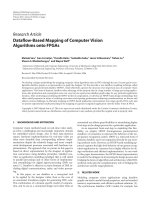
In our work, we have therefore used correlative microscopy image stacks such as
those described in Fig. 1 and we have developed approaches to automatically building
the dendritic arborescence in LM stacks [5, 6], to segmenting intra-neuronal structures
from EM images [1, 4], and to registering the resulting models [3]. Figure 1 depicts
some of these results. In all cases, Statistical Machine Learning algorithms are key to
obtaining good results. Therefore, our challenge is now to develop Domain Adaptation
This work was supported in part by ERC project MicroNano and in part by the Swiss National Science
Foundation.
X
P. Fua and G. Knott
techniques that will allow us to retrain them quickly and without excessive amounts of
additional annotated data when new image data is acquired [2]. For additional details
on this work, we refer the interested reader to the above mentioned publications.
(a)
(b)
(c)
Fig. 1. Correlative Microscopy. (a) Fluorescent neurons in vivo in the adult mouse
brain imaged through a cranial window. (b) Image stack at the 1 μm resolution acquired
using a 2-photon microscope. (c) Image slice of a sub-volume at the 5 nm resolution
above a reconstruction of a neuron, dendrite, and associated organelles.
(a)
(b)
Fig. 2. Automated delineation and segmentation. (a) Dendrites from an LM Stack.
(b) Mitochondria from an EM stack. The colors denote those that are either within a
dendrite or an axon.
Modeling Brain Circuitry over a Wide Range of Scales
XI
References
1. Becker, C., Ali, K., Knott, G., Fua, P.: Learning context cues for synapse segmentation. IEEE
Trans. Med. Imaging (2013)
2. Becker, C., Christoudias, M., Fua, P.: Domain adaptation for microscopy imaging. IEEE
Trans. Med. Imaging (2015)
3. Glowacki, P., Pinheiro, M., Turetken, E., Sznitman, R., Lebrecht, D., Holtmaat, A., Kybic, J.,
Fua, P.: Modeling evolving curvilinear structures in time-lapse imagery. In: Conference on
Computer Vision and Pattern Recognition (2014)
4. Lucchi, A., Smith, K., Achanta, R., Knott, G., Fua, P.: Supervoxel-based segmentation of
mitochondria in EM image stacks with learned shape features. IEEE Trans. Med. Imaging 31
(2), 474–486 (2012)
5. Turetken, E., Benmansour, F., Andres, B., Pfister, H., Fua, P.: Reconstructing loopy curvilinear structures using integer programming. In: Conference on Computer Vision and Pattern
Recognition, June 2013
6. Turetken, E., Benmansour, F., Fua, P.: Automated reconstruction of tree structures using path
classifiers and mixed integer programming. In: Conference on Computer Vision and Pattern
Recognition, June 2012
Contents
Workshop Overview
Overview of the 2015 Workshop on Medical Computer
Vision — Algorithms for Big Data (MCV 2015) . . . . . . . . . . . . . . . . . . . .
Henning Müller, Bjoern Menze, Georg Langs, Albert Montillo,
Michael Kelm, Shaoting Zhang, Weidong Cai, and Dimitris Metaxas
3
Predicting Disease
Information-Theoretic Clustering of Neuroimaging Metrics Related
to Cognitive Decline in the Elderly . . . . . . . . . . . . . . . . . . . . . . . . . . . . . .
Madelaine Daianu, Greg Ver Steeg, Adam Mezher, Neda Jahanshad,
Talia M. Nir, Xiaoran Yan, Gautam Prasad, Kristina Lerman,
Aram Galstyan, and Paul M. Thompson
Relationship Induced Multi-atlas Learning for Alzheimer’s
Disease Diagnosis . . . . . . . . . . . . . . . . . . . . . . . . . . . . . . . . . . . . . . . . . .
Mingxia Liu, Daoqiang Zhang, Ehsan Adeli-Mosabbeb,
and Dinggang Shen
13
24
Atlas Exploitation and Avoidance
Hierarchical Multi-Organ Segmentation Without Registration
in 3D Abdominal CT Images . . . . . . . . . . . . . . . . . . . . . . . . . . . . . . . . . .
Vasileios Zografos, Alexander Valentinitsch, Markus Rempfler,
Federico Tombari, and Bjoern Menze
Structure Specific Atlas Generation and Its Application to Pancreas
Segmentation from Contrasted Abdominal CT Volumes. . . . . . . . . . . . . . . .
Ken’ichi Karasawa, Takayuki Kitasaka, Masahiro Oda,
Yukitaka Nimura, Yuichiro Hayashi, Michitaka Fujiwara,
Kazunari Misawa, Daniel Rueckert, and Kensaku Mori
37
47
Machine Learning Based Analyses
Local Structure Prediction with Convolutional Neural Networks
for Multimodal Brain Tumor Segmentation . . . . . . . . . . . . . . . . . . . . . . . .
Pavel Dvořák and Bjoern Menze
59
XIV
Contents
Automated Segmentation of CBCT Image with Prior-Guided Sequential
Random Forest . . . . . . . . . . . . . . . . . . . . . . . . . . . . . . . . . . . . . . . . . . . .
Li Wang, Yaozong Gao, Feng Shi, Gang Li, Ken-Chung Chen,
Zhen Tang, James J. Xia, and Dinggang Shen
Subject-Specific Estimation of Missing Cortical Thickness Maps
in Developing Infant Brains . . . . . . . . . . . . . . . . . . . . . . . . . . . . . . . . . . .
Yu Meng, Gang Li, Yaozong Gao, John H. Gilmore, Weili Lin,
and Dinggang Shen
72
83
Advanced Methods for Image Analysis
Calibrationless Parallel Dynamic MRI with Joint Temporal Sparsity . . . . . . .
Yang Yu, Zhennan Yan, Li Feng, Dimitris Metaxas, and Leon Axel
Creating a Large-Scale Silver Corpus from Multiple
Algorithmic Segmentations. . . . . . . . . . . . . . . . . . . . . . . . . . . . . . . . . . . .
Markus Krenn, Matthias Dorfer, Oscar Alfonso Jiménez del Toro,
Henning Müller, Bjoern Menze, Marc-André Weber, Allan Hanbury,
and Georg Langs
Psoas Major Muscle Segmentation Using Higher-Order Shape Prior . . . . . . .
Tsutomu Inoue, Yoshiro Kitamura, Yuanzhong Li, Wataru Ito,
and Hiroshi Ishikawa
95
103
116
Poster Session
Joint Feature-Sample Selection and Robust Classification for Parkinson’s
Disease Diagnosis . . . . . . . . . . . . . . . . . . . . . . . . . . . . . . . . . . . . . . . . . .
Ehsan Adeli-Mosabbeb, Chong-Yaw Wee, Le An, Feng Shi,
and Dinggang Shen
Dynamic Tree-Based Large-Deformation Image Registration
for Multi-atlas Segmentation. . . . . . . . . . . . . . . . . . . . . . . . . . . . . . . . . . .
Pei Zhang, Guorong Wu, Yaozong Gao, Pew-Thian Yap,
and Dinggang Shen
127
137
Hippocampus Segmentation from MR Infant Brain Images
via Boundary Regression . . . . . . . . . . . . . . . . . . . . . . . . . . . . . . . . . . . . .
Yeqin Shao, Yanrong Guo, Yaozong Gao, Xin Yang, and Dinggang Shen
146
A Survey of Mathematical Structures for Extending 2D Neurogeometry
to 3D Image Processing . . . . . . . . . . . . . . . . . . . . . . . . . . . . . . . . . . . . . .
Nina Miolane and Xavier Pennec
155
Contents
XV
Efficient 4D Non-local Tensor Total-Variation for Low-Dose CT Perfusion
Deconvolution . . . . . . . . . . . . . . . . . . . . . . . . . . . . . . . . . . . . . . . . . . . .
Ruogu Fang, Ming Ni, Junzhou Huang, Qianmu Li, and Tao Li
168
Author Index . . . . . . . . . . . . . . . . . . . . . . . . . . . . . . . . . . . . . . . . . . . .
181
Workshop Overview
Overview of the 2015 Workshop
on Medical Computer Vision —
Algorithms for Big Data (MCV 2015)
Henning M¨
uller1,2,11(B) , Bjoern Menze3,4 , Georg Langs5,6 , Albert Montillo7 ,
Michael Kelm8 , Shaoting Zhang9 , Weidong Cai10,11 , and Dimitris Metaxas12
1
2
University of Applied Sciences Western Switzerland (HES–SO),
Sierre, Switzerland
University Hospitals and University of Geneva, Geneva, Switzerland
3
Technical University of Munich, Munich, Germany
4
INRIA, Sophia–antipolis, France
5
Medical University of Vienna, Vienna, Austria
6
MIT, Cambridge, MA, USA
7
GE Global Research, Niskayuna, USA
8
Siemens Healthcare, Erlangen, Germany
9
UNC Charlotte, Charlotte, USA
10
University of Sydney, Sydney, Australia
11
Harvard Medical School, Boston, USA
12
Rutgers University, New Brunswick, USA
Abstract. The 2015 workshop on medical computer vision (MCV):
algorithms for big data took place in Munich, Germany, in connection
with MICCAI (Medical Image Computing for Computer Assisted Intervention). It is the fifth MICCAI MCV workshop after those held in 2010,
2012, 2013 and 2014 with another edition held at CVPR 2012 previously. This workshop aims at exploring the use of modern computer
vision technology in tasks such as automatic segmentation and registration, localisation of anatomical features and extraction of meaningful visual features. It emphasises questions of harvesting, organising and
learning from large–scale medical imaging data sets and general–purpose
automatic understanding of medical images. The workshop is especially
interested in modern, scalable and efficient algorithms that generalise
well to previously unseen images. The strong participation in the workshop of over 80 persons shows the importance of and interest in Medical
Computer Vision. This overview article describes the papers presented
at the workshop as either oral presentations or posters. It also describes
the three invited talks that received much attention and a very positive
feedback and the general discussions that took place during workshop.
Keywords: Medical image analysis
Segmentation · Detection
·
Medical computer vision
c Springer International Publishing Switzerland 2016
B. Menze et al. (Eds.): MCV Workshop 2015, LNCS 9601, pp. 3–9, 2016.
DOI: 10.1007/978-3-319-42016-5 1
·
4
H. M¨
uller et al.
1
Introduction
The Medical Computer Vision workshop (MCV) took place in conjunction with
MICCAI (Medical Image Computing for Computer–Assisted Interventions) on
October 9, 2015 in Munich, Germany. This fifth workshop on medical computer vision was organised in connection with MICCAI after the workshops in
2010 [12], 2012 [10], 2013 [11] and 2014 [14] and an additional workshop at CVPR
in 2012. The workshop received 22 submissions and ten papers were accepted as
oral presentations and another 5 papers were accepted as posters. In addition to
these scientific papers three invited speakers presented, linked to the main topics of the workshop, so big data and clinical data intelligence, multi–scale modelling and machine learning approaches for medical imaging with a comparison
of decision forests with deep learning. All these approaches were also strongly
represented at the main MICCAI conference. This article summaries the presentations and posters of the workshop and also the main discussions that took
place during the sessions and the breaks. All papers are presented in the post
workshop proceedings that allowed authors to include the comments that were
received during the workshop into the final versions of their texts.
2
Papers Presented at the Workshop
The oral presentations were separated into four topic areas: papers on predicting
disease, atlas exploitation and avoidance, machine learning–based analysis and
the last session on advanced methods for image analysis.
2.1
Predicting Disease
Daianu et al. [2] identify latent factors that explain how sets of biomarkers
cluster together and how the clusters significantly predict cognitive decline in
Alzheimer’s disease (AD). Meanwhile, to diagnose Alzheimer’s with higher accuracy, Liu et al. [8] employ a multi–atlas strategy which models the relationships
among the atlases and among the subjects and an ensemble AD/MCI (Mild
Cognitive Impairment) classification approach.
2.2
Atlas Exploitation and Avoidance
Zografos et al. [19] present a novel atlas–free approach for simultaneous organ
segmentation using a set of discriminative classifiers trained to learn the multi–
scale appearance of the organs of interest. Karasawa et al. [6] in contrast present
a method to segment the pancreas in contrasted abdominal CT in which only
training examples with similar vascular systems to the target subject are used
to build a structure–specific atlas.
Overview of the 2015 Workshop on Medical Computer Vision
2.3
5
Machine Learning–Based Analysis
Dvorak et al. [3] propose a convolutional neural network to form a local structure
prediction approach for 3D segmentation tasks and apply it for brain tumor
segmentation in MRI. Using a different machine learning strategy Wang et al. [16]
develop a sequential random forest guided by voting based probability maps and
apply it for the automated segmentation of cone–beam computed tomography in
cases of facial deformity. Meng et al. [9] use a different random forest approach
based on regression forests with added capabilities to ensure spatial smoothness
and apply it to impute missing cortical thickness maps in longitudinal studies
of developing infant brains.
2.4
Advanced Methods for Image Analysis
Yu et al. [17] develop an efficient image reconstruction algorithm for parallel
dynamic MRI, which does not require coil sensitivity profiles and models the
correlated pixel intensities across time and across coils using a joint temporal
sparsity.
Krenn et al. [7] use research algorithms that were submitted in the VISCERAL benchmark to run them on non–annotated data sets. Label fusion of
the results of challenge participants then allows to create a so–called silver corpus that has shown to be better than the best system in the competition and
can be useful to train new algorithms. The approach uses relatively simple label
fusion. Inoue et al. [5] use higher order graph cuts to segment the posts major
muscle, a difficult structure in terms of structure contrast. The approach uses
prior knowledge to estimate shapes.
2.5
Poster Session
The poster session took place during the lunch break and allowed all authors to
also present their results in a poster, which is often the most adapted form to
foster discussions among persons working on closely related topics.
In [1], Adeli et al. present an approach for the classification of Parkinson’s
disease patients using MRI data. A joint feature–sample section process is used
to select the most robust subset of features leading to promising results on
synthetic and real databases.
Zhang et al. [18] present an approach to multi–atlas segmentation. To solve
the problem of potentially large anatomical differences between pair–wise registrations, coarse registrations are first obtained in a tree like structure to reduce
the potential misalignment and improve segmentation results.
Shay et al. [15] present a new approach for the segmentation of the hippocampus in MRI infant brains. A boundary regression method is used to deal
with the strong differences that infant brains have compared to adult brains.
A survey of mathematical structures for extending neurogeometry from 2D
to 3D is presented in [13]. Low dose CT images are used with perfusion deconvolution.
6
H. M¨
uller et al.
In [4], Fang et al. present and approach to 4D hemodynamic data analysis
by fusing the local anatomical structure correlation and temporal blood flow
continuation. The approach limits local artefacts and leads to better results
than previous approaches.
3
3.1
Invited Speakers
Volker Tresp
Volker Tresp from Siemens and LMU (Ludwig Maximilians University) Munich,
Germany gave a talk about structured relational learning and the role of knowledge graphs in the capturing and representation of clinical data for large-scale
learning problems. He discussed the role of tensor factorizations in the learning
with graph structured data, and the possible impact on understanding, predicting, and modelling clinical events, and the large amount of linked clinical data
available. The talk highlighted several aspect of big data in clinical environments
and thus the topic of the workshop.
3.2
Pascal Fua
Pascal Fua of the EPFL (Ecole Polytechnique Federal de Lausanne), Switzerland presented impressive results on the use of machine learning techniques in
the delineation of curvilinear structures, and reconstruction of networks such as
neurons in microscopy data. Specifically he discussed approaches that overcome
discontinuities and occlusions, to reconstruct a network despite imperfect data.
A multi scale analysis was used.
3.3
Antonio Criminisi
The talk of Antonio Criminisi titled “Efficient Machine Learning for Medical
Image Analysis” was visited by a large number of persons, as machine learning
and choice of the right methods has really become a corner stone in medical
imaging. Antonio is with Microsoft research in Cambridge, United Kingdom and
he mentioned at the beginning of the talk that he as an expert on decision forests
has taken some time to really ready into the literature on deep learning, one of
the most discussed techniques in general at MICCAI 2015. He thus compared
approaches of deep learning and the quite impressive performance he obtained
with them but also a detailed comparison with random forests to select what
technique might be best in which scenario. Random forests can in his view be
reformulated as a neural network. Stability of results and also the amount of
available training data were mentioned as examples to look into when choosing
a technique. All applications of these techniques were on medical image analysis.
Overview of the 2015 Workshop on Medical Computer Vision
4
7
Discussions at the Workshop
One of the dominating topics at the conference and also at the workshop were
the applied machine learning techniques and particularly the use of convolutional
neural networks in various tasks of imaging such as segmentation, detection and
classification. Choosing the right techniques and tools and then optimizing them
is seen as a key to success.
Many people mentioned large data sets to be analysed as important for getting good results but also the challenges in getting large data sets. Multi-Centre
studies and partly incomplete data sets were another topic discussed and where
solutions would strongly help many of the existing techniques. Using data from
several centers can create larger cohorts but standardization of imaging and meta
data are challenges.
Where many data sets are now available get much annotated data with segmentations or regions of interest remains a challenge. Annotations are expensive
to obtain and the tasks are often containing some subjectivity. In this context
scientific challenges were highlighted as important to share data and also tools
around a common objective.
5
Conclusions
Much positive feedback was given at the end of the workshop on the invited
talks and the scientific presentations. The use of larger data sets and also longitudinal data were seen as important next steps. Quality ground truth and region
annotations were other aspects mentioned to be important and the integration
of image data with other clinical data sources to get more complete clinical
analysis. Much work in medical computer vision is still required for the current
challenges of quantitative medical image analysis and to bring at least a few of
the tools into clinical practice in the foreseeable future.
Acknowledgments. This work was supported by the EU in the FP7 through the
VISCERAL (318068) project.
References
1. Adeli-M, E., Wee, C.Y., An, L., Shi, F., Shen, D.: Joint feature-sample selection
and robust classification for parkinson’s disease diagnosis. In: Menze, B., Langs,
G., M¨
uller, H., Montillo, A., Kelm, M., Zhang, S., Cai, W., Metaxas, D. (eds.)
MICCAI Workshop on Medical Computer Vision. LNCS, vol. 9601, pp. 127–136.
Springer, Heidelberg (2015)
2. Daianu, M., Ver Steeg, G., Mezher, A., Jahanshad, N., Nir, T., Lerman, K., Prasad,
G., Galstyan, A., Thompson, P.: Information-theoretic clustering of neuroimaging metrics related to cognitive decline in the elderly. In: Menze, B., Langs, G.,
M¨
uller, H., Montillo, A., Kelm, M., Zhang, S., Cai, W., Metaxas, D. (eds.) MICCAI Workshop on Medical Computer Vision. LNCS, vol. 9601, pp. 13–23. Springer,
Heidelberg (2015)
8
H. M¨
uller et al.
3. Dvorak, P., Menze, B.: Structured prediction with convolutional neural networks
for multimodal brain tumor segmentation. In: Menze, B., Langs, G., M¨
uller, H.,
Montillo, A., Kelm, M., Zhang, S., Cai, W., Metaxas, D. (eds.) MICCAI Workshop
on Medical Computer Vision. LNCS, vol. 9601, pp. 59–71. Springer, Heidelberg
(2015)
4. Fang, R., Ni, M., Huang, J., Li, Q., Li, T.: A efficient 4d non-local tensor totalvariation for low-dose ct perfusion deconvolution. In: Menze, B., Langs, G., M¨
uller,
H., Montillo, A., Kelm, M., Zhang, S., Cai, W., Metaxas, D. (eds.) MICCAI Workshop on Medical Computer Vision. LNCS, vol. 9601, pp. 168–179. Springer, Heidelberg (2015)
5. Inoue, T., Kitamura, Y., Li, Y., Ito, W., Ishikawa, H.: Psoas major muscle segmentation using higher-order shape prior. In: Menze, B., Langs, G., M¨
uller, H.,
Montillo, A., Kelm, M., Zhang, S., Cai, W., Metaxas, D. (eds.) MICCAI Workshop
on Medical Computer Vision. LNCS, vol. 9601, pp. 116–124. Springer, Heidelberg
(2015)
6. Karasawa, K., Oda, M., Mori, K., Kitasaka, T.: Structure specific atlas generation
and its application to pancreas segmentation from contrasted abdominal CT volumes. In: Menze, B., Langs, G., M¨
uller, H., Montillo, A., Kelm, M., Zhang, S., Cai,
W., Metaxas, D. (eds.) MICCAI Workshop on Medical Computer Vision. LNCS,
vol. 9601, pp. 47–56. Springer, Heidelberg (2015)
7. Krenn, M., Dorfer, M., Jim`enez del Toro, O., Menze, B., M¨
uller, H., Weber, M.A.,
Hanbury, A., Langs, G.: Creating a large-scale silver corpus from multiple algorithmic segmentations. In: Menze, B., Langs, G., M¨
uller, H., Montillo, A., Kelm, M.,
Zhang, S., Cai, W., Metaxas, D. (eds.) MICCAI Workshop on Medical Computer
Vision. LNCS, vol. 9601, pp. 103–115. Springer, Heidelberg (2015)
8. Liu, M., Zhang, D., Shen, D.: Relationship induced multi-atlas learning for
alzheimer’s disease diagnosis. In: Menze, B., Langs, G., M¨
uller, H., Montillo, A.,
Kelm, M., Zhang, S., Cai, W., Metaxas, D. (eds.) MICCAI Workshop on Medical
Computer Vision. LNCS, vol. 9601, pp. 24–33. Springer, Heidelberg (2015)
9. Meng, Y., Li, G., Gao, Y., Lin, W., Gilmore, J., Shen, D.: Subject-specific estimation of missing cortical thickness in dynamic developing infant brains. In: Menze,
B., Langs, G., M¨
uller, H., Montillo, A., Kelm, M., Zhang, S., Cai, W., Metaxas,
D. (eds.) MICCAI Workshop on Medical Computer Vision. LNCS, vol. 9601, pp.
83–92. Springer, Heidelberg (2015)
10. Langs, G., Lu, L., Montillo, A., Tu, Z., Criminisi, A., Menze, B.H. (eds.): MCV
2012. LNCS, vol. 7766. Springer, Heidelberg (2013)
11. Menze, H.B., Langs, G., Montillo, A., Kelm, M., M¨
uller, H., Tu, Z. (eds.): MCV
2013. LNCS, vol. 8331. Springer, Heidelberg (2014)
12. Menze, B.H., Langs, G., Tu, Z., Criminisi, A. (eds.): MICCAI-MCV 2010. LNCS,
vol. 6533. Springer, Heidelberg (2010)
13. Miolane, N., Pennec, X.: A survey of mathematical structures for extending 2d
neurogeometry to 3d image processing. In: Menze, B., Langs, G., M¨
uller, H., Montillo, A., Kelm, M., Zhang, S., Cai, W., Metaxas, D. (eds.) MICCAI Workshop
on Medical Computer Vision. LNCS, vol. 9601, pp. 155–167. Springer, Heidelberg
(2015)
14. M¨
uller, H., Menze, B., Langs, G., Montillo, A., Kelm, M., Zhang, S., Cai, W.T.,
Metaxas, D.: Overview of the 2014 workshop on medical computer vision—
algorithms for big data (MCV 2014). In: Menze, B., Langs, G., Montillo, A., Kelm,
M., M¨
uller, H., Zhang, S., Cai, W.T., Metaxas, D. (eds.) MCV 2014. LNCS, vol.
8848, pp. 3–10. Springer, Heidelberg (2014)
Overview of the 2015 Workshop on Medical Computer Vision
9
15. Shao, Y., Gao, Y., Yang, X., Shen, D.: Hippocampus segmentation from infant
brains via boundary regression. In: Menze, B., Langs, G., M¨
uller, H., Montillo, A.,
Kelm, M., Zhang, S., Cai, W., Metaxas, D. (eds.) MICCAI Workshop on Medical
Computer Vision. LNCS, vol. 9601, pp. 146–154. Springer, Heidelberg (2015)
16. Wang, L., Gao, Y., Shi, F., Li, G., Xia, J., Shen, D.: Automated segmentation of
CBCT image with prior-guided sequential random forest. In: Menze, B., Langs,
G., M¨
uller, H., Montillo, A., Kelm, M., Zhang, S., Cai, W., Metaxas, D. (eds.)
MICCAI Workshop on Medical Computer Vision. LNCS, vol. 9601, pp. 72–82.
Springer, Heidelberg (2015)
17. Yu, Y., Yan, Z., Metaxas, D., Axel, L.: Calibrationless parallel dynamic mri with
joint temporal sparsity. In: Menze, B., Langs, G., M¨
uller, H., Montillo, A., Kelm,
M., Zhang, S., Cai, W., Metaxas, D. (eds.) MICCAI Workshop on Medical Computer Vision. LNCS, vol. 9601, pp. 95–102. Springer, Heidelberg (2015)
18. Zhang, P., Wu, G., Gao, Y., Yap, P.T., Shen, D.: Dynamic tree-based largedeformation image registration for multi-atlas segmentation. In: Menze, B., Langs,
G., M¨
uller, H., Montillo, A., Kelm, M., Zhang, S., Cai, W., Metaxas, D. (eds.)
MICCAI Workshop on Medical Computer Vision. LNCS, vol. 9601, pp. 137–145.
Springer, Heidelberg (2015)
19. Zografos, V., Menze, B., Tombari, F.: Hierarchical multi-organ segmentation without registration in 3D abdominal ct images. In: Menze, B., Langs, G., M¨
uller, H.,
Montillo, A., Kelm, M., Zhang, S., Cai, W., Metaxas, D. (eds.) MICCAI Workshop
on Medical Computer Vision. LNCS, vol. 9601, pp. 37–46. Springer, Heidelberg
(2015)
Predicting Disease
Information-Theoretic Clustering
of Neuroimaging Metrics Related to Cognitive
Decline in the Elderly
Madelaine Daianu1,2(&), Greg Ver Steeg3, Adam Mezher1,
Neda Jahanshad1, Talia M. Nir1, Xiaoran Yan2, Gautam Prasad1,
Kristina Lerman3, Aram Galstyan3, and Paul M. Thompson1,2,4
1
Imaging Genetics Center, Mark and Mary Stevens Institute for Neuroimaging
and Informatics, University of Southern California, Marina del Rey, CA, USA
2
Department of Neurology, UCLA School of Medicine, Los Angeles, CA, USA
3
USC Information Sciences Institute, Marina del Rey, CA, USA
4
Departments of Neurology, Psychiatry, Radiology, Engineering, Pediatrics,
and Ophthalmology, University of Southern California, Los Angeles, CA, USA
Abstract. As Alzheimer’s disease progresses, there are changes in metrics of
brain atrophy and network breakdown derived from anatomical or diffusion
MRI. Neuroimaging biomarkers of cognitive decline are crucial to identify, but
few studies have investigated how sets of biomarkers cluster in terms of the
information they provide. Here, we evaluated more than 700 frequently studied
diffusion and anatomical measures in 247 elderly participants from the
Alzheimer’s Disease Neuroimaging Initiative (ADNI). We used a novel unsupervised machine learning technique - CorEx - to identify groups of measures
with high multivariate mutual information; we computed latent factors to
explain correlations among them. We visualized groups of measures discovered
by CorEx in a hierarchical structure and determined how well they predict
cognitive decline. Clusters of variables significantly predicted cognitive decline,
including measures of cortical gray matter, and correlated measures of brain
networks derived from graph theory and spectral graph theory.
Keywords: Machine learning Á Diffusion weighted imaging
connectivity Á Spectral graph theory Á Gray matter
Á
Brain
1 Introduction
Neuroimaging offers a broad range of predictors of cognitive decline in aging and
Alzheimer’s disease, and it is vital to find out how different predictors relate to each
other, and what common and distinctive information each set of predictors provides. In
neurodegenerative conditions such as Alzheimer’s disease, standard MRI techniques
can be used to detect gray and white matter loss in the brain, and fluid space expansions
that index these changes. A variant of MRI – diffusion weighted imaging (DWI) – is
increasingly used to reveal white matter microstructure abnormalities not detectable
with standard MRI. Despite the greater information available from DWI, we know far
© Springer International Publishing Switzerland 2016
B. Menze et al. (Eds.): MCV Workshop 2015, LNCS 9601, pp. 13–23, 2016.
DOI: 10.1007/978-3-319-42016-5_2
14
M. Daianu et al.
less about the microstructural changes that accompany cortical changes, and which
diffusion-derived metrics change the most with disease. Recently, DWI has been added
to major neuroimaging initiatives to better understand changes in white matter integrity
and connectivity.
An important question in diffusion MRI is which DWI metrics best predict cognitive
decline or differentiate between healthy elderly people and patients with Alzheimer’s
disease. It is also important to compare diffusion-derived measures to more standard
anatomical measures of brain atrophy (such as gray matter volume measures); combining metrics may improve the prediction of cognitive decline. Here, we assessed a
variety of DWI measures including standard ones based on the diffusion tensor –
fractional anisotropy, mean, radial and axial diffusivity (FA, MD, RD and AxD). We
computed these measures from 57 distinct white matter regions of interest (ROIs). We
also assessed measures of brain connectivity, including network metrics (including
nodal degree, efficiency, and path length, among others), and more exotic metrics from
spectral graph theory – a branch of mathematics less frequently applied in the context of
Alzheimer’s disease [1, 2] but widely used to analyze network topology, as well as
bottlenecks and information flow in graphs and networks. Brain connectivity measures
describe the level of connectedness among various pairs of brain regions (such as
cortical regions); these are further detailed in the Methods section.
We implemented a novel unsupervised Correlation Explanation method (called
CorEx) [3–5] to construct a hierarchical network that quantitatively and visually
characterizes relationships among a large set of variables. CorEx does this by learning
low-dimensional representations that reflect correlations among variables. We estimated the significance of each group of DWI measures as detected by CorEx for
predicting cognitive decline. To do this, we attempted to predict three widely used
cognitive decline scores – (1) the Mini Mental State Examination (MMSE), (2) the
Alzheimer’s disease Assessment Scale-cognitive subscale (ADAS-Cog), and (3) the
global Clinical Dementia Rating Sum of Boxes scores (CDR-SOB).
2 Methods
2.1
Participants and Diffusion-Weighted Brain Imaging
We analyzed diffusion-weighted images (DWI) from 247 participants scanned as part of
the Alzheimer’s Disease Neuroimaging Initiative (ADNI): 52 healthy controls, 29 with
subjective memory complaints (SMC), 79 with early mild cognitive impairment (eMCI),
40 with late mild cognitive impairment (lMCI) and 47 with Alzheimer’s disease. ADNI
is a large multi-site longitudinal study to evaluate biomarkers of Alzheimer’s disease at
sites across North America. Table 1 shows the demographics of the participants,
including age, sex, and cognitive decline scores (MMSE, ADAS-Cog, CDR-SOB)
broken down by diagnosis. All participants underwent MRI scans of the brain, on
3-Tesla GE Medical Systems scanners, at 16 sites across North America. Standard
anatomical T1-weighted IR-FSPGR (inverse recovery fast spoiled gradient recalled
echo) sequences were collected (256 × 256 matrix; voxel size = 1.2 × 1.0 × 1.0 mm3;
TI = 400 ms, TR = 6.984 ms; TE = 2.848 ms; flip angle = 11°) in the same session as the