British journal of dermatology volume 148 issue 3 2003 doi 10 1046 j 1365 2133 2003 05067 x j i ross a m snelling e carnegie p coates w j cunliffe antibiotic resistant acne lessons f
Bạn đang xem bản rút gọn của tài liệu. Xem và tải ngay bản đầy đủ của tài liệu tại đây (239.26 KB, 12 trang )
British Journal of Dermatology 2003; 148: 467–478.
Clinical and Laboratory Investigations
Antibiotic-resistant acne: lessons from Europe
J.I.ROSS, A.M.SNELLING,* E.CARNEGIE, P.COATES, W.J.CUNLIFFE,†
V . B E T T O L I , ‡ G . T O S T I , ‡ A . K A T S A M B A S , § J . I . G A L V A N P E R E´ Z D E L P U L G A R , –
O . R O L L M A N , * * L . T O¨ R O¨ K , †† E . A . E A D Y A N D J . H . C O V E
Division of Microbiology, School of Biochemistry and Molecular Biology, University of Leeds, Leeds LS2 9JT, U.K.
*Department of Biomedical Sciences, University of Bradford, U.K.
†Department of Dermatology, Leeds General Infirmary, U.K.
‡Department of Dermatology, University of Ferrara, Ferrara, Italy
§Department of Dermatology, A.Sygros Hospital, Athens, Greece
–Clinic of Dermatology, Malaga, Spain
**Department of Dermatology, Akademiska Hospital, Uppsala, Sweden
††Department of Dermatology, County Hospital, Kecskeme´t, Hungary
Accepted for publication 17 June 2002
Summary
Background Propionibacterium acnes and P. granulosum are widely regarded as the aetiological
agents of inflammatory acne. Their proliferation and metabolism are controlled using lengthy
courses of oral and ⁄ or topical antibiotics. Despite numerous reports of skin colonization by
antibiotic-resistant propionibacteria among acne patients, accurate prevalence data are available
only for the U.K.
Objectives To determine the prevalence of skin colonization by antibiotic-resistant propionibacteria
among acne patients and their contacts from six European centres.
Methods Skin swabs were collected from 664 acne patients attending centres in the U.K., Spain,
Italy, Greece, Sweden and Hungary. Phenotypes of antibiotic-resistant propionibacteria were
determined by measuring the minimum inhibitory concentrations (MIC) of a panel of tetracycline
and macrolide, lincosamide and streptogramin B (MLS) antibiotics. Resistance determinants were
characterized by polymerase chain reaction (PCR) using primers specific for rRNA genes and
erm(X), followed by nucleotide sequencing of the amplified DNA.
Results Viable propionibacteria were recovered from 622 patients. A total of 515 representative
antibiotic-resistant isolates and 71 susceptible isolates to act as control strains were characterized
phenotypically. The prevalence of carriage of isolates resistant to at least one antibiotic was lowest
in Hungary (51%) and highest in Spain (94%). Combined resistance to clindamycin and
erythromycin was much more common (highest prevalence 91% in Spain) than resistance to the
tetracyclines (highest prevalence 26Æ4% in the U.K.). No isolates resistant to tetracycline were
detected in Italy, or in Hungary. Overall, there were strong correlations with prescribing patterns.
Prevalence of resistant propionibacteria on the skin of untreated contacts of the patients varied from
41% in Hungary to 86% in Spain. Of the dermatologists, 25 of 39 were colonized with resistant
propionibacteria, including all those who specialized in treating acne. None of 27 physicians
working in other outpatient departments harboured resistant propionibacteria.
Conclusions The widespread use of topical formulations of erythromycin and clindamycin to treat
acne has resulted in significant dissemination of cross-resistant strains of propionibacteria.
Resistance rates to the orally administered tetracycline group of antibiotics were low, except in
Sweden and the U.K. Resistant genotypes originally identified in the U.K. are distributed widely
throughout Europe. Antibiotic-resistant propionibacteria should be considered transmissible
between acne-prone individuals, and dermatologists should use stricter cross-infection control
measures when assessing acne in the clinic.
Correspondence: Dr J.H.Cove. E-mail:
Ó 2003 British Association of Dermatologists
467
468
J . I . R O S S et al.
Key words: clindamycin, erythromycin, Propionibacterium acnes, resistance, tetracyclines
Acne responds slowly to antibiotic therapy; typical
courses of treatment last several months. Topical and
oral antibiotics are widely prescribed and the selective
pressure resulting from over 30 years of long-term
prescribing is considerable. Propionibacterium acnes and
P. granulosum develop resistance to macrolide antibiotics via point mutations in the ribosomal binding site
(23S rRNA)1 and uniquely use a similar target site
protection mechanism (point mutation in 16S rRNA)
to reduce susceptibility to tetracyclines.2 Previous
investigations classified erythromycin-resistant propionibacteria from the U.K. into four phenotypic classes
based on their patterns of cross-resistance to a panel of
macrolide–lincosamide–streptogramin B (MLS) antibiotics.3,4 Resistance groups I, III and IV were shown to
be associated with point mutations in the peptidyl
transferase region of 23S rRNA at Escherichia coliequivalent bases 2058, 2057 and 2059, respectively.1
The corynebacterial transposon Tn5432 that carries
erm(X) encoding an erythromycin ribosomal methylase
has recently been reported in P. acnes and this
represents the first example of acquisition of a potentially mobile antibiotic resistance determinant by cutaneous propionibacteria.4 Briefly, erm(X) gives rise to
resistance to all the MLS antibiotics and corresponds to
phenotypic resistance group II. Propionibacteria with
the group I phenotype also have resistance to the MLS
antibiotics and reduced susceptibility to the macrolide
josamycin and to the lincosamide clindamycin. Those
identified as belonging to group IV have resistance to
macrolides and reduced susceptibility to clindamycin
and streptogramins.
In the mid-1970s researchers in the U.S.A. did not
detect antibiotic-resistant propionibacteria on the skin
of a large cohort of acne patients,5 but by 1979 the
situation had changed. Resistance to the macrolide,
erythromycin and the lincosamide, clindamycin has
been reported among cutaneous propionibacteria from
Europe, the U.S.A., Australasia and the Far East.6,7
There are fewer reports of propionibacterial resistance
to the tetracyclines. The prevalence of antibiotic-resistant propionibacteria on the skin of outpatients attending the acne clinic at Leeds General Infirmary rose
steadily from 1991 to a peak of 64% in 1997.8 Reported
resistance rates from other countries are lower than
this.6 Many investigators tested single randomly selected isolates obtained using a non-selective culture
medium to assess the prevalence of resistant strains, a
technique that we demonstrate results in significant
under-reporting.6 In this study, we used direct plating
on to antibiotic-containing medium in order to determine the true prevalence of antibiotic-resistant strains.
Our primary aims were to compare the prevalence of
skin colonization by antibiotic-resistant propionibacteria among acne patients in six European centres with
different prescribing patterns and to examine the
dissemination of the different propionibacterial resistance genotypes across Europe. These data were used
to quantify the extent of the problem. A secondary
objective was to assess the spread of resistant strains
among the patients’ close contacts, including dermatologists specializing in the treatment of acne. Finally,
by pooling the data from all centres, it was possible to
test for direct relationships between current antibiotic
treatments and the carriage rate of antibiotic-resistant
propionibacteria and also to examine the effect of
antibiotic treatment on population densities of antibiotic-resistant propionibacteria.
Methods
Subjects
In order to obtain a ÔsnapshotÕ of typical acne patients
in each centre, eligibility criteria were kept to a
minimum. Dermatologists were asked to provide at
least 100 patients over a period of 5 days. This number
was based on a sample size calculation that showed
that 96 patients per centre were needed to detect a
20% difference in the prevalence of resistance (a ¼ 5%,
1–b ¼ 80%), assuming that the average rate of colonization was 50% (derived from 1999 Leeds data).
Patients were included whether currently on or off
treatment and there were no exclusions on the basis of
treatment type. Ethical approval was obtained locally
where necessary (Sweden, Italy and Hungary). Patients
under 12 years were not sampled.
Identical case report forms were used at each site to
record demographic details, including age, sex, acne
severity (using the scale of Burke and Cunliffe9),
treatment history, and duration, and response to,
current therapy. The required information was
obtained from hospital notes as well as by talking to
patients and dermatologists. In four of the six countries,
close contacts were sampled in order to determine
whether selective pressure extended beyond treated
Ó 2003 British Association of Dermatologists, British Journal of Dermatology, 148, 467–478
ANTIBIOTIC-RESISTANT ACNE IN EUROPE
patients. The majority of contacts (93%) lived at the
same address as the patient, 87% were patients’
parents and none had received antibiotics for any
indication in the previous 12 months.
Sampling methods
Cutaneous propionibacterial isolates were collected
from the face of acne patients attending dermatology
clinics at: the General Infirmary at Leeds, U.K.; Hospital
St Anna, Ferrara, Italy; Akademska Hospital, Uppsala,
Sweden; County Hospital, Kecshmet, Hungary; A.
Sgryos Hospital, Athens; and a local practice in Malaga,
Spain. At every location, the dermatologist responsible
for recruiting patients and any colleagues were also
screened for carriage of antibiotic-resistant propionibacteria. At two sites (Sweden and the U.K.), physicians
working in other areas of the hospital were also
screened. The procedure used to collect samples of skin
bacteria was identical for all participants.
Applying firm pressure, the surface of the entire face
was rubbed with a transport swab moistened in wash
fluid (0Æ075 mol L)1 sodium phosphate buffer, pH 7Æ9)
containing 0Æ1% Triton-X 100. Outside the U.K.,
samples were collected by a microbiologist from
J.H.Cove’s laboratory at Leeds University. At the Leeds
site, samples were collected by designated members of
the dermatology nursing staff trained in this procedure
and processed immediately. Swabs from outside the
U.K. were placed into tubes of Amies medium (Sterilin,
Stone, Staffs, U.K.) prior to transfer at 4 °C to Leeds by
overnight courier.
Microbiological methods
Swabs were used to inoculate plates containing selective
concentrations of tetracycline (5 lg mL)1), minocycline (5 lg mL)1), erythromycin (0Æ5 lg mL)1) and
clindamycin (0Æ5 lg mL)1) as well as antibiotic-free
control plates, always inoculated last. The base medium
was TYEG agar (Oxoid, Basingstoke, U.K.) containing
2% tryptone, 1% yeast extract agar, 0Æ5% glucose and
2 lg mL)1 furazolidone to inhibit the growth of staphylococci. After 7 days’ anaerobic incubation at
37 °C, a semiquantitative method was used to estimate
propionibacterial population densities by recording the
level of growth on isolation plates. Bacterial growth was
assigned a score of 0–5+ where 5+ denoted confluent
growth, 4+ denoted > 200 colonies to semiconfluent
growth, 3+ indicated 51–200 colonies, 2+ indicated
11–50 colonies, and 1+ indicated £ 10 colonies.8 It
469
should be emphasized that this method is semiquantitative and was used in this study as it can be employed
in situations that are unsuitable for the quantitative
sampling method of Williamson and Kligman.10
For every patient who yielded viable organisms, one
randomly selected isolate from the non selective
medium was subcultured and its susceptibility to
tetracycline (10 lg), erythromycin (5 lg) and clindamycin (2 lg) was assessed using antibiotic impregnated discs. P. acnes NCTC (National Collection of Type
Cultures) 737 and P. granulosum NCTC 11865 were
used as fully susceptible control strains. Resistance was
defined as a zone diameter of less than 15 mm.
Resistant colonies were saved from the antibiotic
containing plates. Where more than one colony type
was present, all were saved. Strains from individual
patients growing on more than one resistance plate
and therefore giving rise to multiple isolates were
identified after minimum inhibitory concentration
(MIC) profile, species determination (as described by
Marples and McGinley11) and visual comparison of
colony morphology. Using these criteria, duplicate
strains were removed from the study.
Antibiotics
Antibiotics were purchased from Sigma (Poole, U.K.),
except the following, which were provided by the
manufacturers: pristinamycin IA (Rhone-Poulenc
Rorer, Collegeville, PA, U.S.A.), josamycin (Novartis,
Kundl, Austria) and azithromycin (Pfizer, Sandwich,
U.K.). Antibiotics were dissolved in ethanol with the
exception of clindamycin hydrochloride, tetracycline
hydrochloride, doxycycline hydrochloride and minocycline hydrochloride (water) and pristinamycin IA
(dimethyl sulphoxide).
Minimum inhibitory concentration determination
MICs were determined by agar dilution on Wilkins
Chalgren agar (Oxoid) as described by the National
Committee for Clinical Laboratory Standards
(U.S.A.).12 Antibiotics used in MIC determinations
were: erythromycin, tylosin, spiramycin, josamycin,
azithromycin, clindamycin, pristinamycin IA, tetracycline hydrochloride, doxycycline hydrochloride and
minocycline hydrochloride. Inocula contained 105
colony-forming units per 1 lL spot delivered by a
multipoint inoculator (Denley, Billinghurst, U.K.). MICs
were recorded after 3 days’ incubation at 37 °C as the
lowest concentration yielding no growth or a barely
Ó 2003 British Association of Dermatologists, British Journal of Dermatology, 148, 467–478
470
J . I . R O S S et al.
visible haze as determined by the unaided eye. Type
strains P. acnes (NCTC 737) and P. granulosum (NCTC
11865) were included as controls.
Polymerase chain reaction amplification and sequencing of
the 23S and 16S rRNA genes
Genomic DNA was extracted from propionibacteria and
PCR amplification of the DNA encoding the 23S and
16S rRNA was as described previously.1,2 PCR amplicons were purified using the Wizard PCR purification
system (Promega, Madison, WI, U.S.A.). 23S amplicons
were sequenced across the peptidyl transferase region.1
The DNA encoding the 16S rRNA was sequenced
across a 400-bp region, including helix 34.2 Sequencing reactions were performed with an ABI PRISM Dye
Terminator Cycle Sequencing Ready Reaction Kit
(Perkin Elmer Applied Biosystems, Warrington, U.K.),
and determined at the Automated DNA Sequencing
Facility, University of Leeds. The erm(X) resistance
determinant was detected as previously described.4
Data analysis
Data was held in a Microsoft AccessÒ database and
analysed using StatviewÒ (Abacus Concepts, Berkeley,
CA, U.S.A.). The prevalence of skin colonization by
antibiotic-resistant propionibacteria at each site was
calculated as the percentage of patients with propionibacterial growth on one or more antibiotic-containing
plates out of those with growth on the non selective
plate. Patients who yielded no viable propionibacteria
were excluded from the prevalence data as absence of
growth, while it may have been a true treatment effect,
could also have been due to loss of viability during
transport. Such patients were included in all other
summary statistics.
The significance of differences in prevalence rates
between sites were explored using v2 and among
patients on different antibiotic-based treatment regimens were computed using Fisher’s exact test. Differences in population density indicated by growth scores
were computed using the Mann–Whitney U-test. Twotailed tests with a significance level of 5% were used.
Results
Treatment histories
The study was conducted between October 1999 and
February 2001, with Spain the first country to be
enrolled and Greece the last. The target of 100
patients was reached in four countries but not in
Spain or Hungary. In Spain, the practice was singlehanded and in Hungary the catchment area and clinic
population was small. Viable propionibacteria were
recovered from 622 of 664 (93Æ7%) patients sampled.
The patient populations at each location were broadly
similar with fewer males than females in all six
centres, and mean ages varying from 20Æ8 years in
Italy to 24Æ1 years in Sweden (Table 1). Few of the
patients sampled had never used any acne treatment
prior to the study, but the Greek centre had the
highest proportion of such patients (11%). However,
there was considerable variation in treatment practices with only 18% of patients currently on antibiotic
therapy when sampled in Hungary (the lowest)
compared with 84% in Spain (the highest). Similarly,
fewer Hungarian patients (38%) were receiving any
kind of acne therapy at the time of sampling,
compared with patients in all other countries. Treatment histories revealed that Hungarian patients
received fewer prescribed acne medications than
patients elsewhere, whereas Spanish patients received
far more, including a number of adjunctive therapies
such as face masks and peeling agents. Moreover,
Spanish patients had been given 3Æ4 times as many
different antibiotic-containing medications as patients
in Hungary. Antibiotics were the most commonly
prescribed treatment type everywhere except Greece,
where they were less commonly prescribed than
benzoyl peroxide, topical retinoids and oral isotretinoin (Table 1). Topical erythromycin (alone or in
combination) was the most frequently prescribed
antibiotic in Italy, Spain and the U.K. and the most
widely prescribed overall, but was not used at all in
Greece (a local prescribing choice of the centre taking
part in the study); it is not licensed to treat acne in
Sweden (national policy). Minocycline was the most
commonly prescribed oral antibiotic but is not
licensed to treat acne in Sweden (national policy)
where tetracycline was used instead. Oral tetracycline
was not among current therapies in Spain, Italy or
Hungary. The most commonly used combination
therapies are summarized in Table 2. Combination
therapy was the norm in the Spanish centre, with
one in two treatment regimens based on topical
erythromycin plus benzoyl peroxide. In contrast,
regimens based on topical erythromycin or clindamycin plus a topical retinoid were the commonest
combinations in Greece and Italy. In the U.K., Spain
and Greece, combination regimens based on an oral
Ó 2003 British Association of Dermatologists, British Journal of Dermatology, 148, 467–478
Ó 2003 British Association of Dermatologists, British Journal of Dermatology, 148, 467–478
1Æ0
39
Clindamycin
(7, 10Æ3%)
Benzoyl
peroxide
(7, 10Æ3%)
Topical
erythromycin
(5, 7Æ4%)
Oral isotretinoin
(3, 4Æ4%)
1Æ3
67
Topical retinoids
(62, 41Æ3%)
Benzoyl
peroxide
(39, 26%)
Oral isotretinoin
(39, 26%)
None
identified
1. Topical retinoid plus benzoyl
peroxide ± clindamycin
(n ¼ 35)
This regimen was also used in Greece (n ¼ 4).
2. Any oral tetracycline plus topical
retinoid ± benzoyl peroxide
(n ¼ 28)
3. Any oral tetracycline plus
topical clindamycin
(n ¼ 8)
Hungary
Greece
a
Italy
59
Topical erythromycin
(47, 36Æ7%)
Benzoyl
peroxide
(42, 32Æ8%)
Topical
retinoids
(28, 21Æ9%)
Minocycline
(17, 13Æ3%)
2Æ0
3Æ9
128
12–45
20Æ8 (6Æ9)
41 (32%)
0Æ1–4b
11 (9%)
1 (0Æ8%)
77 (60%)
Topical retinoids
(16, 17Æ4%)
14
Benzoyl peroxide
(72, 78Æ3%)
Topical
erythromycin
(67, 72Æ8%)
Minocycline
(17, 18Æ5%)
3Æ4
7Æ4
92
13–35
22Æ1 (5Æ2)
25 (32%)
0Æ1–4b
14 (15%)
0
77 (84%)
Spain
Centre
1. Benzoyl peroxide and topical
erythromycin ± topical
retinoid (n ¼ 47)
2. Minocycline or doxycycline
and topical erythromycin
(n ¼ 14)
1. Topical retinoid plus
topical erythromycin
(n ¼ 19)
2. Minocycline plus
benzoyl peroxide
(n ¼ 10)
3. Oral isotretinoin
plus clindamycin
(n ¼ 5)a
Italy
Spain
Burke and Cunliffe scale.9 bConverted from other scales. cAntibiotics for acne are available over the counter in this country.
Clindamycin
(26, 17Æ3%)
2Æ0
3Æ1
Hungary
68
13–54
21Æ1 (8Æ8)
23 (34%)
0Æ1–4Æ5
5 (7%)
4 (6%)
12 (18%)
150
12–47
21Æ3 (5Æ6)
44 (29%)
0Æ1–5Æ5
8 (5%)
17 (11%)
53 (35%)
Table 2. Combined treatment regimens used for at least five patients in each centre
a
Fourth most-common current treatment
(number and percentage on treatment)
Third most-common current treatment
(number and percentage on treatment)
Number of patients
Age range
Mean age in years (SD)
Number (%) of males
Acne severity rangea
Number (%) with no viable bacteria
Number (%) never treated for acne
Number (%) on antibiotic therapy
when sampled
Average number of acne treatments
per patient
Average number of antibiotics
per patient
Number of contacts sampled
Most common current treatment
(number and percentage on treatment)
Second most-common current treatment
(number and percentage on treatment)
Greecec
Table 1. Characteristics of acne patients and most common current acne treatments at each centre
Sweden
U.K.
Topical retinoid
plus oral
tetracycline
(n ¼ 6)
Sweden
Combined
U.K.
179
Benzoyl peroxide
(182, 27Æ4%)
Topical
retinoids
(145, 21Æ8%)
Topical
erythromycin
(141, 21Æ2%)
Oral isotretinoin
(70, 10Æ5%)
1Æ9
3Æ9
664
12–59
22Æ0 (7Æ5)
231 (35%)
0Æ1–5Æ5b
42 (6%)
25 (4%)
318 (48%)
Minocycline or trimethoprim
plus topical erythromycin
and ⁄ or a topical retinoid
(n ¼ 16)
Benzoyl peroxide
(18, 17%)
N⁄A
Trimethoprim
(23, 21Æ7%)
Topical
erythromycin
(21, 19Æ8%)
Topical retinoids
(21, 19Æ8%)
N⁄A
Clindamycin
(21, 17Æ5%)
Topical
retinoids
(18, 15%)
Tetracycline
(17, 14Æ2%)
Oral isotretinoin
(11, 9Æ2%)
2Æ5
4Æ4
106
12–49
22Æ5 (7Æ8)
49 (46%)
0Æ1–3Æ5
0
1 (1%)
65 (61%)
1Æ5
3Æ1
120
12–59
24Æ1 (9Æ7)
49 (41%)
0Æ1–4
4 (3%)
2 (2%)
34 (28%)
ANTIBIOTIC-RESISTANT ACNE IN EUROPE
471
472
J . I . R O S S et al.
Table 3. Treatment histories of patients sampled
No. (%) of patients currently or previously treated with:
Sample
site
Greece
Hungary
Italy
Spain
Sweden
U.K.
Topical erythromycin Oral macrolides Topical clindamycin Oral minocycline Other tetracyclines Any antibiotic Oral isotretinoin
5
12
93
89
0
36
(3Æ3%)
(17Æ6%)a
(72Æ7%)
(96Æ7%)
(34%)
7
9
13
2
17
40
(4Æ7%)
(13Æ2%)
(11Æ7%)c
(2Æ2%)
(14Æ2%)
(37Æ8%)
80
27
37
17
72
11
(53Æ3%)
(39Æ7%)
(28Æ9%)
(18Æ5%)
(60%)
(10Æ4%)
45
0
72
47
0
62
(30%)
(56Æ3%)d
(51Æ1%)
(58Æ5%)
49
14
9
41
86
52
(32Æ7%)
(20Æ6%)b
(7Æ0%)
(44Æ6%)
(71Æ7%)e
(49Æ1%)
102
50
121
91
106
101
(68%)
(73Æ5%)
(94Æ5%)
(98Æ9%)
(88Æ3%)
(94Æ3%)
53
17
16
11
43
27
(35Æ3%)
(25%)
(12Æ5%)
(12%)
(35Æ8%)
(25Æ5)
All ZinerytÒ (a combination of zinc acetate and erythromycin). bMainly doxycycline. cMainly azithromycin. dShort course therapy (2 months
maximum). eAll oral tetracycline.
a
antimicrobial (a tetracycline or, in the U.K., trimethoprim) with topical erythromycin or clindamycin
were sometimes prescribed. The majority of patients
had been treated with an antibiotic for their acne
(Table 3). The antibiotic patients were most likely to
have received was topical erythromycin in Spain and
Italy, topical clindamycin in Greece and Hungary,
oral tetracycline in Sweden and minocycline in the
U.K.
Prevalence of antibiotic-resistant propionibacteria isolated
from acne patients in six European centres
Resistant propionibacteria were found on the facial skin
of acne patients in all six countries studied. Prevalence
rates were lowest in Hungary (50Æ8%) and highest in
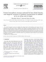
Spain (93Æ6%, Fig. 1). Combined resistance to clindamycin and erythromycin was much more common
(highest prevalence 91% in Spain) than resistance to
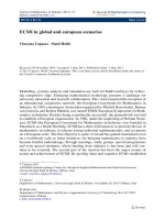
Figure 1. Comparison of prevalence of skin colonization by antibiotic-resistant propionibacteria among patients in six European centres as
determined by direct plating, and testing a randomly chosen colony from the non selective medium. Key: bar chart (a) shows the prevalence of
erythromycin resistance; (b) shows the prevalence of clindamycin resistance; (c) shows the prevalence of tetracycline resistance; and (d) shows the
prevalence of resistance to any one of the antibiotics tested. The proportion of colonized patients is expressed as a percentage of the number of
patients from whom viable propionibacteria were recovered as follows: Greece, n ¼ 142; Hungary, n ¼ 63; Italy, n ¼ 117; Spain, n ¼ 78;
Sweden, n ¼ 116; U.K., n ¼ 106. The upper limit of the bar (darker) shows the rate as determined by direct plating, while the lower bar (lighter)
shows the rate as determined by testing a randomly chosen colony from the non-selective medium. ***P < 0Æ0001, **P < 0Æ001, *P < 0Æ05,
compared with the U.K. rate as determined by direct plating (derived using v2). All other values show no significant difference from U.K. rates.
Ó 2003 British Association of Dermatologists, British Journal of Dermatology, 148, 467–478
ANTIBIOTIC-RESISTANT ACNE IN EUROPE
the tetracyclines (highest prevalence 26Æ4% in the
U.K.). No isolates resistant to tetracycline were detected
in Hungary or Italy (Fig. 1). Prevalence rates for
erythromycin and clindamycin-resistant propionibacteria were significantly elevated in Greece and Spain
compared with the U.K. In contrast, prevalence rates for
tetracycline-resistant isolates were significantly lower at
all sites outside the U.K. No minocycline-resistant
propionibacteria were found in any of the samples.
Resistance rates were seriously underestimated when
randomly selected isolates from the non-selective plates
were screened for resistance using antibiotic impregnated discs, the method used in many previous studies
(Fig. 1). The ratio of the prevalence rate determined by
random selection of a colony vs. direct plating varied
between 0Æ6 (the best, in Spain), and 0Æ12 (the worst,
in Hungary)—an eightfold reduction in the apparent
prevalence of resistant isolates. Recoveries of viable
473
propionibacteria on the non selective medium were
similar for all six centres.
The effect of treatment on the prevalence of resistance and
the population density of antibiotic-resistant propionibacteria
Treatment effects on the prevalence of resistance
were explored by pooling data from all six sites.
When the current treatment regimen included any
tetracycline, patients were significantly more likely to
be colonized with tetracycline-resistant organisms
compared with untreated patients (Table 4). The most
selective agent appeared to be minocycline. However, the highest prevalence of tetracycline-resistant
propionibacteria was detected among patients receiving oral therapy with a non tetracycline antibiotic.
The most likely reason for this is that in the U.K. a
Table 4. Effect of current treatment regimen on the prevalence and population density (growth score) of tetracycline-resistant and erythromycinresistant propionibacteria
No. of patients
treated with
tetracyclines (n)
Tetracycline (40)
Minocycline (57)
Doxycycline (17)
Any tetracycline (114)
Any other oral antibiotic (33)c
No treatment (196)
All patients (622)
No. of patients
treated with MLS
antibiotics (n)
Topical erythromycin (124)d
Topical clindamycin (71)
Any oral macrolide (10)e
Any MLS antibiotic (202)
Any oral antibiotic (147)
No treatment (196)
All patients (622)
Population density
(median growth score)
of propionibacteriab
No. of patients (%)
colonized with
tetracycline-resistant
isolates, TETR
P value
(Fisher’s
exact test)a
TETR
Susceptible and resistant
P value
(Mann–Whitney)a
5 (12Æ5%)
10 (17Æ5%)
2 (11Æ8%)
17 (14Æ9%)
8 (24Æ2%)
13 (6Æ6%)
58 (9Æ35)
NS
0Æ02
NS
0Æ03
0Æ004
comparator
N⁄A
3
3
3
3
2
3
3
3
3
3
3
3
4
3
NS
0Æ01
NS
0Æ017
0Æ0013
comparator
N⁄A
No. of patients (%)
colonized with
erythromycin-resistant
isolates, ERYR
93
52
6
149
81
119
387
(75%)
(73Æ2%)
(60%)
(73Æ4%)
(55Æ1%)
(60Æ7%)
(62Æ2%)
Population density
(median growth score)
of propionibacteriab
P value
(Fisher’s
exact test)a
ERYR
0Æ01
NS
NS
0Æ007
NS
comparator
N⁄A
4
2
2
3
4
3
3
Susceptible and resistant
4
3
2Æ5
3
3
4
3
P value
(Mann–Whitney)a
0Æ003
NS
NS
0Æ006
NS
comparator
N⁄A
MLS, macrolide–lincosamide–streptogramin B-resistant strains. TETR, tetracycline-resistant propionibacteria; ERY, erythromycin-resistant propionibacteria. aVersus no treatment. Differences in prevalence rates between treatments were tested using Fisher’s exact test. Differences between
growth scores of TETR and ERYR propionibacteria obtained in each treatment group were compared with those of antibiotic sensitive and resistant
propionibacteria isolated on non selective plates from the untreated group using the Mann–Whitney U-test. A significance level of 5% was used
with two-tailed tests. bThe median values are shown for patients colonized with antibiotic-resistant P. acnes. cMost of these patients were from the
U.K. (24 of 33) where patients with tetracycline-resistant floras are deliberately switched to trimethoprim. Ten of the 13 patients with tetracyclineresistant organisms on a non-tetracycline antibiotic were U.K. patients on trimethoprim. The others were two patients from Italy on azithromycin
and one from Sweden on oral erythromycin. dTwo patients were being treated concomitantly with topical erythromycin and topical clindamycin.
e
One patient was being treated concomitantly with oral erythromycin and topical clindamycin.
Ó 2003 British Association of Dermatologists, British Journal of Dermatology, 148, 467–478
474
J . I . R O S S et al.
high proportion of patients who carried tetracyclineresistant propionibacteria were switched from tetracycline treatment to trimethoprim. When the current
therapy included an MLS antibiotic, patients were
significantly more likely to be colonized by erythromycin-resistant propionibacteria compared with untreated patients (Table 4).
Treatment effects on the population density indicated
by the measure of growth score of antibiotic-resistant
propionibacteria were explored by pooling data from all
six sites (Table 4). The population density of tetracycline-resistant propionibacteria was elevated significantly among patients taking any tetracycline and in
the minocycline-treated patients. The growth scores of
erythromycin-resistant isolates were also increased
significantly among patients receiving treatment with
an MLS antibiotic, and were most elevated in patients
using topical erythromycin.
When data from all six centres were combined,
current treatment regimens, including benzoyl peroxide, reduced neither the prevalence (P ¼ 0Æ97) nor the
population density (P ¼ 0Æ62) of erythromycin-resistant isolates compared with other regimens. However,
when data from the centre in Spain were omitted, the
reductions in both prevalence (P ¼ 0Æ006) and population density (P ¼ 0Æ002) became highly significant.
Carriage of antibiotic-resistant propionibacteria by
untreated contacts of acne patients
Carriage rates of resistant propionibacteria on the skin
of untreated close contacts of the patients were 41% in
Hungary, 51% in Italy, 70% in Greece and 86% in
Spain. Twenty-five of 39 dermatologists (64%) were
also colonized on the face with resistant propionibacteria, including all those who specialized in treating
acne. In contrast, none of 27 physicians working in
other outpatient departments harboured resistant
propionibacterial isolates.
Phenotypic and genetic analysis of antibiotic-resistant
propionibacteria
A total of 515 antibiotic-resistant propionibacteria
were isolated from 664 patients and 39 dermatologists.
The susceptibilities of the 515 resistant isolates to 12
antibiotics, including seven MLS antibiotics were
determined by agar dilution together with 71 fully
susceptible isolates (12 per country but only 11
available from Spain). P. acnes was the most commonly
isolated resistant organism (65% of strains) with
P. granulosum (34% of strains) less commonly seen.
P. avidum only accounted for 1% of resistant strains.
Resistance to erythromycin and clindamycin with
tetracycline susceptibility was the most common profile, with 80% of strains demonstrating this phenotype.
This was also the most common profile in every
country tested. MIC values for erythromycin ranged
from 4 to > 2048 lg mL)1 (mode > 2048 lg mL)1).
Clindamycin MIC values were between 1 and
> 512 lg mL)1 (mode 128 lg mL)1). Combined resistance to erythromycin, clindamycin and tetracycline
accounted for 12Æ5% of strains, mostly from the U.K.
and Sweden. Resistance to tetracycline alone was
uncommon (1Æ4% of strains). No tetracycline-resistant
strains were isolated from Italy or Hungary.
Tetracycline resistance and base mutations
Tetracycline MICs of strains resistant to tetracycline
were in the range 8–64 lg mL)1 (mode 32 lg mL)1).
All of these strains were more susceptible to doxycycline (MIC 1–16 lg mL)1) and minocycline (0Æ5–
4 lg mL)1). Partial sequences across the helix 34 region
of the 16S rRNA gene were determined for a total of 20
tetracycline-resistant strains (at least three from each
country). In 19 of 20 a single base change, G fi C at E.
coli equivalent base 1058, was identified. In contrast
none of three sensitive strains from each of Sweden,
Spain, Greece and the U.K. possessed this base change.
Classification of macrolide–lincosamide–streptogramin
B-resistant strains
The 508 isolates that were resistant to MLS antibiotics
were classified into resistance groups I–IV4 based on
their resistance patterns to eight MLS antibiotics. At
least three isolates from each country that were
assigned to groups I and IV were sequenced across
the peptidyl transferase region of 23S rRNA, and the
presence of mutations at E. coli equivalent base 2058
or 2059, respectively, was confirmed. The numbers of
strains exhibiting each phenotype from each country is
displayed in Table 5. Table 6 shows the range of MIC
values to a panel of eight MLS antibiotics for each
phenotype given in Table 5. As expected, the majority
of isolates belonged to phenotypic classes associated
with a 2058 or 2059 rRNA base mutation with group I
(2058) the most common in all countries tested (64–
80% of strains resistant to MLS antibiotics). No strains
were assigned to group III (2057 base mutation).
Forty-five of 486 erythromycin-resistant isolates with
Ó 2003 British Association of Dermatologists, British Journal of Dermatology, 148, 467–478
ANTIBIOTIC-RESISTANT ACNE IN EUROPE
475
Table 5. Phenotypic resistance groups of cutaneous propionibacteria resistant to macrolide–lincosamide–streptogramin B-resistant strain antibiotics
Phenotypic resistance groupsa
Country
No. of
resistant
strains
Greece
Hungary
Italy
Spain
Sweden
U.K.
Total
130
47
97
59
100
75
508
Group I
(2058
mutation)
99
36
79
38
65
51
367
Group IV
(2059
mutation)
Group II
erm(X)
(75Æ6%)
(76Æ6%)
(81Æ4%)
(64Æ4%)
(65%)
(68%)
(72%)
19
6
3
8
7
2
45
(14Æ5%)
(12Æ7%)
(3Æ1%)
(13Æ6%)
(7Æ0%)
(2Æ7%)
(8Æ9%)
5
3
10
13
2
17
52
(3Æ8%)
(6Æ4%)
(10Æ3%)
(22Æ0%)
(2Æ0%)
(22Æ7%)
(10Æ2%)
Group V
CLNR only
0
0
3
0
19
0
22
Unclassifiable
resistance
phenotypes
(0%)
(0%)
(3Æ1%)
(0%)
(19Æ0%)
(0%)
(4Æ3%)
8
2
2
0
7
5
24
(6Æ1%)
(4Æ2%)
(2Æ1%)
(0%)
(7Æ0%)
(6Æ7%)
(4Æ7%)
CLNR, Clindamycin-resistant propionibacteria. aThe minimum inhibitory concentration ranges used to define the phenotypic resistance groups are
as defined previously.4
Table 6. Minimum inhibitory concentrations (MICs) of macrolide–lincosamide–streptogramin B-resistant strain antibiotics for erythromycinsusceptible and -resistant propionibacteria4
MIC (lg L)1)
Resistance
group4
(no. of isolates)
23S rRNA base mutation ⁄
resistance gene
I (367)
II (45)
III (0)
IV (52)
V (22)
Susceptible (71)
2058
erm(X)
2057
2059
Unknown
None
ERY
TEL
AZM
TYL
SPI
JOS
CLN
PRS
512 ‡ 2048
> 2048
1–2
> 2048
£ 0Æ125
£ 0Æ125
0Æ5–4
> 512
£ 0Æ03
0Æ5–2
£ 0Æ03
£ 0Æ03
256 ‡ 512
> 512
£ 0Æ25
‡ 512
£ 0Æ25
£ 0Æ25
128 ‡ 512
> 512
£2
‡ 512
£2
£2
1–256
> 512
£2
‡ 512
£2
£2
0Æ5–128
> 512
£ 0Æ125
‡ 512
£ 0Æ125
£ 0Æ125
4–512
‡ 512
£ 0Æ5
1–64
2–4
£ 0Æ5
8 ‡ 256
‡ 256
1–8
2–128
1–16
1–16
ERY, erythromycin; TEL, telithromycin (HMR 3647); AZI, azithromycin; TYL, tylosin; SPI, spiramycin; JOS, josamycin; CLN, clindamycin; PRS,
pristinamycin IA.
at least two from each country were found to carry the
recently described erm(X) resistance determinant4 and
were uniformly resistant at high level (MICs
‡ 512 lg mL)1) to all MLS antibiotics tested.
Twenty-two strains, mainly from Sweden (20
strains) had raised MICs to clindamycin only (MIC 2–
4 lg mL)1). The genetic basis of this resistance is not
known. Strains with this phenotype have been assigned
to the new resistance group V (Table 5).
Twenty-four isolates (4Æ7% of the total) displayed
miscellaneous cross-resistance patterns and could not
be classified into any group. Sequence analysis of
selected isolates revealed no mutations in the peptidyl
transferase region of 23S rRNA. These strains were not
studied further (Table 5).
Discussion
Prevalence of antibiotic-resistant propionibacteria isolated
from six European centres
The aim of this study was to estimate the size of the
resistance problem in Europe and to link prescribing
behaviour to resistance patterns. Our findings confirm for acne what we know from other infections—that while propionibacterial resistance does
not respect national boundaries, local antibiotic use
does indeed influence the distribution of resistant
isolates. Skin colonization by antibiotic-resistant propionibacteria was common in all six centres and
overall two-thirds of patients were colonized with
resistant strains. Unfortunately, prevalence data for
other countries have not been collected using uniform methodology, and resistance rates have often
been estimated by screening isolates from a nonselective medium.6 Population densities of resistant
isolates were invariably lower or equal to those of
the total propionibacterial population (data not
shown) so that selecting single colonies at random
from non-selective plates underestimates resistance.
We urge anyone wishing to study propionibacterial
resistance to use direct plating on to breakpoint
concentrations of antibiotics as the means of detecting resistant isolates otherwise they are likely to be
falsely reassured by low but inaccurate resistance
rates.
Ó 2003 British Association of Dermatologists, British Journal of Dermatology, 148, 467–478
476
J . I . R O S S et al.
Analysis of treatment histories and prescribing habits
has shed some light on drivers of resistance. Summary
statistics show that oral tetracyclines prescribed for
acne promote propionibacterial resistance to them.
Although the evidence confirms minocycline as a
driver, numbers treated with other tetracyclines were
too small to confirm or refute the selectivity of these
agents. We were unable to detect any propionibacteria
from any centre with minocycline resistance. We
advise extreme caution when interpreting bacterial
growth on minocycline-containing media as the drug is
unstable during prolonged incubation at 37 °C. Occasionally isolates appeared on minocycline-containing
plates, but in every case were subsequently shown in
MIC determinations to be susceptible to minocycline.
However, MIC testing revealed that some tetracyclineresistant isolates show reduced susceptibility to minocycline (£ 4 lg mL)1) as has been previously shown
for isolates of P. acnes from the U.K. and elsewhere.
To date, minocycline-resistant propionibacteria (MIC
8–16 lg mL)1) have been detected only in the U.S.A.7
Paradoxically, patients on treatment with non tetracycline oral antimicrobials at the time of sampling were
the most likely to be colonized by tetracycline-resistant
propionibacteria. In the U.K. centre at least, it is
standard practice to switch patients unresponsive to
therapy with tetracyclines to a different oral regimen
(such as trimethoprim), and this strategy may have led
to this unexpected finding. The results also show that
resistance to erythromycin and clindamycin is promoted by treatment with an MLS antibiotic, with the
selectivity of topical erythromycin clearly demonstrated. There was also more resistance to erythromycin in
topical clindamycin treated patients, although this
increase compared with untreated patients just failed
to reach statistical significance (P ¼ 0Æ06). Because
most patients had been treated with more than one
course of antibiotics, the resistance status of the
patients when they were sampled was influenced by
both past and current treatments. Even among patients
not on treatment when sampled, a majority were
colonized by resistant isolates.
We can draw some additional conclusions with
respect to drivers of resistance in propionibacteria. In
Greece, patients were less likely to be prescribed an
antibiotic for their acne than anywhere else. Despite
this, resistance rates were second only to Spain. The
most commonly used antibiotic in Greece was topical
clindamycin and topical erythromycin was very little
used. These observations suggest that topical clindamycin drives resistance to itself and to erythromycin.
This would be expected as both mutational and
acquired resistance confers cross-resistance to both
antibiotics. There is one caveat; antibiotics are freely
available in Greece without prescription, and nonrecorded use of other agents may have contributed to
the high rates of resistance observed.
The Hungarian centre was the most isolated in
geographical terms and patients there had fewest
opportunities for travel outside national borders. Fewer
patients were undergoing treatment when sampled and
they were less likely to have been treated at any time
with an antibiotic for their acne. This reduced exposure
to selective pressure was reflected both in lower
prevalence rates of resistant organisms and also in
their lower population densities on the skin (data not
shown). Tetracyclines are rarely prescribed in Hungary
and resistance to tetracyclines was not detected.
Resistance to tetracyclines was also not detected in
Italy despite the high usage of minocycline. Courses of
minocycline for acne at this site were restricted to
2 months by national guidelines, which may limit the
selectivity of the drug. Other tetracyclines were only
infrequently prescribed for acne and national usage of
tetracyclines for all indications is the lowest in the
European Union.13
In Spain, patients were almost always prescribed an
antibiotic, most commonly topical erythromycin, and
cumulatively they had received the greatest number of
courses of antibiotics for their acne. Unsurprisingly,
erythromycin resistance rates and population densities
of resistant organisms were highest in Spain. Benzoyl
peroxide was invariably coprescribed with erythromycin in the Spanish centre. A combined formulation is
available in most European countries but not in Spain.
As a broad-spectrum bactericidal agent, benzoyl peroxide should have acted as an antiresistance agent, and
prescribing it together with antibiotics makes good
sense on theoretical grounds.14 Why it appears to have
reduced resistance rates outside but not within Spain is
not easily explained, although variation in compliance
may have been an issue. Owing to the national high
usage of MLS antibiotics for a variety of indications,
selective pressure associated with non acne prescribing
may also have exacerbated the erythromycin resistance
problem in Spain. Conversely, rates of resistance to
tetracyclines were very low despite high usage for acne
treatment.
Sweden is well known for its restrictive policies
regarding the licensing and use of antibiotics. It has the
lowest usage rate of MLS antibiotics in the European
Union.13 Few antibiotics are licensed for acne
Ó 2003 British Association of Dermatologists, British Journal of Dermatology, 148, 467–478
ANTIBIOTIC-RESISTANT ACNE IN EUROPE
treatment. The most commonly used antiacne antibiotic is topical clindamycin. Oral tetracycline and
occasionally oral erythromycin are also used. The
results from Sweden add further support to the
interpretation (based on the Greek data) that topical
clindamycin drives resistance both to itself and to
erythromycin.
The overall resistance rate in Sweden was virtually
identical to the U.K., where all possible antibioticcontaining products can be prescribed but where few
patients had been treated with topical clindamycin.
Propionibacterial resistance rates have been monitored
in the U.K. centre for the past 10 years. They peaked in
1997 when 64% of patients were colonized with one or
more resistant strains.8 Since then, prescribing practices have been modified to reduce use of topical
erythromycin and clindamycin. The U.K. is the only
centre to use trimethoprim, normally considered thirdline therapy for acne. These changes halted the steady
rise in resistance rates, which to date remain below the
1997 peak.
Carriage of antibiotic-resistant propionibacteria by the
contacts of acne patients
Concordance between resistance rates among patients
and their contacts suggests that selective pressure
extends to contacts and that resistant strains may be
transferred between them. Untreated siblings and even
offspring of acne patients may be colonized de novo at
puberty with resistant isolates. Within a family it is
easy to understand how isolates can be transferred
between individuals, but we must not overlook the role
of the dermatologist. All the dermatologists whose
patients were sampled in this study were colonized on
the face by erythromycin ⁄ clindamycin-resistant propionibacteria. However, none of the physicians working
elsewhere in the hospital who were sampled were
colonized. This raises the very distinct possibility that
dermatologists may transfer resistant isolates from
their own or from other patients’ skin to previously
uncolonized patients during clinic visits.
Phenotypic and genetic analysis of antibiotic-resistant
propionibacteria
Selection of ribosomal mutations leading to resistance
to the MLS antibiotics occurred in patients at all the
centres tested despite differences in the treatments
used. It seems clear that the use of oral or topical
erythromycin drives the selection of ribosomal muta-
477
tions, but mutations are also common in Sweden and
Greece where topical clindamycin is used extensively
with only sparing use of erythromycin. The transposon
based erm(X) resistance determinant accounted for
8Æ9% of MLS-resistant isolates and was detected in all
six countries. This resistance determinant gives greater
protection against clindamycin than the ribosomal
mutations and we may speculate that the use of topical
clindamycin selects for erm(X). We may expect the
prevalence of the transposon to increase if topical
treatments continue to be used widely. In Sweden, but
not Greece, otherwise susceptible strains with raised
MIC values for clindamycin have emerged (assigned to
phenotypic group V). This low-level resistance would
be of little protection against the high surface concentrations of clindamycin achieved by topical treatment;
however, it may confer a selective advantage in skin
areas not directly treated.
Implications for the future management of acne
It has been argued that the most likely effect of
resistance is to reduce the clinical efficacy of antibioticbased treatment regimens below that which would
occur in patients with fully susceptible floras.15,16 The
extent of this reduction will depend upon many factors,
including the route of administration and compliance.
Antibiotics have represented one of the cornerstones of acne management for over 30 years. Most
doctors consider antibiotics necessary, representing
the most powerful agents against inflammatory
lesions in patients for whom oral isotretinoin is not
appropriate. Direct anti-inflammatory activity has
been ascribed to them. There are several learning
points from this study. Resistance in the target
organisms is widespread, and should be considered
as a possible cause of unsatisfactory improvement.
Although acne itself is not infectious, resistant
propionibacteria may be transmissible between susceptible individuals. Doctors who routinely palpate
patients’ skin to assess acne severity should use crossinfection control measures to avoid transferring
resistant isolates between patients.
Propionibacterial resistance to the tetracyclines is
not a significant issue in most countries. Taken in
isolation our results suggest that tetracyclines should
be first-line and topical erythromycin and clindamycin
second-line antibiotics for acne. However, oral antibiotics select for the overgrowth of resistant bacteria at
all body sites supporting a resident microflora. The
consequences of a switch to increased prescribing of
Ó 2003 British Association of Dermatologists, British Journal of Dermatology, 148, 467–478
478
J . I . R O S S et al.
tetracyclines may be more resistance to multiple antibiotics in bacterial species other than propionibacteria.
Another strategy to minimize and overcome resistance
would be to prescribe topical combination therapies
based on an antibiotic with a broad-spectrum antibacterial agent—only benzoyl peroxide and zinc are
available for acne therapy at the present time. Some
dermatologists in our study already employ such
regimens. Selective pressure can also be reduced by
keeping antibiotic courses short and by not using
antibiotics for maintenance therapy.
5
6
7
8
9
Acknowledgments
10
We thank Dermik Laboratories Inc (Aventis) and the
Leeds Foundation for Dermatological Research for
providing financial support, Katerina Mourelatos and
Jennifer Lewis for assisting with sample collection in
Greece and Hungary.
11
12
References
1 Ross JI, Eady EA, Cove JH et al. Clinical resistance to erythromycin
and clindamycin in cutaneous propionibacteria isolated from
acne patients is associated with mutations in 23S rRNA. Antimicrob Agents Chemother 1997; 41: 1162–5.
2 Ross JI, Eady EA, Cove JH, Cunliffe WJ. 16S rRNA mutation
associated with tetracycline resistance in a gram-positive bacterium. Antimicrob Agents Chemother 1998; 42: 1702–5.
3 Eady EA, Ross JI, Cove JH et al. Macrolide–lincosamide–streptogramin B (MLS) resistance in cutaneous propionibacteria:
definition of phenotypes. J Antimicrob Chemother 1989; 23: 493–
502.
4 Ross JI, Eady EA, Carnegie E, Cove JH. Detection of transposon
Tn5432-mediated macrolide–lincosamide–streptogramin B (MLS
13
14
15
16
B) resistance in cutaneous propionibacteria from six European
cities. J Antimicrob Chemother 2002; 49: 165–8.
Leyden JJ. Antibiotic resistant acne. Cutis 1976; 17: 593–6.
Cooper AJ. Systematic review of Propionibacterium acnes resistance
to systemic antibiotics. Med J Aust 1998; 169: 259–61.
Ross JI, Snelling AM, Eady EA et al. Phenotypic and genotypic
characterization of antibiotic-resistant Propionibacterium acnes
isolated from acne patients attending dermatology clinics in
Europe, the U.S.A., Japan and Australia. Br J Dermatol 2001;
144: 339–46.
Coates P, Vyakrnam S, Eady EA et al. Prevalence of antibiotic
resistant propionibacteria on the skin of acne patients: 10-year
surveillance data and snapshot distribution study. Br J Dermatol
2002; 146: 840–8.
Burke BM, Cunliffe WJ. The assessment of acne vulgaris—the
Leeds technique. Br J Dermatol 1984; 111: 83–92.
Williamson P, Kligman AM. A new method for the quantitative
investigation of cutaneous bacteria. J Invest Dermatol 1965; 45:
498–503.
Marples RR, McGinley KJ. Corynebacterium acnes and other anaerobic diptheroids from human skin. J Med Microbiol 1974; 7:
349–57.
National Committee for Clinical Laboratory Standards. Methods
for Antimicrobial Susceptibility Testing of Anaerobic Bacteria;
Approved Standard, 4th edn. NCCLS publication no. M11-A4.
Villanova, PA: National Committee for Clinical Laboratory
Standards, 1997.
Cars O, Mo¨lstad S, Melander A. Variation in antibiotic use in the
European Union. Lancet 2001; 357: 1851–3.
Farmery MR, Jones CE, Eady EA et al. In vitro activity of azelaic
acid, benzoyl peroxide and zinc acetate against antibiotic-resistant propionibacteria from acne patients. J Dermatol Treat 1994; 5:
63–5.
Leyden JJ, McGinley KL, Cavalieri A et al. Propionibacterium acnes
resistance to antibiotics in acne patients. J Am Acad Dermatol
1983; 8: 41–5.
Eady EA, Cove JH, Holland KT, Cunliffe WJ. Erythromycin
resistant propionibacteria in antibiotic treated acne patients:
association with therapeutic failure. Br J Dermatol 1989; 121:
51–7.
Ó 2003 British Association of Dermatologists, British Journal of Dermatology, 148, 467–478