The mycota v, plant relationships 2nd ed k esser, h deising (springer, 2009)
Bạn đang xem bản rút gọn của tài liệu. Xem và tải ngay bản đầy đủ của tài liệu tại đây (9.38 MB, 405 trang )
The Mycota
Edited by
K. Esser
The Mycota
I
Growth, Differentiation and Sexuality
1st edition ed. by J.G.H.Wessels and F. Meinhardt
2nd edition ed. by U. Kües and R. Fischer
II
Genetics and Biotechnology
Ed. by U. Kück
III
Biochemistry and Molecular Biology
Ed. by R. Brambl and G. Marzluf
IV
Environmental and Microbial Relationships
1st edition ed. by D. Wicklow and B. Söderström
2nd edition ed. by C.P. Kubicek and I.S. Druzhinina
V
Plant Relationships
1st edition ed. by G. Carroll and P. Tudzynski
2nd edition ed. by H.B. Deising
VI
Human and Animal Relationships
1st edition ed. by D.H. Howard and J.D. Miller
2nd edition ed. by A.A. Brakhage and P.F. Zipfel
VII
Systematics and Evolution
Ed. by D.J. McLaughlin, E.G. McLaughlin, and P.A. Lemke†
VIII
Biology of the Fungal Cell
Ed. by R.J. Howard and N.A.R. Gow
IX
Fungal Associations
Ed. by B. Hock
X
Industrial Applications
1st edition ed. by H.D. Osiweacz
2nd edition ed. by M. Hofrichter and R. Ullrich
XI
Agricultural Applications
Ed. by F. Kempken
XII
Human Fungal Pathogens
Ed. by J.E. Domer and G.S. Kobayashi
XIII
Fungal Genomics
Ed. by A.J.P. Brown
XIV
Evolution of Fungi and Fungal-like Organisms
Ed. by J. Wöstemeyer
XV
Physiology and Genetics: Selected Basic and Applied Aspects
Ed. by T. Anke and D. Weber
The Mycota
A Comprehensive Treatise
on Fungi as Experimental Systems
for Basic and Applied Research
Edited by K. Esser
V
Plant Relationships
2nd Edition
Volume Editor:
H.B. Deising
Series Editor
Professor Dr. Dr. h.c. mult. Karl Esser
Allgemeine Botanik
Ruhr-Universität
44780 Bochum, Germany
Tel.: +49 (234)32-22211
Fax.: +49 (234)32-14211
e-mail:
Volume Editor
Professor Dr. Holger B. Deising
Naturwissenschaftliche Fakultät III
Institut für Agrar- und Ernährungswissenschaften
Phytopathologie und Pflanzenschutz
Ludwig-Wucherer-Str. 2
06099 Halle (Saale), Germany
Tel.: +49 345 5522660
Fax: +49 345 5527120
e-mail:
Library of Congress Control Number: 2008937452
ISBN 978-3-540-87406-5
e-ISBN 978-3-540-87407-2
ISBN 3-540-58006-9 (Part A)
ISBN 3-540-62018-4 (Part B) 1st ed.
This work is subject to copyright. All rights are reserved, whether the whole or part of the material is concerned, specifically the
rights of translation, reprinting, reuse of illustrations, recitation, broadcasting, reproduction on microfilm or in any other way, and
storage in data banks. Duplication of this publication or parts thereof is permitted only under the provisions of the German Copyright Law of September 9, 1965, in its current version, and permissions for use must always be obtained fromSpringer-Verlag.
Violations are liable for prosecution under the German Copyright Law.
springer.com
© Springer-Verlag Berlin Heidelberg 1997, 2009
The use of general descriptive names, registered names, trademarks, etc. in this publication does not imply, even in the absence of
a specific statement, that such names are exempt from the relevant protective laws and regulations and therefore free for general
use.
Cover design: Erich Kirchner and WMXDesign GmbH, Heidelberg, Germany
Printed on acid-free paper
5 4 3 2 1 0
Karl Esser
(born 1924) is retired Professor of General Botany and Director
of the Botanical Garden at the Ruhr-Universität Bochum (Germany). His scientific work focused on basic research in classical
and molecular genetics in relation to practical application. His
studies were carried out mostly on fungi. Together with his collaborators he was the first to detect plasmids in higher fungi. This
has led to the integration of fungal genetics in biotechnology. His
scientific work was distinguished by many national and international honors, especially three honorary doctoral degrees.
Holger B. Deising
(born 1956) studied agricultural sciences and botany at the
University of Kiel, Germany. His PhD thesis focused on nitrate
reduction by Sphagnum species. After graduating in 1987 he
worked on the infection structures of plant-pathogenic rust fungi
and he qualified as a lecturer at the University of Konstanz in
1996. In 1997 he became a Full Professor for Phytopathology and
Plant Protection at Martin-Luther-University Halle-Wittenberg.
His research stays include McMaster University (Hamilton, ON,
Canada), the University of Georgia (Athens, GA, USA), and Purdue University (West Lafayetta, IN, USA). His scientific interest is
directed to various aspects of fungus–plant interactions, with special focus on the differentiation and function of fungal infection
structures and pathogenicity factors in the causal agent of maize
anthracnose and stalk rot, Colletotrichum graminicola. Another
area of research includes molecular mechanisms conferring fungicide resistance in several plant-pathogenic fungi.
Series Preface
Mycology, the study of fungi, originated as a subdiscipline of botany and was a descriptive discipline, largely neglected as an experimental science until the early years of this
century. A seminal paper by Blakeslee in 1904 provided evidence for selfincompatibility, termed “heterothallism”, and stimulated interest in studies related to the control
of sexual reproduction in fungi by mating-type specificities. Soon to follow was the
demonstration that sexually reproducing fungi exhibit Mendelian inheritance and that
it was possible to conduct formal genetic analysis with fungi. The names Burgeff, Kniep
and Lindegren are all associated with this early period of fungal genetics research.
These studies and the discovery of penicillin by Fleming, who shared a Nobel Prize
in 1945, provided further impetus for experimental research with fungi. Thus began a
period of interest in mutation induction and analysis of mutants for biochemical traits.
Such fundamental research, conducted largely with Neurospora crassa, led to the one
gene: one enzyme hypothesis and to a secondNobel Prize for fungal research awarded to
Beadle and Tatum in 1958. Fundamental research in biochemical genetics was extended
to other fungi, especially to Saccharomyces cerevisiae, and by the mid-1960s fungal
systems were much favored for studies in eukaryotic molecular biology and were soon
able to compete with bacterial systems in the molecular arena.
The experimental achievements in research on the genetics andmolecular biology
of fungi have benefited more generally studies in the related fields of fungal biochemistry, plant pathology,medicalmycology, and systematics. Today, there ismuch interest in
the geneticmanipulation of fungi for applied research. This current interest in biotechnical genetics has been augmented by the development of DNA-mediated transformation systems in fungi and by an understanding of gene expression and regulation at the
molecular level. Applied research initiatives involving fungi extend broadly to areas of
interest not only to industry but to agricultural and environmental sciences as well.
It is this burgeoning interest in fungi as experimental systems for applied as well as
basic research that has prompted publication of this series of books under the title The
Mycota. This title knowingly relegates fungi into a separate realm, distinct from that of
either plants, animals, or protozoa. For consistency throughout this Series of Volumes
the names adopted for major groups of fungi (representative genera in parentheses) are
as follows:
Pseudomycota
Division:
Division:
Oomycota (Achlya, Phytophthora, Pythium)
Hyphochytriomycota
Eumycota
Division:
Division:
Division:
Chytridiomycota (Allomyces)
Zygomycota (Mucor, Phycomyces, Blakeslea)
Dikaryomycota
viii
Subdivision:
Class:
Class:
Subdivision:
Class:
Class:
Series Preface
Ascomycotina
Saccharomycetes (Saccharomyces, Schizosaccharomyces)
Ascomycetes (Neurospora, Podospora, Aspergillus)
Basidiomycotina
Heterobasidiomycetes (Ustilago, Tremella)
Homobasidiomycetes (Schizophyllum, Coprinus)
We have made the decision to exclude from The Mycota the slime molds which, although
they have traditional and strong ties to mycology, truly represent nonfungal forms
insofar as they ingest nutrients by phagocytosis, lack a cell wall during the assimilative
phase, and clearly show affinities with certain protozoan taxa.
The Series throughoutwill address three basic questions:what are the fungi,what do
they do, andwhat is their relevance to human affairs? Such a focused and comprehensive
treatment of the fungi is long overdue in the opinion of the editors.
A volume devoted to systematics would ordinarily have been the first to appear
in this Series. However, the scope of such a volume, coupled with the need to give
serious and sustained consideration to any reclassification of major fungal groups, has
delayed early publication. We wish, however, to provide a preamble on the nature of
fungi, to acquaint readers who are unfamiliar with fungi with certain characteristics
that are representative of these organisms and which make them attractive subjects for
experimentation.
The fungi represent a heterogeneous assemblage of eukaryotic microorganisms. Fungal metabolism is characteristically heterotrophic or assimilative for organic carbon and
some nonelemental source of nitrogen. Fungal cells characteristically imbibe or absorb,
rather thaningest,nutrients andtheyhave rigid cellwalls.The vastmajorityof fungi are
haploid organisms reproducing either sexually or asexually through spores. The spore
forms and details on theirmethod of production have been used to delineate most fungal
taxa.Although there is amultitude of spore forms, fungal spores are basically only of two
types: (i) asexual spores are formed followingmitosis (mitospores) and culminate vegetative growth, and (ii) sexual spores are formed following meiosis (meiospores) and are
borne in or upon specialized generative structures, the latter frequently clustered in a
fruit body. The vegetative forms of fungi are either unicellular, yeasts are an example, or
hyphal; the latter may be branched to form an extensive mycelium.
Regardless of these details, it is the accessibility of spores, especially the direct
recovery of meiospores coupled with extended vegetative haploidy, that have made
fungi especially attractive as objects for experimental research.
The ability of fungi, especially the saprobic fungi, to absorb and grow on rather simple
and defined substrates and to convert these substances, not only into essential metabolites
but into important secondarymetabolites, is also noteworthy.Themetabolic capacities of
fungi have attracted much interest in natural products chemistry and in the production of
antibiotics and other bioactive compounds. Fungi, especially yeasts, are important in fermentation processes. Other fungi are important in the production of enzymes, citric acid
and other organic compounds as well as in the fermentation of foods.
Fungi have invaded every conceivable ecological niche. Saprobic forms abound,
especially in the decay of organic debris. Pathogenic forms exist with both plant and
animal hosts. Fungi even grow on other fungi. They are found in aquatic as well as soil
environments, and their spores may pollute the air. Some are edible; others are poisonous. Many are variously associated with plants as copartners in the formation of lichens
and mycorrhizae, as symbiotic endophytes or as overt pathogens. Association with animal systems varies; examples include the predaceous fungi that trap nematodes, the
microfungi that grow in the anaerobic environment of the rumen, the many insectas-
Series Preface
ix
sociated fungi and themedically important pathogens afflicting humans. Yes, fungi are
ubiquitous and important.
There are many fungi, conservative estimates are in the order of 100,000 species,
and there are many ways to study them, from descriptive accounts of organisms found
in nature to laboratory experimentation at the cellular and molecular level. All such
studies expand our knowledge of fungi and of fungal processes and improve our ability
to utilize and to control fungi for the benefit of humankind.
We have invited leading research specialists in the field of mycology to contribute
to this Series. We are especially indebted and grateful for the initiative and leadership
shown by theVolumeEditors in selecting topics and assembling the experts.We have all
been a bit ambitious in producing these Volumes on a timely basis and therein lies the
possibility of mistakes and oversights in this first edition.We encourage the readership
to draw our attention to any error, omission or inconsistency in this Series in order that
improvements can be made in any subsequent edition.
Finally, we wish to acknowledge the willingness of Springer-Verlag to host this
project, which is envisioned to require more than 5 years of effort and the publication
of at least nine Volumes.
Bochum, Germany
Auburn, AL, USA
April 1994
KARL ESSER
PAUL A. LEMKE
Series Editors
Addendum to the Series Preface
In early 1989, encouraged by Dieter Czeschlik, Springer-Verlag, Paul A. Lemke and I
began to plan The Mycota. The first volume was released in 1994, 12 volumes followed in
the subsequent years, and two more volumes (Volumes XIV and XV) will be published
within the next few years. Unfortunately, after a long and serious illness, Paul A. Lemke
died in November 1995. Thus, it was my responsibility to proceed with the continuation
of this series, which was supported by JoanW. Bennett for Volumes X–XII.
The series was evidently accepted by the scientific community, because several
volumes are out of print. Therefore, Springer-Verlag has decided to publish completely
revised and updated new editions of Volumes I, II, III, IV, V, VI, VIII, and X. I am glad
that most of the volume editors and authors have agreed to join our project again.I
would like to take this opportunity to thank Dieter Czeschlik, his colleague, Andrea
Schlitzberger, and Springer-Verlag for their help in realizing this enterprise and for
their excellent cooperation for many years
Bochum, Germany
May 2008
KARL ESSER
Volume Preface to the Second Edition
Joseph G.H. Wessels, in a review on fungal growth and morphogenesis, described fungi
as the natural complement to plant life. He speculated that plants could probably have
arisen without animals evolving, but raised doubts whether plants could have ever
have evolved without the advent of fungi. The intimacy of trans-kingdom relationships
between fungi and plants could not have been circumscribed any clearer.
The first edition of volume V Plant Relationships was been published in The Mycota
series more than ten years ago. In their preface George Carroll and Paul Tudzynski ,the
editors, commented on the large number of fungal and vascular plant species (respectively estimated to be in the order of 106 and 300–350×103) and emphasized the enormous number of interactions displayed by fungi and plants. The large number of fungi
and plants reflects the complexity of the mechanisms of the interactions between them.
Therefore, only examples can be given of fungal lifestyles and their interactions with
plants, either mutualistic or pathogenic.
Significant methodological progress has been made in almost all areas of mycological research (e.g. transcriptomics, proteomics, metabolomics) since the first edition
was published. For example, molecular genetics has experienced strong support from
various genome sequencing efforts. To date, more than 50 fungal and several oomycete
genomes have been sequenced and genome-wide gene expression profiling, functional
screens for genes in yeasts or bacteria, the labelling of gene products, and the efficiency
of targeted inactivation of genes have improved. These advances were accompanied by
improvements in light and electron microscopy which, together with the utilization of
molecular tools, allowed a proportional development in our knowledge of the cell biology of fungal interactions. Last but not least, increased sensitivies in the detection of
biomolecules through analytical chemistry enabled our understanding of the chemical
basis of both mutualistic and pathogenic fungus–plant interactions.
The second edition of The Mycota, volume V, reflects the substantial progress made
in various areas of fungus–plant interactions. Organized in three parts, i.e. profiles in
pathogenesis and mutualism, mechanisms of pathogenic and mutualistic interactions,
and plant response to pathogen ingress, this book provides an overview of fundamental
aspects of fungal lifestyles.
Chapters 1–5 focus on different fungal systems, characterize their profiles, and give
examples both for pathogenic Phytophthora species belonging to the Oomycota and for
fungi with biotrophic, necrotrophic, or mutualistic lifestyles.
Chapters 6–16 focus on mechanisms of the interactions, with detailed discussions
on specific aspects, such as spore release and distribution (which are of critical importance to the success of pathogens), the basis of the specificity of fungus–plant interactions, and signal transduction. Protein secretion, the role played by cell wall-degrading
enzymes and toxins, and the induction of programmed cell death represent further
areas of research that significantly helped our understanding fungal pathogenicity.
The last four chapters of this section, 13–16, describe in detail mechanisms of mutualism, i.e. mycorrhizal and endophytic interactions, as well as interactions between
fungi and algae in lichens. Finally, chapters 17 and 18 describe the response of plants
xiv
Volume Preface to the Second Edition
towards attacking pathogenic fungi and focus on signal perception and transduction
and the mechanisms of defense.
Forty-six internationally acknowledged scientists specialized in different areas of
eukaryotic microbiology (mycology), microbial genetics and genomics, plant pathology, plant molecular biology, and plant genetics contributed to this volume. I would like
to express my gratitude to all authors for their tremendous efforts, to the series editor,
Karl Esser, who provided many helpful comments, and to Springer-Verlag for continuous assistance and patience.
I hope that the second edition of Plant Relationships, volume V in The Mycota series,
will be appreciated by a wide variety of professional biologists, as it allows one to keep
pace with the rapidly developing field of fungal research. In addition, the text and highquality illustrations may provide a source for teaching at graduate level; and the different chapters can be used by young researchers as a helpful introduction to the relevant
literature on fungus–plant interactions.
Halle (Saale), Germany
October 2008
HOLGER B. DEISING
Volume Editor
Volume Preface to the First Edition
The number of fungal species has been loosely estimated to be on the order of 1 million,
while the number of vascular plants is known with considerably greater certainty to lie
between 300000 and 350000. Clearly, any volume which purports to deal with interactions between these two vast assemblages of organisms must do so concisely and selectively. In the chapters to foIlow, we have made no attempt to be allinclusive, but rather
have chosen examples from which general conclusions about fungus/plant interactions
might be drawn. The materials presented here come from the core literature on plant
pathology and from research on fungal mutualisms and on evolutionary biology. A
variety of approaches are evident: biochemistry, molecular biology, cellular fine structure, genetics, epidemiology, population biology, ecology, and computer modeling. The
frequent overlap of such approach es within single reviews has resulted in a rich array
of insights into the factors which regulate fungus/plant interactions. In these chapters,
such interactions have also been considered on a variety of scales, both geographic and
temporal, from single plant cells to ecosystems, from interactions which occur within
minutes of contact to mechanisms which have presumably evolved during the course of
several hundred million years.
Volume V consists of two parts: Volume V, Part A, and Volume V, Part B. While section headings provide signposts, we wish to make the rationale for the organization
of these volumes absolutely dear. Part A begins with a brief introduction to both volumes. A series of reviews follows (Chaps. 1-6) which deal with the temporal sequence of
events from the time fungal spores make contact with a host plant until the point where
fungal hyphae are either firmly ensconced within a host or the attempted infections
have been repulsed. Chapters 7-12 deal with metabolic interactions between host and
fungus within the host plant after infection and particularly with the roles played by
low molecular weight fungal metabolites such as toxins and phytohormones in pathogenic as weIl as mutualistic associations.
Chapters 1-8 of Part B are grouped in a section labeled, “Profiles in Pathogenesis
and Mutualism”; here, interactions between fungi and host plants are explored in a
variety of important model systems. These reviews focus less on processes per se and
more on the specific fungi or groups of fungi as examples of pathogens or mutualists
on plants. Chapters 9-12 of Part B move from discussions of physiological interactions
between individuals to considerations of interactions at an expanded geographic scale,
within populations of plants. Here, Chapter 9 provides a treatment of dassical plant
epidemiology, while Chapter 11 provides the same focus for mutualistic mycorrhizal
associations. Chapter 10 covers the fuzzy area between population biology and microevolution in a genus of ubiquitous and pleurivorous pathogens; Chapter 12 ofters much
the same approach for mutualistic endophytes of grasses.
Chapters 13-16 of Part B ofter a view of an expanded temporal scale and consider
the evolution of plant/fungus interactions. Chapter 13 considers the flexibility of the
fungal genome, the ultimate substrate on which evolutionary forces must act. Chapter
14 discusses the evolutionary relationships between pathogenic and mutualistic fungi
xvi
Volume Preface to the First Edition
in one situation which has been particularly weIl worked out, the clavicipitaceous endophytes of grasses. Chapter 15 considers the evolutionary interplay between fungi and
plants as illuminated through the use of mathematical and computer-driven models.
The final chapter in the volume (Chap. 16) deals with overall evolution of fungal parasitism and plant resistance and provides an appropriate coda for this series of essays.
Who is the audience for these volumes? Who might and will read them with profit?
Basic literacy in mycology, in particular, and in modern biology, in general, has been
assumed as a background for these chapters, and they clearly are not intended for the
biological novice. However, we do expect that these volumes will be appreciated by a
wide variety of professional biologists including, for example: teachers of upper division courses in general mycology engaged in the valiant (but often futile) attempt to
keep their lectures up-to-date; graduate students contemplating literature reviews in
connection with a thesis project; nonmycologists who wish to know wh at the fungi
might have to ofter in the way of model systems for the study of some fundamental
aspect of host/parasite interactions; evolutionary biologists who have just become
aware that fungi offer advantages in studying the evolutionary consequences of asexual
reproduction. These, and many others, will read these chapters with pleasure. On the
whole we are very pleased with the contributions presented here and believe they will
prove informative and useful as entrees into the literature on fungus/plant interactions
for some years to come.
Eugene, Oregon, USA
Münster, Germany
March 1997
GEORGE CARROLL
PAUL TUDZYNSKI
Volume Editors
Contents
Profiles in Pathogenesis and Mutualism
1 Cellular and Molecular Biology of Phytophthora–Plant Interactions . . . . . . . .
ADRIENNE R. HARDHAM, WEIXING SHAN
3
2 Botrytis cinerea: Molecular Aspects of a Necrotrophic Life Style . . . . . . . . . . .
PAUL TUDZYNSKI, LEONIE KOKKELINK
29
3 Profiles in Pathogenesis and Mutualism: Powdery Mildews. . . . . . . . . . . . . . . .
CHRISTOPHER JAMES RIDOUT
51
4 The Uredinales: Cytology, Biochemistry, and Molecular Biology . . . . . . . . . . .
RALF T. VOEGELE, MATTHIAS HAHN, KURT MENDGEN
69
5 The Sebacinoid Fungus Piriformospora indica:
an Orchid Mycorrhiza Which May Increase Host
Plant Reproduction and Fitness . . . . . . . . . . . . . . . . . . . . . . . . . . . . . . . . . . . . . . .
PATRICK SCHÄFER, KARL-HEINZ KOGEL
99
Mechanisms of Pathogenic and Mutualistic Interactions
6 Biomechanics of Spore Release in Phytopathogens. . . . . . . . . . . . . . . . . . . . . . .
NICHOLAS P. MONEY, MARK W.F. FISCHER
7 Gene for Gene Models and Beyond: the Cladosporium
fulvum–Tomato Pathosystem . . . . . . . . . . . . . . . . . . . . . . . . . . . . . . . . . . . . . . . . .
PIERRE J.G.M. DE WIT, MATTHIEU H.A.J. JOOSTEN,
BART H.P.J. THOMMA, IOANNIS STERGIOPOULOS
8 The cAMP Signaling and MAP Kinase Pathways
in Plant Pathogenic Fungi . . . . . . . . . . . . . . . . . . . . . . . . . . . . . . . . . . . . . . . . . . . .
RAHIM MEHRABI, XINHUA ZHAO, YANGSEON KIM, JIN-RONG XU
9 The Secretome of Plant-Associated Fungi and Oomycetes. . . . . . . . . . . . . . . . .
SOPHIEN KAMOUN
10 From Tools of Survival to Weapons of Destruction:
The Role of Cell Wall-Degrading Enzymes in Plant Infection . . . . . . . . . . . . . .
ANTONIO DI PIETRO, Ma ISABEL GONZÁLEZ RONCERO
Ma CARMEN RUIZ ROLDÁN
115
135
157
173
181
xviii
Contents
11 Photoactivated Perylenequinone Toxins in Plant Pathogenesis. . . . . . . . . . . . .
MARGARET E. DAUB, KUANG-REN CHUNG
201
12 Programmed Cell Death in Fungus–Plant Interactions . . . . . . . . . . . . . . . . . . .
AMIR SHARON, ALIN FINKELSHTEIN
221
13 The Ectomycorrhizal Symbiosis: a Marriage of Convenience . . . . . . . . . . . . . .
FRANCIS MARTIN, ANDERS TUNLID
237
14 Establishment and Functioning of Arbuscular Mycorrhizas . . . . . . . . . . . . . . .
PAOLA BONFANTE, RAFFAELLA BALESTRINI, ANDREA GENRE, LUISA LANFRANCO
259
15 Epichloë Endophytes: Clavicipitaceous Symbionts of Grasses. . . . . . . . . . . . . .
CHRISTOPHER L. SCHARDL, BARRY SCOTT, SIMONA FLOREA, DONGXIU ZHANG
275
16 Lichen-Forming Fungi and Their Photobionts. . . . . . . . . . . . . . . . . . . . . . . . . . .
ROSMARIE HONEGGER
307
Plant Response
17 Signal Perception and Transduction in Plants . . . . . . . . . . . . . . . . . . . . . . . . . . .
WOLFGANG KNOGGE, JUSTIN LEE, SABINE ROSAHL, DIERK SCHEEL
337
18 Defence Responses in Plants . . . . . . . . . . . . . . . . . . . . . . . . . . . . . . . . . . . . . . . . . .
CHIARA CONSONNI, MATT HUMPHRY, RALPH PANSTRUGA
363
Biosystematic Index . . . . . . . . . . . . . . . . . . . . . . . . . . . . . . . . . . . . . . . . . . . . . . . . . . . .
387
Subject Index . . . . . . . . . . . . . . . . . . . . . . . . . . . . . . . . . . . . . . . . . . . . . . . . . . . . . . . . . .
393
List of Contributors
RAFFAELLA BALESTRINI
(e-mail: )
Dipartimento di Biologia Vegetale, Università di Torino and Istituto Protezione Piante-CNR,
Viale Mattioli 25, 10125 Torino, Italy
PAOLA BONFANTE
(e-mail: )
Dipartimento di Biologia Vegetale, Università di Torino and Istituto Protezione Piante-CNR,
Viale Mattioli 25, 10125 Torino, Italy
KUANG-REN CHUNG
(e-mail: krchung@ufl.edu)
Research and Education Center, University of Florida, Lake Alfred, FL 33850, USA
CIARA CONSONNI
(Tel.: +49 221 5062 316, Fax: +49 221 5062 353)
Max-Planck Institute for Plant Breeding Research, Department of Plant–Microbe
Interactions, Carl-von-Linné-Weg 10, 50829 Köln, Germany
MARGARET E. DAUB
(e-mail: , Tel.: +1 919 513 3807, Fax: +1 919 515 3436)
Department of Plant Biology, North Carolina State University, Raleigh, NC 27695-7612,
USA
PIERRE J.G.M. DE WIT
(e-mail: , Tel.: +31 317 483130/483410, Fax: +31 317 483412)
Laboratory of Phytopathology, Wageningen University, and Wageningen Centre for
Biosystems Genomics, Binnenhaven 5, 6709 PD Wageningen, The Netherlands
ANTONIO DI PIETRO
(e-mail: , Tel.: +34 957 218981, Fax: +34 957 212072)
Departamento de Genética, Universidad de Córdoba, Campus de Rabanales, Edificio
Gregor Mendel, 14071 Córdoba, Spain
ALIN FINKELSHTEIN
(e-mail: )
Department of Plant Sciences, Tel Aviv University, Tel Aviv 69978, Israel
MARK W.F. FISCHER
Department of Chemistry and Physical Science, College of Mount St Joseph, Cincinnati,
OH 45233, USA
SIMONA FLOREA
(e-mail: , Tel.: +1 859 257 7445, Fax: +1 859 323 1961)
Department of Plant Pathology, 201F Plant Science Building, 1405 Veterans Drive,
Lexington, KY 40546-0312, USA
xx
List of Contributors
ANDREA GENRE
(e-mail: )
Dipartimento di Biologia Vegetale, Università di Torino and Istituto Protezione
Piante-CNR, Viale Mattioli 25, 10125 Torino, Italy
MA ISABEL GONZÁLEZ RONCERO
(Tel.: +34 957 218981, Fax: +34 957 212072)
Departamento de Genética, Universidad de Córdoba, Campus de Rabanales,
Edificio Gregor Mendel, 14071 Córdoba, Spain
MATTHIAS HAHN
Phytopathologie, Fachbereich Biologie, Technische Universität Kaiserslautern,
67663 Kaiserslautern, Germany
ADRIENNE R. HARDHAM
(e-mail: , Tel.: +61 2 6125 4168, Fax: +61 2 6125 4331)
Plant Cell Biology Group, Research School of Biological Sciences, The Australian
National University, Canberra, ACT 2601, Australia
ROSMARIE HONEGGER
(e-mail: , Tel.: +41 1 634 82 43, Fax: +41 1 634 82 04)
Institute of Plant Biology, University of Zürich, Zollikerstrasse 107, 8008 Zürich,
Switzerland
MATT HUMPHRY
(Tel.: +49 221 5062 316, Fax: +49 221 5062 353)
Max-Planck Institute for Plant Breeding Research, Department of Plant–Microbe
Interactions, Carl-von-Linné-Weg 10, 50829 Köln, Germany
MATTHIEU H.A.J. JOOSTEN
(Tel.: +31 317 483130/483410, Fax: +31 317 483412)
Laboratory of Phytopathology, Wageningen University, and Wageningen Centre
for Biosystems Genomics, Binnenhaven 5, 6709 PD Wageningen, The Netherlands
SOPHIEN KAMOUN
(e-mail: , Tel.: +44 1603 450410)
The Sainsbury Laboratory, Colney Lane, Norwich, NR1 3LY, United Kingdom
YANGSEON KIM
(Tel.: +1 765 496 6918, Fax: +1 765 496 6918)
Department of Botany and Plant Pathology, Purdue University, West Lafayette,
IN 47907, USA
WOLFGANG KNOGGE
(e-mail: )
Leibniz Institute of Plant Biochemistry, Department of Stress and Developmental
Biology, Weinberg 3, 06120 Halle (Saale), Germany
KARL-HEINZ KOGEL
(e-mail: , Tel.: +49 641 9937491,
Fax: +49 641 9937499)
Interdisciplinary Research Centre for BioSystems, Land Use and Nutrition, Institute
of Phytopathology and Applied Zoology, Justus Liebig University, Heinrich-Buff-Ring
26–32, 35392 Giessen, Germany
LEONIE KOKKELINK
(Tel.: +49 251 8324998, Fax: +49 251 8321601)
Institut für Botanik, Westf. Wilhelms-Universität, Schlossgarten 3, 48149 Münster,
Germany
List of Contributors
xxi
LUISA LANFRANCO
(e-mail: )
Dipartimento di Biologia Vegetale, Università di Torino and Istituto Protezione
Piante-CNR, Viale Mattioli 25, 10125 Torino, Italy
JUSTIN LEE
(e-mail: )
Leibniz Institute of Plant Biochemistry, Department of Stress and Developmental
Biology, Weinberg 3, 06120 Halle (Saale), Germany
FRANCIS MARTIN
(e-mail: , Tel.: +33 383 39 40 80, Fax: +33 383 39 40 69)
UMR 1136, INRA-Nancy Université, Interactions Arbres/Microorganismes,
INRA-Nancy, 54280 Champenoux, France
KURT MENDGEN
(Tel.: +49 7531 88 4305, Fax: +49 7531 88 3035)
Lehrstuhl Phytopathologie, Fachbereich Biologie, Universität Konstanz,
78457 Konstanz, Germany
RAHIM MEHRABI
(Tel.: +1 765 496 6918, Fax: +1 765 496 6918)
Department of Botany and Plant Pathology, Purdue University, West Lafayette,
IN 47907, USA
NICHOLAS P. MONEY
(e-mail: , Tel.: +1 513 529 2140)
Department of Botany, Miami University, Oxford, OH 45056, USA
RALPH PANSTRUGA
(e-mail: , Tel.: +49 221 5062 316, Fax: +49 221 5062 353)
Max-Planck Institute for Plant Breeding Research, Department of Plant–Microbe
Interactions, Carl-von-Linné-Weg 10, 50829 Köln, Germany
CHRISTOPHER J. RIDOUT
(e-mail: )
John Innes Centre, Norwich Research Park, Norwich, NR4 7UH, United Kingdom
SABINE ROSAHL
(e-mail: )
Leibniz Institute of Plant Biochemistry, Department of Stress and Developmental
Biology, Weinberg 3, 06120 Halle (Saale), Germany
MA CARMEN RUIZ ROLDÁN
(Tel.: +34 957 218981, Fax: +34 957 212072)
Departamento de Genética, Universidad de Córdoba, Campus de Rabanales, Edificio
Gregor Mendel, 14071 Córdoba, Spain
PATRICK SCHÄFER
(e-mail: , Tel.: +49 641 9937494,
Fax: +49 641 9937499)
Interdisciplinary Research Centre for BioSystems, Land Use and Nutrition, Institute
of Phytopathology and Applied Zoology, Justus Liebig University, Heinrich-Buff-Ring
26–32, 35392 Giessen, Germany
CHRISTOPHER L. SCHARDL
(e-mail: , Tel.: +1 859 257 7445, Fax: +1 859 323 1961)
Department of Plant Pathology, 201F Plant Science Building, 1405 Veterans Drive,
Lexington, KY 40546-0312, USA
xxii
List of Contributors
DIERK SCHEEL
(e-mail: )
Leibniz Institute of Plant Biochemistry, Department of Stress and Developmental
Biology, Weinberg 3, 06120 Halle (Saale), Germany
BARRY SCOTT
(e-mail: )
Institute of Molecular BioSciences, Massey University, Palmerston North 5321,
New Zealand
WEIXING SHAN
Plant Cell Biology Group, Research School of Biological Sciences, The Australian
National University, Canberra, ACT 2601 Australia; current address: Shaanxi Key
Laboratory for Molecular Biology of Agriculture and Department of Plant Pathology,
Northwest A&F University, Yangling, Shaanxi 712100, P.R. China
AMIR SHARON
(e-mail: )
Department of Plant Sciences, Tel Aviv University, Tel Aviv 69978, Israel
IOANNIS STERGIOPOULOS
(Tel.: +31 317 483130/483410, Fax: +31 317 483412)
Laboratory of Phytopathology, Wageningen University, Binnenhaven 5, 6709 PD
Wageningen,
The Netherlands
BART H.P.J. THOMMA
(Tel.: +31 317 483130/483410, Fax: +31 317 483412)
Laboratory of Phytopathology, Wageningen University, and Wageningen Centre
for Biosystems Genomics, Binnenhaven 5, 6709 PD Wageningen, The Netherlands
PAUL TUDZYNSKI
(e-mail: , Tel.: +49 251 8324998, Fax: +49 251 8321601)
Institut für Botanik, Westf. Wilhelms-Universität, Schlossgarten 3,
48149 Münster, Germany
ANDERS TUNLID
Department of Microbial Ecology, Ecology Building, Lund University,
SE 223 62 Lund, Sweden
RALF T. VOEGELE
(e-mail: , Tel.: +49 7531 88 4305, Fax: +49 7531 88 3035)
Lehrstuhl Phytopathologie, Fachbereich Biologie, Universität Konstanz, 78457
Konstanz, Germany
JIN-RONG XU
(e-mail: , Tel.: +1 765 496 6918, Fax: +1 765 496 6918)
Department of Botany and Plant Pathology, Purdue University, West Lafayette,
IN 47907, USA
DONGXIU ZHANG
(e-mail: Dongxiu , Tel.: +1 859 257 7445, Fax: +1 859 323 1961)
Department of Plant Pathology, 201F Plant Science Building, 1405 Veterans Drive,
Lexington, KY 40546-0312, USA
XINHUA ZHAO
(Tel.: +1 765 496 6918, Fax: +1 765 496 6918)
Department of Botany and Plant Pathology, Purdue University, West Lafayette,
IN 47907, USA
1
Cellular and Molecular Biology of Phytophthora–Plant Interactions
ADRIENNE R. HARDHAM1, WEIXING SHAN1,2
CONTENTS
I.
II.
III.
IV.
V.
VI.
Introduction . . . . . . . . . . . . . . . . . . . . . . . . . . . . . .
Establishing Plant Infection . . . . . . . . . . . . . . . . .
A. Targeting Preferred Infection Sites . . . . . . .
B. Attaching to the Plant Surface . . . . . . . . . . .
C. Penetration of the Host Surface . . . . . . . . . .
D. Nutrient Acquisition to Support
Pathogen Growth and Reproduction . . . . . .
Phytophthora Effectors and Elicitors
of the Plant Defence Response . . . . . . . . . . . . . . .
A. Extracellular Phytophthora Effectors. . . . . .
1. Inhibitors of Plant Enzymes. . . . . . . . . .
2. Phytophthora Elicitins . . . . . . . . . . . . . .
3. Cellulose Binding Proteins . . . . . . . . . . .
B. Phytophthora Effectors Translocated
into the Host Cytoplasm . . . . . . . . . . . . . . . .
C. Secreted elicitors of the plant
defence response . . . . . . . . . . . . . . . . . . . . . . .
1. The Gp42 Cell Wall Transglutaminase . . .
2. Necrosis and Ethylene Inducing
Proteins . . . . . . . . . . . . . . . . . . . . . . . . . . .
The Plant’s Response to Potential Pathogens . . .
A. The Products of Plant Resistance Genes
Recognize Phytophthora Elicitors. . . . . . . . . .
B. Subcellular Reorganization Within
Plant Cells During the Defence Response . . .
1. Strengthening the Cell Wall Barrier
to Prevent Pathogen Penetration . . . . . .
2. Cytoskeletal Reorganization Plays
a Key Role in Facilitating
Penetration Resistance . . . . . . . . . . . . . .
3. Actin-Based Polarized Transport
and Secretion at the Infection Site. . . . .
Counterdefence: Suppressing Plant Defences . . .
Conclusions . . . . . . . . . . . . . . . . . . . . . . . . . . . . . . .
References. . . . . . . . . . . . . . . . . . . . . . . . . . . . . . . . .
3
4
4
6
7
11
12
12
12
13
14
14
15
15
15
15
15
16
16
16
18
18
19
20
I. Introduction
There are over 60 species of Phytophthora and
many are aggressive plant pathogens that cause
1
Plant Cell Biology Group, Research School of Biological Sciences,
The Australian National University, Canberra, ACT 2601, Australia;
e-mail:
2
Current address: Shaanxi Key Laboratory for Molecular Biology
of Agriculture and Department of Plant Pathology, Northwest
A&F University, Yangling, Shaanxi 712100, P.R. China
extensive losses in agricultural crops, horticulture
and natural ecosystems (Erwin and Ribeiro 1996).
Some species, such as P. infestans the causal agent
of late blight of potato and P. sojae the cause of soybean root rot, have a limited host range. Others, such
as P. cinnamomi and P. nicotianae, have extremely
broad host ranges, with both of these pathogens
infecting over 1000 different plant species (Erwin
and Ribeiro 1996; Hardham 2005). The genus
Phytophthora belongs to the class Oomycetes and
is now grouped with a variety of other protists
within the Stramenopile cluster (Adl et al. 2005;
Harper et al. 2005; Yoon et al. 2002). The Stramenopiles
also include the coloured algae, the diatoms and
the apicomplexans (i.e. malarial parasites). One
of the distinguishing structural characteristics of
organisms classified within the Stramenopiles is
possession of flagella adorned with tubular tripartite hairs called mastigonemes (Barr 1992; Patterson and Sogin 1992). Modern molecular analyses
of gene sequences have strengthened evidence
of the close phylogenetic relationships between
these different groups of Stramenopile organisms
(Gunderson et al. 1987; Van de Peer et al. 1996) and
have provided a basis for informative comparative
studies of infection strategies.
Species of Phytophthora produce biflagellate,
asexual spores called zoospores and, in most cases,
these motile zoospores are instrumental in initiating plant infection. The zoospores are formed
within a multinucleate cell called a sporangium
that subsequently cleaves to form and release the
uninucleate zoospores (Hardham and Hyde 1997).
Phytophthora sporangium superficially resemble
fungal conidia and during vegetative growth
Phytophthora species form hyphae whose appearance and life style are also similar to those of
fungi. These similarities in morphology and mode
of nutrient acquisition between Phytophthora and
fungi are accompanied by similarities in aspects
of their infection strategies (Hardham 2007). In
both cases, the development of disease requires
the pathogen to establish initial contact with a
Plant Relationships, 2nd Edition
The Mycota V
H. Deising (Ed.)
© Springer-Verlag Berlin Heidelberg 2009
4
Adrienne R. Hardham and Weixing Shan
potential host, to attach onto and then to penetrate
the host surface and to obtain nutrients from the
plant in order to grow and reproduce. Most Phytophthora species are necrotrophs that obtain the
nutrients they need from dead or dying plant cells.
However, while none are true biotrophs, a number
of species are hemibiotrophs that initially establish
a biotrophic relationship with their host plant
before turning to a necrotrophic life style.
The establishment of infection through the
activity of a motile Phytophthora spore contrasts
with the situation in most fungi, however, there are
clear parallels in the mechanisms employed by Phytophthora and fungi, in plant penetration and colonization. Both groups of organisms secrete effector
molecules required for pathogenicity, including cell
wall degrading enzymes and proteins that are
transported into the plant cell cytoplasm (see Chap.
9). In susceptible hosts, these effectors successfully
orchestrate colonization of the plant. In resistant
hosts, on the other hand, these effectors or other elicitors trigger plant defence and there are strong similarities in the plant’s response to attempted invasion
by Phytophthora or fungi. In this chapter, we explore
current understanding of the cellular and molecular
basis of the interactions between plants and their
Phytophthora pathogens, focusing on key aspects
of Phytophthora pathogenicity, plant recognition of
Phytophthora invasion and plant defence responses.
II. Establishing Plant Infection
A. Targeting Preferred Infection Sites
Species of Phytophthora may arrive at and initiate
infection of potential host plants as hyphae,
sporangia or zoospores (Fig. 1.1). These three cell
types differ in the distances they may travel before
reaching a host. Dissemination through hyphal
growth restricts the spread of disease to the vicinity
of a pre-existing infection site. In contrast, production of caducous sporangia that detach from
the mycelial mass may facilitate pathogen dispersal
over large distances if the sporangia are blown
in the wind, as is believed to have occurred for
P. infestans during the spread of the late blight disease through Europe in the 1840s (Aylor 2003; Erwin
and Ribeiro 1996). Not all species of Phytophthora,
however, produce caducous sporangia. Sporangia
may germinate either directly through production
of hyphae or indirectly though cleavage of their
multinucleate cytoplasm and subsequent release
of uninucleate motile zoospores. Zoospores swim
at speeds of up to about 200 μm/s and can cover
distances of several centimetres. The movement of
zoospores may allow infection at nearby sites as
occurs, for example, when zoospores of P. infestans
are released from sporangia that have landed on
the surface of a leaf. Zoospores may also allow the
pathogen to spread over much greater distances
if they get into water that is moving through the
environment. The zoospores of soil-borne Phytophthora species, for example, may be carried
downhill in streams or water-logged soils.
Phytophthora zoospores are able to swim
through the action of two flagella that emerge from
the centre of a groove along the ventral surface of
the spore (Fig. 1.1A). The zoospore flagella are
typical eukaryotic flagella based on a microtubular
axoneme that consists of nine microtubule doublets surrounding a central pair of microtubules
(Hardham 1987a). The axonemal microtubules are
connected by protein complexes that form radial
spokes and a variety of other linkages; flagellar function is achieved by the sliding of adjacent
microtubule doublets relative to their neighbours,
a process powered by the mechanochemical protein, dynein (Silflow and Lefebvre 2001).
Being able to swim enhances the chance that
the zoospores initiate disease because they are
chemotaxis and electrotactically attracted to potential infection sites on the surface of host plants (Gow
2004; Tyler 2002). In general, these tactic responses
appear to be non-specific in that the zoospores
move towards both host and non-host plants, being
attracted by gradients of compounds such as
sugars and amino acids diffusing from the plant
surface (Carlile 1983). Specific recognition of a chemoattractant produced by a host plant is, however,
known to occur (Tyler et al. 1996). Two isoflavones
secreted by soybean roots attract zoospores of the
soybean pathogen, P. sojae, but not zoospores of
several Phytophthora species that do not cause
disease on soybean (Morris and Ward 1992). Specificity of attraction in terms of targeting zoospore
movement to particular locations on the plant is
also known. For instance, zoospores of a number of
soil-borne species swim to the root elongation zone
rather than the root cap or root hair regions (Carlile 1983; Van West et al. 2002). Zoospores also swim
towards wounds and may show auto-aggregation
phenomena. There is evidence that targeting to different regions of a root surface may involve electrotaxis, a process in which the cells are able to detect
and swim towards anodic or cathodic regions of the
Cellular and Molecular Biology of Phytophthora–Plant Interactions
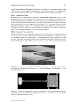
Fig. 1.1. Phytophthora zoospores and cysts. A P. nicotianae
zoospore showing emergence of the two flagella (arrowheads) from the centre of the ventral groove. B P. nicotianae
zoospore showing the water expulsion vacuole (arrow) at
the anterior end of the cell. C P. nicotianae cysts. D P. nico-
root (Van West et al. 2002). At an even finer spatial
scale, zoospores of foliar pathogens may be preferentially attracted to stomata and zoospores of root
pathogens may target the grooves between adjacent epidermal cells (Fig. 1.1E; Gees and Hohl 1988;
Hardham 2001, 2005; Judelson and Blanco 2005).
Like the zoospores of other Stramenopiles, Phytophthora
zoospores are said to have heterokont flagella because
the two flagella have different morphologies. The anteriorly
directed flagellum is shorter than the posterior flagellum
and possesses two rows of tubular hairs called mastigonemes about 1 μm in length (Hardham 1987a). Observations of zoospore motility suggest that the anterior
flagellum pulls the cell forward while the posterior flagellum acts like a rudder, occasionally bending to change the
swimming direction. Both flagella form quasi-sinusoidal
5
tianae cysts 2 h after germination. E Scanning electron
micrograph of P. nicotianae cysts that have targeted and settled in the grooves between the epidermal cells of a tobacco
(Nicotiana tobacum) root. Material secreted by the spores
coats the cyst and nearby plant surface. Bars 10 μm
waves that emanate from the base of the flagella and propagate to their tip. This form of beating of the anterior flagellum would normally propel the cell backwards but the two
rows of rigid mastigonemes reverse the thrust of flagellar
beat, causing the zoospore to be pulled forwards (Cahill et
al. 1996; Jahn et al. 1964). Until recently, there has been little
information on the nature of the components that make up
the mastigonemes of Phytophthora or other stramenopile
species. The first advances arose from immunocytochemical studies using monoclonal antibodies directed towards
zoospore surface molecules that revealed that the shaft of
P. nicotianae mastigonemes is made of a 40-kDa glycoprotein
(Robold and Hardham 1998). Amino acid sequence data
have now been obtained following immunoprecipitation
purification of the mastigoneme protein and these data
used to clone the corresponding gene (M. Arikawa,
T. Suzaki, L.M. Blackman and A.R. Hardham, unpublished data). The results indicate that the Phytophthora
6
Adrienne R. Hardham and Weixing Shan
mastigoneme protein Pn14B7 is related to the Sig1 and
Ocm1 proteins recently cloned from two algal Stramenopile,
Scytosiphon lomentaria and Ochromonas danica, respectively
(Honda et al. 2007; Yamagishi et al. 2007).
As yet we have little information on the identity of
zoospore proteins involved in the reception of chemotaxis
or electrotaxis signals, however, recent studies of P. infestans
genes encoding the α-subunit of a trimeric G-protein (Dong
et al. 2004; Latijnhouwers et al. 2004) and a bZIP transcription
factor (Blanco and Judelson 2005) indicate that both these
proteins play a role in zoospore motility. Silencing of these
two genes inhibits zoospore motility by causing the cells to
turn more frequently or spin in tight circles. Unfortunately
it has not been possible to use these mutants to assess the
contribution of zoospore motility and taxis to pathogen
virulence because silencing the genes also produced aberrations during infection structure development. Regulation
of flagellar activity is known to involve controls of cytoplasmic Ca2+ concentration and two calcium-binding proteins,
calmodulin and centrin, have been localized within the
flagella apparatus of P. cinnamomi zoospores (Gubler et al.
1990; Harper et al. 1995). Genes encoding centrin, a dynein
light chain protein and a radial spoke protein were recently
cloned from P. cinnamomi and are currently being further
characterized (R. Narayan, L.M. Blackman and A. R. Hardham, unpublished data).
Phytophthora zoospores are not surrounded
by a cell wall and their outer surface is that of the
plasma membrane (Hardham 1987b). Water from
their surroundings enters the zoospores down its
chemo-osmotic gradient and, in order to maintain
cell volume and homeostasis, must be pumped
out of the cell. Zoospores achieve this through the
operation of a contractile vacuole (often called a
water expulsion vacuole; Fig. 1.1B) that consists
of a reticulate spongiome surrounding a central
bladder (Mitchell and Hardham 1999; Patterson
1980). It is not known exactly how contractile vacuoles function in any protist but it is believed that
H+-pumping ATPases power the accumulation of
solutes within the spongiome, accompanied by the
passive influx of water (Stevens and Forgac 1997).
Localization of vacuolar H+-ATPase in the spongiome
of P. nicotianae zoospores is consistent with this
hypothesis (Mitchell and Hardham 1999). Water is
believed then to be transferred from the spongiome to the bladder which periodically fuses with
the plasma membrane and contracts to expel the
accumulated water.
Phytophthora zoospores are able to swim for
many hours utilizing endogenous energy stores,
thought to be predominantly polysaccharides
(such as mycolaminarins) and lipids (Bimpong
1975; Wang and Bartnicki-Garcia 1974). They
inherit many, if not the majority, of their proteins
from the sporangium and early inhibitor studies
suggested that mRNA and protein synthesis were
not required for zoospore function (Penington
et al. 1989). However, more recently labelling studies
have shown that new proteins are synthesized in P.
infestans zoospores (Krämer et al. 1997) and proteomic analyses have identified polypeptides that
are more abundant in zoospores than in any other
stage of the life cycle of P. palmivora (Shepherd
et al. 2003). In addition, transcriptome and other
studies have identified genes that are preferentially
expressed in Phytophthora zoospores (Ambikapathy et al. 2002; Connolly et al. 2005; Judelson and
Blanco 2005; Škalamera et al. 2004). Proteins synthesized in zoospores may function during this
motile phase or they may be required to function in
the cysts that are formed by zoospore encystment.
For example, one gene that is highly expressed in
P. nicotianae zoospores is that encoding Δ1-pyrroline-5-carboxylate reductase, an enzyme involved
in proline biosynthesis (Ambikapathy et al. 2002).
High levels of proline may be required for osmoregulation in the wall-less Phytophthora zoospores as
they are in some other protists (Steck et al. 1997).
In contrast, cell wall degrading enzymes (e.g.
cellulase) encoded by genes identified in the transcriptome study (Škalamera et al. 2004) may be
synthesized in readiness for secretion by germinated cysts during plant invasion.
B. Attaching to the Plant Surface
Having reached the surface of a potential host plant,
Phytophthora zoospores adjust their swimming
pattern so that the ventral surface faces the plant
(Hardham and Gubler 1990). While they maintain
this orientation, the zoospores encyst (Fig. 1.1C, E).
This is a rapid process during which the two flagella
are detached, rendering the spores non-motile, and
material is secreted from three different categories of
spherical vesicles in the zoospore peripheral cytoplasm (Fig. 1.1E). A network of cortical cisternae also
fragments, apparently fusing with the plasma membrane, possibly thereby bringing about a rapid and
wholesale change in the composition of the spore
plasma membrane (Hardham 1989). The material that is secreted during zoospore encystment
includes adhesion proteins that firmly attach the
spores to the plant surface (Hardham and Gubler
1990). Attachment of pathogen spores or other cells
to the surface of their hosts is an important aspect of
the infection process (Epstein and Nicholson 1997;
Cellular and Molecular Biology of Phytophthora–Plant Interactions
Tucker and Talbot 2001). Not only does it prevent
the pathogen being dislodged before it penetrates
the plant, but the close contact also aids the reception of signals that guide pathogen growth and that
trigger the development of specialized infection
structures. Strong adhesion also facilitates host
penetration by hyphae or appressoria.
The secretion of adhesive and other proteins
from encysting zoospores is complete within about
2 min and a cellulosic cell wall capable of withstanding
cell turgor is formed within 5–10 min (Hardham and
Gubler 1990). As the cell wall forms, the pulsing of the
contractile vacuole slows down and become undetectable (Mitchell and Hardham 1999). Zoospore
encystment is triggered by a range of physical and
chemical factors and there is evidence for a role of
cell surface receptors and of the phospholipase D
signal transduction pathway in induction of this
process (Bishop-Hurley et al. 2002; Hardham and
Suzaki 1986; Latijnhouwers et al. 2002).
The regulated secretion triggered during
zoospore encystment involves exocytosis of the
contents of the so-called large peripheral, dorsal
and ventral vesicles (Fig. 1.2; Hardham 1995, 2005;
Hardham and Hyde 1997; Škalamera and Hardham
2006). Material released from the dorsal vesicles
includes a high molecular weight glycoprotein
that forms a mucilage-like covering that coats the
cysts and the nearby plant surface (Figs. 1.1E, 1.2A,
B; Gubler and Hardham 1988). This material may
protect the young cysts from physical or chemical
damage but evidence to support this hypothesis
has not yet been obtained. Material released from
the ventral vesicles includes a 220-kDa adhesive
protein, named Vsv1, that attaches the cyst to the
plant (Fig. 1.2C, D). Cloning of the gene encoding Vsv1 in P. cinnamomi has revealed that, apart
from short N- and C-terminal sequences, the bulk
of the PcVsv1 protein is composed of 47 copies of
a domain approximately 50 amino acids in length
that shows homology to thrombospondin type 1
repeats found in a number of adhesive extracellular
matrix proteins in animals and secreted adhesins
in apicomplexan malarial parasites (Adams and
Tucker 2000; Robold and Hardham 2005; Tomley and
Soldati 2001). Homologues of the PcVsv1 adhesive
occur in other Phytophthora species and in species
of Pythium, Plasmopara and Albugo, suggesting
that the Vsv1 protein may be a spore adhesive used
throughout the plant pathogenic Oomycetes.
Until recently, studies of the large peripheral vesicles in
the zoospore cortex indicated that their contents were not
7
secreted during encystment but that the vesicles moved
away from the plasma membrane and became randomly
distributed within the cyst cytoplasm (Gubler and Hardham
1990). However, in zoospore transcriptome studies in
P. nicotianae, cloning of a gene encoding a complement
control protein has given rise to evidence that some of the
contents of large peripheral vesicles are secreted during
encystment (Škalamera and Hardham 2006). Evidence for
the selective secretion of PnCcp proteins from the large
peripheral vesicles comes from double immunolabelling
of PnCcp and Lpv proteins at both the light and electron
microscope levels (Fig. 1.2E–I). In motile zoospores both
proteins are localized to the large peripheral vesicles but
in young cysts, PnCcp proteins are absent from the vesicles
and instead coat the cyst surface. In mammals, proteins containing complement control protein modules play
a number of roles in signalling and adhesion (King et al.
2003). Their role in the infection of plants by Phytophthora
zoospores remains to be elucidated.
In addition to the adhesives secreted by
zoospores during their encystment, other Phytophthora genes encoding putative adhesives that may
function in hyphae or germinated cysts have been
cloned and characterized. Hyphae and cysts of P.
nicotianae (formerly P. parasitica) have been shown
to synthesize and secrete a 34-kDa glycoprotein,
termed CBEL, that contains two cellulose-binding domains (Séjalon-Delmas et al. 1997; Villalba
Mateos et al. 1997). Silencing of the expression
of the CBEL gene interferes with adhesion of the
hyphae to cellophane membranes and with morphogenetic changes normally induced by contact
with cellulose in vitro (Gaulin et al. 2002). Silencing of CBEL expression does not have a great effect
on pathogenicity on host tobacco plants. Another
family of secreted proteins that may play a role
in adhesion of germinated cysts is the Car proteins (cyst-germination-specific acid repeat) from
P. infestans (Görnhardt et al. 2000). The Car proteins
contain multiple copies of an octapeptide repeat, a
motif found in mammalian mucin proteins (Guyonnet Duperat et al. 1995). Car genes are expressed
during cyst germination and appressorium differentiation and the Car proteins are secreted onto the
germling surface. By analogy with the functions of
mammalian mucins, the Car proteins have been
hypothesized as playing roles in protecting the
germlings from desiccation or physical damage
and in germling adhesion (Görnhardt et al. 2000).
C. Penetration of the Host Surface
Zoospores may carry mRNA transcripts and
proteins that function in the cysts that are formed