Nghiên cứu đặc điểm giải phẫu hệ thống cân cơ nông vùng mặt và mối liên quan với thần kinh mặt trên người việt trưởng thành tt tiếng anh
Bạn đang xem bản rút gọn của tài liệu. Xem và tải ngay bản đầy đủ của tài liệu tại đây (558.65 KB, 24 trang )
1
INTRODUCTION
In modern eras, statistical community-based studies show that
facial traumas, usually inflicting damage to the superficial
musculoaponeurotic system (SMAS) and leaving the patients with
disfiguring scars, has been becoming more prevalent in traffic,
occupational and domestic accidents along with socioeconomic
developments. Besides, the aging process also decreases the skin
elasticity and therefore leads to the appearance of striae, wrinkles and
fat disposition beneath the facial SMAS.
In this day and age, plastic surgery has been being extensively
developed and researched all around the world. Furthermore,
surgeons’ aptitude has ameliorated more than ever before, and they
have achieved many great feats in restoring aesthetics and physical
functions to the patients. However, intervention to facial structures
can be occasionally limited and sometimes iatrogenic damage such as
facial paralysis, rupture of the parotid duct, etc., can be attributed to
surgeons’ restricted knowledge and expertise, especially about the
aspect of clinical anatomical landmarks of the SMAS.
There has been many in-depth studies on the SMAS and its
relation to important facial structures such as the parotid duct, the
facial nerve, the superficial temporal artery, etc., but their results are
contradictory still. On the other hand, SMAS studies are scarce in
Vietnam and the SMAS is simply depicted as a superficial facial
fascia.
Based on the aforementioned arguments, I’ve decided to
conduct a “Study on the anatomical characteristics of the facial
SMAS and its relation to the facial nerve on Vietnamese adults”
2
with two main objectives: (1) To investigate the anatomical
characteristics of the SMAS and (2) to identify the gross and microsco
Figure relation of the facial nerve branches to the SMAS.
New contributions of the study:
1. It always has SMAS layer in each side of face and devides
that side into three parts: upper, middle and lower part. Its shape likes
number 3 with the upper vacancy is orbicularis oculi and the lower
one is orbicularis oris, the area of SMAS is affected by size of the
superficial muscles such as frontalis, orbicularis oculi and orbicularis
oris. We noticed the thickeness of the upper temporal septum, anguli
oculi lateralis, zygomatic, masseteric and mandibular ligaments,
which tighten SMAS.
2. At the place ligaments attach, there always have the branches
of facial nerve run into the ligaments, such as frontal branch runs
under temporal septum, orbital branch runs into anguli oculi lateralis,
buccal branches run into zygomatic ligament, masseter and
mandibular branch runs into mandibular ligament, these are the
protective mechanism of the branches of facial nerrve, that is why
surgician need to be careful when performing process in the SMAS.
Outline:
Our study has 125 pages, including 4 main chapters;
introduction: 2 pages; Chapter 1- Literature review: 36 pages (2 tables,
26 figures); Chapter 2- Subjects and Methods: 22 pages (6 tables, 24
figures); Chapter 3-Results: 36 pages (19 tables, 47 figures); Chapter
4- Discussion: 26 pages (6 tables, 12 figures); Conclusion: 2 pages;
Recommendations: 1 page. This study referenced from 120 research
3
papers (comprising 12, 108 papers in Vietnamese and English
respectively).
CHAPTER 1
LITERATURE REVIEW
1.1.2. Basic principles of the facial layers
Figure 1.2. The facial layers
* Source: from Mendelson B.C. (2009) [22]
1.1.3. Basic structures of five facial layers
1.1.3.1. Skin
Skin depth can vary depending on regional function, thinnest
skin is located in the eyelid, as opposed to foreheads and nasal tip [26].
1.1.3.2 Subcutaneous layer
The subcutaneous layer attaches more tightly to the skin above
than to the underlying tissue, which is similar to the tree model of
fibrous tissues and ligaments. The fibres go deeper they will unify to
form large fibres and thereby increase in size and decrease in quantity,
along with fewer fat tissues. As a consequence, dissecting the
subcutaneous layer will be easier for the deeper layers [16], [30], [31].
1.1.3.3. Superficial musculoaponeurotic system (SMAS)
The SMAS, first described in 1976, the muscular part of this
system is predominant in several regions in the third layer while in
other parts of the face the aponeurotic part dominates. Fibrous tissues
4
of the three outer layers are the SMAS, being the deepest layer of this
single unified unit. In the midface and the lower third of the face, this
mixed structure still persists despite being ambiguous [6], [41]. The
third layer exists in a multilayered form, in which the flat part
comprises the superficial layer covering the anterior aspect of the face:
frontalis muscle makes up the upper third and the orbicularis muscle
form the middle third of the face, while the platysma muscle,
extending from the lower third to the lateral face [43], [44].
1.1.3.4. Fascial spaces
The anatomical complexity of this fourth layer will become
much more comprehensible if we understand the arrangement of these
structures, which follow this principle [7]: they all lie above the bones
as this layer originally forms the virtual spaces and the immobolized
region; the virtual spaces are functional regions, each of which has
distinct borders and has minimal coupling; internal arrangement of the
retaining ligaments further reinforces the borders of the virtual spaces
and facilitates the identification of different regions; the muscles lie
within the deep fascial layers and attach to the underlying bones at the
borders; there is always a continuous line, being an extension of the
retaining ligaments, running along the circumference of the bone
cavities.
1.1.3.5 Deep fascia
Has the same structure as the periosteum, but instead is a mobile
membrane which lies over the supraperiosteal fat. It runs beneath the
origin of the deep muscles and the retaining ligaments.
1.2. Concepts and studies on the structure of retaining ligaments,
ligamentous attachments and septa of the face.
5
1.2.1. McGregor’s patch
The term ”McGregor’s patch”, zygomatic ligaments or
zygomatic cutaneous ligaments were all synonyms that were used in
the past. In 1959, ”McGregor’s patch” was described as ” an area of
fibrous attachment between the anterior margin of the parotid
fascia and the dermis of the skin of the cheek” [47]. When the
”McGregor’s patch” is found, there are 3 important anatomical
structures which go through the parotideomasseteric fascia: the
transverse facial artery, the parotid duct and the zygomatic branch of
the facial nerve [49].
1.2.3. The masseteric retaining ligaments
Özdemir R. et al hypothesized that the fibrous attachments can
originate from the anterior border of the masseter, 1-2 cm posterior to
the anterior border or even in the middle part of the muscle. The
variability of the origins of the masseteric retaining ligaments may
relate to the corresponding structure of the intermingling region
between the masseteric fascia and the size of the parotid fascia [7],
[15], [19].
1.2.4. Zygomatic retaining ligaments
Funas D.W. described the zygomatic retaining ligaments as
tough fibres which originate from the inferior border of the zygomatic
arch and then extend to the anterior aspect of the junction between the
arch and the body of the zygoma [3], [6].
1.2.5. Orbicularis retaining ligaments
Muzaffar A.R. et al recorded the presence of a septum-like
structure of the ligaments, whose origin is the periosteum of the lateral
border of inferior orbital rim which lies closely to orbital septum.
6
These fibres then attach deeply to the orbicularis oculi and have
unclear borders [57].
1.2.6. Temporalis retaining ligaments
Knize D.M. documented and described a 6-mm-wide region
having fibrous attachments locating medially to the superior temporal
line at the level of the galea aponeurotica and the periosteum is
attached to the underlying bones; they also named a stout ligament
found cranially to the superior orbital rim and at the distal end of the
ligamentous attachments region to be the ”orbicularis retaining
ligaments” [9], [59]. Moss C.J. et al have studied and classified the
types of the ligamentous attachments of the temporal region into:
septa, ligamentous attachments and the thickened region surrounding
the orbital rim namely the thickening of the periorbital septum [10].
1.2.7. Mandibular ligaments
The mandibular retaining ligaments originate from the anterior
third of the mandibular body and have fibres which perforate the
inferior aspect of the depressor anguli oris muscle to tether directly to
the skin. Furnas D.W. also documented these fibres consist of two
layers laying 2-3 mm apart and traveling parallel to each other and
were observed approximately 1 cm above the mandibular body [3].
1.4. Studies on the relation of the facial nerve to the facial layers
1.4.2. Characteristics of the facial nerve branching pattern
According to Davis R.A. et al classification [80], there are 6
patterns: type I absence of an anastomosis between the temporofacial
division and cervicofacial division; type II anastomosis among the
branches of the temporofacial division only; type III single
anastomosis among the branches of the temporofacial division and
7
cervicofacial division; type IV combination of type II and III. Also
called “multi loops” due to the presence of multiple anastomoses of
different branches; type V: double anastomosis between the
temporofacial division and cervicofacial division; type VI multiple
complex anastomosis between the two divisions.
Figure 1.24. Six basic types of the branching pattern of the facial
nerve according to the classification of Davis R.A. et al.
* Source: from Myint K. (1992) [67].
1.4.2.1. Temporal branch
Being the uppermost branch of the temporofacial nerve, it
traverses the superior margin at the point where the temporal hairline
descends and intersects the zygomatic arch. It also emerges 2 cm
above the tragus and then traverses the zygomatic arch; runs beneath
the facial fasciae, caudally to the arch. The temporal branch divides
into 3 branches: the auricular, the frontal and the orbicularis branches
[70].
1.4.2.2. Mandibular branch
The mandibular branch is located behind the facial artery, and
20% of the cases were observed to have this branch traveling along
the inferior border of the mandibular body but it’s never found 1 cm
below the inferior border. It’s rarely damaged during cervical surgery,
8
parotid gland removal surgery, mandibular angle osteotomy, facelift
surgery and other submandibular surgeries [80], [85], [96].
1.5. Domestic studies on the SMAS, ligaments and facial nerve.
In Vietnam,
the terminology of
retaining ligaments,
attachments, septa, and superficial musculoaponeurotic layer are
unsatisfactorily defined and not many studies have been conducted on
these structures [31], [81], [83].
CHAPTER 2
SUBJECTS AND METHODS
2.1. Subjects
We performed facial dissections on 30 Vietnamese adult
cadavers, which have been treated with formaldehyde. Selection
criteria of participants:
1. Vietnamese adult cadavers are above 18 year old
2. The participants have not been operated any ENT surgeries
3. The participants don’t have any deformation or any tumor on the
face and neck region
4. The participants do not have any abnormal structure on the
otolaryngology region.
Facial dissection was performed on skin layers, ligaments and
facial nerves. 20 tissues with the size of 1cm2 were collected for
histological analysis. The sampling location was depended on
anatomical location of branches of the facial nerves related to SMAS
2.3. Research method
2.3.1. Macroscopic analysis of the facial layers, ligaments, cavities
and the facial nerve
9
We dissected the nerve VII with facial ligaments, and measure
the dimensions of the SMAS
2.3.1.2 Macroscopic indicators
+ Qualitative analysis:
Description of the insertion of ligament of anguli oculi lateralis,
zygomatic ligament, masseteric ligament, mandibular ligament.
Identification of the facial nerves with the facial layers and ligaments;
hence choose the appropriate histological analysis location
+ Quantitative analysis:
- Measurement the depth of the skin, subcutaneous layer:
eyelids, parotid gland, temporal region, frontal, mental, nasal tip.
Measurement dimensions of SMAS layers
2.3.2. Microscopic analysis of the facial layers, ligaments, cavities
and the facial nerve
We collect 20 tissues, the size of 1cm2 and sliced into each
sampling of 3-4micrometers which stained by H-E, H-SG method
CHAPTER 3
RESULTS
3.1. Anatomical characteristics of the SMAS and the borders
between regions
3.1.3. The superficial musculoaponeurotic system
3.1.3.2. The SMAS
* Shape of the SMAS
There are two main types, if the area of the orbicularis oculi and
orbicularis oris muscle are large, then the area of the SMAS in the
middle layer is decreased as well as the superior and inferior notches
are deeper (point P and I don’t coincide) (see Figure 3.5).
10
Figure 3.5. SMAS type 1
* Source: sample of specimen H. No 1162013
- If the area of orbicularis oculi and orbicularis oris muscle are
small, the area of the SMAS will be increased, as well as the 2 superior
and inferior notches are shallower (point P & I coincide) (F. 3.6).
Figure 3.6. SMAS type 2
* Source: sample of specimen L. No 862011
*Dimensions of the SMAS
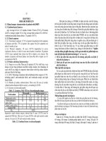
Table 3.7. Dimensions of the SMAS
Dimensions (mm)
Right-sided
Left-sided
(n=15)
(n=15)
p-value
JD
84,60 11,59
79,27 9,93
0,050
Upper third of
IJ
45,07 11,798
38,3 10,35
0,008
the face
ID
67,63 8,199
63,90 9,05
0,093
HI
62,17 8,20
59,60 5,33
0,261
Middle third of
HG
107,93 13,60
103,80 11,69
0,140
the face
HE
79,90 10,75
83,03 9,84
0,298
11
IO
74,70 9,37
77,03 9,04
0,272
FO
44,00 13,06
43,47 10,76
0,878
Lower third of
FE
61,67 14,11
63,87 13,63
0,656
the face
AB
76,27 21,06
83,27 21,16
0,166
CO
23,8 9,17
21,3 5,69
0,368
CE
30,33 7,28
30,67 7,92
0,900
- Comment: The width of the SMAS: the upper facial part (IJ)
40 - 45mm, the middle facial part (HG) 100 - 110mm; the lower facial
part (FE) 60 - 65mm. The height of the SMAS: the upper part (ID) 60
- 70mm; the middle part (IO) 75 - 80mm; the lower part (CO) 20 25mm. Distance from tragus to anguli oculi lateralis (HI) is about
60mm, it is shorter than distance from tragus to angle of the mouth
(HE) 70 - 80mm.
3.1.4. The Superficial Musculoaponeurotic System
3.1.4.1. Ligaments, fibrous tissue
* Ligaments of anguli oculi lateralis
We noticed that in 30 samples, there are connective tissue
which link SMAS to the fifth layer from the anguli oculi lateralis.
Figure 3.8. Angulus oculis latoralis and zygomatic ligament
* Source: sample T. code 952012
* Zygomatic ligament
In 30 samples, from tragus along to the upper border of zygoma,
there are connective tissue which link to hypodermis.
12
Figure 3.11. Zygomatic ligament (Mc Gregor’s patch)
* Source: sample H. code 1162013
* Masseteric Ligament
In 30 samples, there are connective tissue running along the
anterior border of masseter to hypodermis with ramus marginalis
mandibulae nervi facialis.
Figure 3.12. Masseteric ligaments
* Source: Sample H. code 1092013
* Mandibular ligament
We noticed that in 30 samples, there are connective tissue
attached from the anterior border of masseter to the body of mandible,
and link to subcutaneous tissue, which relate to ramus marginalis
mandibulae nervi facialis.
Figure 3.13. Mandibular ligament
* Source: Sample T. code 952012
13
3.2. The relationship between SMAS and branches of facial nerve
3.2.2. Investigating the macro and microanatomy correlation
between SMAS and the branches of facial nerve
3.2.2.1. Investigating the path way of frontal branch
* Frontal branch runs into ligaments of angulus oculi lateralis
The investigated area includes SMAS, frontal branch and
orbicularis oculi (the square in Figure 3.33)
Fig 3.32. Frontal branch runs into lig. of angulus oculi lateralis
* Source: sample T. code 972012
Fig 3.33. The frontal branch runs into lig. of anguli oculi lateralis
* Source: sample T. code 1072013 in the right
We discovered that frontal branch runs into ligaments of anguli
oculi lateralis and relates to vessels.
* Frontal branch runs into frontalis
The investigated area includes SMAS, frontal branch and
frontalis (the square in Figure 3.35)
14
Figure 3.34. Frontal branch runs into frontalis
* Source: sample N. code 1192013
Figure 3.35. Frontal branch runs into frontalis
* Source: sample H. code 1182013 in the right
- Frontal branch along with vessels run between frontalis
bundle
3.2.2.2. Temporal branch of facial nerve
* Temporal branch in fossa temporalis
The macro investigated area includes SMAS, temporal branch
of facial nerve (the square in Figure 3.38)
Figure 3.36. Temporal branch in fossa temporalis
* Source: sample N. code 1192013
We realized that temporal branch runs between muscle layers.
15
3.2.2.3. Zygomatic branch of facial nerve
* The triple Mc Gregor zygomatic branch, saliva gland tube and
transverse artery of face
The macro investigated area includes SMAS, masseter,
zygomatic branch, and parotid gland tube (the square in Figure 3.40)
Figure 3.40. The triple Mc Gregor
* Source: sample T. code 972012
Figure 3.41. The triple Mc Gregor
* Source: sample T. code 1072013 in the right
We noticed that there have arteries, nerves, parotid gland tube.
* Zygomatic branch crosses ligament of masseter
The macro investigated area includes SMAS, ligament,
zygomatic branch and under massester 2mm (the square in Fig 3.42)
16
Figure 3.42. Zygomatic branch crosses ligament of masseter
* Source: sample H. code 1162013
Figure 3.43. Zygomatic branch crosses ligament of masseter
* Source: sample T. code 1292014 in the right
We noticed that there has zygomatic branch, around that are
masseter and ligaments.
3.2.2.4. Mandibular branch of facial nerve
* Mandibular branch goes along with facial artery
The macro investigated area includes mandibular branch,
ligaments, arteries, doesn't include SMAS (the square in Fig 3.46)
Figure 3.46. Mandibular branch goes along with facial artery
* Source: sample N. code 1192013
Figure 3.47. Mandibular branch crosses facial artery
* Source: sample T. code 1072013 in the left
17
We discovered that the upper left corner of this sample is facial
artery, the lower left corner is mandibular branch.
CHAPTER 4
DISCUSSION
4.1. Anatomic characteristics of SMAS
4.1.1.3. SMAS
* The shape of SMAS
We noticed that the larger the orbicularis oculi and orbicularis
oris are, the smaller the area of SMAS is and the deeper the upper and
lower vacancy are (it means point P and I don’t duplicate).
The width of the medium layer of SMAS is the largest while the
upper layer is smallest - equals ⅖ the medium layer, next is the lower
layer (equals ⅗ the medium layer). The height of the upper and
medium layer is the same (the medium layer is a little bit longer),
while the lower layer is about ⅓ the length of the other. Moreover, the
length of two lines dividing the layers of SMAS from tragus to anguli
oculi lateralis(60mm) is shorter than the length of the line from tragus
to angle of mouth (70 - 80mm). That result shows that the parameters
of SMAS is specific for the race. In the past, the incision located to
the posterior of PAF, from that, SMAS is lifted out of PAF, however,
the technique was really hard because the surgician had to separate
two layers of PAF. Nowadays, the incision runs along SMAS away
from the groove in front of ears about 25 - 30mm toward the area out
of PAF. This incision can go through the mobile SMAS, which is the
roof of anterior space of masseter, so that the surgician lift SMAS from
the floor easily. It makes the surgician do the process more quickly,
more certainly and more safely because the branches of facial nerve
18
are not placed in the surgery field. We noticed that there are two main
factors affecting to the result: the first factor is the incision, which
relates to the position of the upper branch of mandibular branch.
Our findings point out that there are two main factors affecting
the outcome of the surgery: The first factor is the location of the
incision related to the upper branch of the mandibular nerve. The
anterior SMAS incision is located overlying space, which is beyond
the mandibular branch situated within the PAF. It has been proved by
the histological photographic illustrating the relation of the
mandibular branch and the facial layers. The second factor is the
mobility of the inferior border of the premasseter space. The
traditional posterior SMAS incision is extended inferiorly behind the
angle of the mandible to where the SMAS and platysma are adherent
to the sternomanoid fascia. When used the anterior SMAS incision, it
can be ended 15mm above the lower border of mandible and situated
anterior to the upper mandibular branch. The mobility of the platysma
together with the ready displacement of the mandibular branch would
minimize the risk of a traction neurapraxia. The lower mandibular and
the cervical branch would remain posterior to the angle of the
mandible to be located inferior to the mandible and outside the
premasseter space when they travel forward on the underside of the
platysma. Due to the short length of the anterior SMAS incision, the
mandibular branch remains outside the operative fields at all times.
4.1.1.4. Facial ligaments, fibrous tissues and spaces
* The zygomatic ligaments or McGregor’s patch
It is undoubtedly obvious that the zygomatic ligaments
originate from the periosteum of the zygoma inferior to the orbicularis
19
retaining ligament to the upper pole of the masseter, as described by
Mendelson B.C. et al [22]. This ligament will correspond to a central
fixation point and will integrate with some fat chambers [104], [105].
This ligament is weaker and often can be disrupted by blunt finger
dissection. We also recognize the presence of 3 structures: the
transverse facial artery, parotid duct, and zygomatic branch of the
facial nerve. This ligament has varied measurements in width and
thickness, and form the letter “L” with the masseteric ligaments. This
finding is also matched with Özdemir R. et al [15] who said that the
surface area of the zygomatic ligament is larger, and that the
dimensions of these structures are varied in each cadaver.
Nevertheless, it is considerable to determine the reference points so as
to provide correlated comparisons. Therefore, in this study, we rather
focus on the presence and the locations of these ligaments than
measure their dimensions.
* Masseteric ligaments
We find out that along the masseter muscle arise the ligaments
adjoining the SMAS to the masseter muscle. These ligaments don’t
have defined borders but situate along the masseter muscle with
different thicknesses. This finding is correspondent to the study of
Furnas D.W. [41]. The zygomatic ligaments and masseteric ligaments
are often described together. Differed from the description of Stuzin
J.M. et al [7] that zygomatic and masseteric ligaments formed roughly
a letter “T”, the zygomatic ligaments are indeed very strong in the
inferior of the zygomaticus minor muscle. We realize that the images
of these two ligaments are similar to the description of Mendelson
with an inverted “L” shape. The horizontal limb is the zygomatic
20
ligament located at the angle of the “L”, which is lateral to the
zygomaticus major muscle, extends medially across the origin of the
facial muscles. The vertical limb is formed by the masseteric
ligaments [106], [107].
* Ligaments of anguli oculi lateralis
We realize the presence of ligaments of anguli oculi lateralis
linking the SMAS with the fifth layer at the lateral canthus, where the
infraorbital nerve always run through. This ligament is different from
the thickening of the skin at the latheral canthus, which is named
“retrator anguli oculi lateralis” linked indirectly with the orbicularis
retaining ligament through the orbicularis oris deep fascia, cartilage
lashes, creating a unique anatomical structure [6], [35], [108], [109].
* Mandibular ligament
There exist the fibers of mandibular ligament attached to the
SMAS along the anterior border of the masseter muscle and inserted
into the inferior border of the mandible. It also bears a close
correlation to the mandibular nerve.
4.2. Relationship of the nerve branches to facial layers - ligaments
4.2.2. Relationship of the nerve branches to the facial ligaments
4.2.2.1. The temporal branch with facial ligaments
* Microscopic comparative analysis of temporal branch with
facial layers
The temporal branch of the facial nerves is originated from the
parotid gland under the name of frontotemporal trunk which is
covered by parotid fascia, and then transverse upwardly into the fat
layer that can be dissected easily from the SMAS. This trunk is then
divided into the temporal and the zygomatic branch after leaving the
21
parotid gland from 1 – 2cm. The temporal branch has a numerous of
divisions, travelling inward and matching with the reference points on
the skin. It has been proved by histology through the histological
samples on the zygomatic arch. Covering the temporal branch is a
unified fascia that can be identified easily on clinical, and it has been
proved histologically to be a separate layer.
On the zygomatic arch, the temporal branch is located near the
periosteum and is covered by a separated fascia from the SMAS.
When this branch crosses anteriorly to the zygomatic arch, the
periosterum is replaced by the superficial layer of the deep temporal
fascia and temporoparetalis fascia 2cm superior to the zygomatic arch,
where the nerve seems to transfer to temporoparetalis fascia in order
to join with the anterior branch of the superficial temporal artery [79].
We can affirm that there is a separate fascia detaching the nerve from
the deep temporal fascia. This fascia has not been named, or being
called “unknown fascia” which can be renamed to “temporoparotid
fascia”. At the zygomatic arch, the nerve lies beneath this fascia and
leans on the periosterum of the zygomatic arch.
4.2.2.2. The orbital branch, relationship with facial ligaments
The orbital branch enters the muscle from the deep layer [80]
and connect with each other [69] to form a plexus of nerves
approaching orbicularis oculi muscle fibers. When carrying out
procedures in this region, the risk of injuring orbital branch is small.
Provided that the dissection is proper the nerves would not be affected.
4.2.2.4. Relationship of the zygomatic and the buccal branch with
facial ligaments
22
The zygomatic retaining ligaments are anatomical landmarks
for the zygomatic facial nerve branches, which is located just inferior
to the lateral side of the orbitomalar ligament. Furnas D.W. considered
the region immediately inferior to the zygomatic ligament to be a
danger zone due to its closeness to zygomatic nerve branches. He was
the first to demonstrate that the zygomatic branch passed in a deep
plane just inferior to the zygomatic ligament. Another division of the
zygomatic branch passes just inferior to or penetrates the upper
masseteric ligaments lower than 1 mm and deep to the deep fascia,
then penetrates the deep fascia distal to the ligament [3]. Hence, the
distance of 1 cm just immediately inferior to the zygomatic ligament
is relatively safe (except that the cases of the upper zygomatic nerves
divide a more superficial branch that travels more superficial to the
zygomaticus major muscle, which account for 5% to 9%). And
Alghoul also concluded that the zygomatic nerve often pierces
through ligaments, which the rate of 27% for the zygomatic ligament
and 66% for the masseteric ligament [6].
After exiting the parotid gland, the branches travel on the
surface of the masseter muscle, until 2cm behind the anterior border,
they branch out superficially to innervate the orbicularis oris. The
region that can injure these branches has the anterior border of the
posterior side of the zygomatic arch and the posterior border of the
anterior side of the masseter muscle. In this region, there are many
perforators from the deep layers to the superficial ones in order to
supply the skin, which is perpendicular to the dissection angle of the
surgeons [119], hence it stands a risk of bleeding when dissecting
[120]. The masseteric ligaments are anatomical landmarks for the
23
buccal branches of the facial nerves. The branches penetrate the deep
fascia, distal to the masseteric ligaments and to the buccal fat pad.
Consequentially, liberating the masseteric retaining ligaments in the
plane inferior to the SMAS may expose the buccal fat pad and cause
it to be herniated, together with the buccal branch of the facial nerves
lying more superficially [6].
4.2.2.5. Relationship of the marginal mandibular branch with facial
ligaments
The marginal mandibular branch is extremely vulnerable, and
could be divided into two parts based on the location of the facial
artery [97]: posterior to the facial artery, the nerve travels above the
inferior border of the mandible (81%) or below the inferior border of
the mandible (19%) but not lower than 1cm; anterior to the facial
artery, the nerve travels above the superior border of the mandible.
Therefore, when dissecting the skin, we should pay more attention to
an important landmark of the facial artery. There are cases when the
platysma is thin, particularly in the elderly or people who have gone
through face-lifting. This layer would be difficult to differentiate as it
is fibrosis or being torn, being taken out during the first surgery [82].
Langevin C.J. et al describe the relationship between the marginal
mandibular branches and the mandibular ligament. This branch is
found just posterior to the mandibular ligament [60].
CONCLUSION
1. The anatomical characteristic of the SMAS
The horizontal dimension of the SMAS of the upper face (IJ)
40 - 45mm; mid face (HG) 100 - 110mm; lower face (FE) 60 - 65mm.
The height of the SMAS: the upper face (ID) 60 - 70mm; the mid face
24
(IO) 75 - 80mm; the lower face (CO) 20 - 25mm. The upper horizontal
of the SMAS: from the external auditory meatus to the lateral canthus
(HI)
60mm; The lower horizontal of the SMAS from the external
auditory meatus to the oral commissure (HE) 70 - 80mm. We point
out the presence of the ligaments, the lateral canthus tendon with the
presence of the orbital branch travelling in this ligament, the
zygomatic, the masseteric and the mandibular ligaments.
2. Relationship of the facial nerves to the SMAS
The temporal branch travels anteriorly to the external auditory
meatus, inferior to the PAF, to the lateral canthus tendon, to the SMAS
and the orbicularis oculi muscle. The infraorbital branch passes above
the zygomatic bone and then behind the orbicularis oculi muscle. The
temporal branch runs in the temporal space, behind the SMAS and
temporoparetalis fascia, the zygomatic branch runs inside the parotid
gland, piercing through the zygomatic ligament. The marginal
mandibular crosses the facial artery superior to the mandible,
penetrating into the mandibular ligament.
RECOMMENDATIONS
1. The study uses fresh cadavers so as to assure the SMAS’s
standards: the size, form, ligament structure, spaces of the SMAS.
2. Integration between the clinical study while performing
facial and cranial surgery and histological study using the digital
microscope.
3. Building a system of naming and glossary for the SMAS.