Nghiên cứu giải phẫu ứng dụng kết xương đinh nội tủy kín có chốt điều trị gãy 13 dưới và đầu dưới xương chày tt tiếng anh
Bạn đang xem bản rút gọn của tài liệu. Xem và tải ngay bản đầy đủ của tài liệu tại đây (356.2 KB, 14 trang )
MINISTRY OF EDUCATION
MINISTRY OF DEFENSE
VIETNAM MILITARY MEDICAL UNIVERSITY
NGUYEN VIET DUNG
ANATOMICAL STUDY OF APPLYING THE TIBIAL PLATEAU WITH
CLOSED INTERLOCKING INTRAMEDULLARY NAIL TO TREAT THE
FRACTURES OF 1/3 LOWER TIBIA AND LOWER END OF THE TIBIA
Specialization: Surgical
Code: 9720104
SUMMARY OF PHD. THESIS IN MEDICINET
HANOI – 2019
This thesis was conducted at:
LIST OF PUBLICATIONS FROM THE THESIS
Vietnam Military Medical University
1.
Nguyen Viet Dung, Nguyen Tien Binh, Vu Nhat Dinh, Nguyen Hai An
(2018).“Evaluation of results of tibial plateau with interlocking intramedullary
nail and fibular plateau with screw fixation in treatment of fractures of the
distal third tibia below both distal leg bones”, Vietnamese medicine journal,
Vol. 470, September, Issue No.1, p.67-71.
2.
Supervisors:
2. Prof. Nguyen Tien Binh, MD, PhD
1. Assoc. Prof. Vu Nhat Dinh, MD, PhD
Nguyen Viet Dung, Nguyen Tien Binh, Vu Nhat Dinh, (2018). “Study of tibia
anatomy in Vietnamese aldults, appication in treatment of lower and distal
tibia fracture by intra medullary with locking nail”, Military medicine and
pharmacy journal, Issue No.9/2018.
Peer-review 1:
Peer-review 2:
Peer-review 3:
The thesis will be defensed at Council of Vietnam Military Medical University
at …… …… …… 2019
24
1
INTRODUCTION
LIMITATIONS OF THE TOPIC
Fracture in 1/3D segment - the lower end of the tibia is one of the bone
fractures that are difficult to treat (due to broken bone characteristics and
The number of patients returning to fix bone fixation facilities in our hospital
anatomical structure). Methods of treatment: Cast wrapping, external fixation,
is not much (due to different reasons and subjective reasons). So we have not
continuous pulling, splinting screw, locking plate & screw, interlocking
encountered difficult cases to remove bone fixation to share with scientists and
intramedullary nailing ... in which each method has its own advantages and
colleagues.
disadvantages.
Bone fixation tools in our study are not common, so it is difficult for other
medical facilities to apply this method widely.
Treatment of the tibial stem fracture with intramedullary nail has been applied
in the world and in Vietnam from the 1960s of the 20th century to the present. The
In order to be able to apply this method of bone combination effectively, it
tibial plateau with interlocking intramedullary nail is the top-choice “gold
requires medical facilities to have a digital X-ray machine, complete equipment,
standard”, because the nail fixes the fracture area firmly, without causing bulge and
and a team of qualified and experienced surgeons.
exposing the nail in the event of the opened incision. For Fracture in 1/3D segment
- the lower end of the tibia (wide flared medullary canal, thin bone wall), the tibial
RECOMMENDATIONS
plateau with interlocking intramedullary nail has the risk of bone axis misalignment
or unstable fixed fracture (especially when there is also a fibula fracture). In order
From the research practice of the subject we find that the usual intramedullary
to contribute to completing the technique of treating fractures in this position and
interlocking nail size for Vietnamese people is: 9mm x 320mm; 9mm x 300mm;
still ensuring good results, minimizing complications, promoting the advantages of
9mm x 340mm; 9mm x 280mm; 8mm x 320mm; 8mm x 300mm; 8mm x 280mm;
intramedullary nails, we conducted to study the topic titled “Anatomical study of
10mm x 320mm; 10mm x 300mm; 10mm x 340mm. So the length of the nail is
applying the tibial plateau with closed interlocking intramedullary nail to treat
over 340mm, we have not met. Nails with a diameter greater than 10mm and
the fractures of 1/3 lower tibia and lower end of the tibia”.
smaller than 8mm we also do not use in this study.
When surgery does not have C.arm, the surgeon must have experience to
reduce complications for the patient.
There should be a policy for patients to get back to the medical facility of
bone fixation to remove bone fixation facilities. Thus patients will be monitored
and treated more effectively.
Strengthening communication and education for patients to see the benefits of
practicing rehabilitation after surgery.
With the objectives:
1. Surveying some anatomical features of tibia on X-ray images of adults,
applications in surgery of the tibial plateau with closed interlocking intramedullary
nail to treat the fractures of 1/3 lower tibia and lower end of the tibia.
2. Evaluating the results of surgery of the tibial plateau with closed
interlocking intramedullary nail to treat the fractures of 1/3 lower tibia and lower
end of the tibia.
New contributions of the thesis
The tibial plateau with the interlocking intramedullary nail to treat Fracture in
1/3D segment - the lower end of the tibia, if it can firmly fix the fracture, there are
2
23
many more advantages when internally fixing bones with screw. This is a
CONCLUSION
minimally invasive surgical technique suitable for the current trend. The difficulties
Studying a number of anatomical characteristics of 228 tibia of 114
of fixing bones with interlocking intramedullary nail in this position is the distal
Vietnamese adults (67 males, 47 females) and the results of treatment of 63 cases
end of the intramedullary nail must pass through the long enough fracture area to
of closed fracture 1/3D, outer tibial plateau joint with or without attached low
be fixed with the the anti-rotating screw, anti-angled screw without opening the
fibula fractures, we give some conclusions as follows:
fracture area.
1. Characteristics of tibia anatomy of the Vietnamese adult group
The research topic is practical and further study the characteristics of tibia of
- Features of tibia anatomy:
Vietnamese adults on digital X-ray film: Identifying size indicators, important
+ The average tibial absolute length of the study group: 36.28 ± 2.30cm.
anatomical landmarks, expected nail length, nail diameter, screw size in order to
+ The tibial body length of the study group: 23.84 ± 1.91cm.
help fit patients to limit seizures, complications during surgery and promote the
+ The absolute length and length of the tibia body is proportional to the height.
advantages of intramedullary interlocking nailing.
Younger group with average tibial length longer than the elderly group.
The structure of the thesis
+ The size of the baseball bone marrow, wide at 2 ends, gradually narrowed in
The dissertation has: 115 pages, including parts: Introduction: 2 pages;
position 1/3G - D (in the range of 8 - 10cm from the joint of the slug baseball). The
Chapter 1: Literature overview: 30 pages; Chapter 2: Rearch subjects and methods:
narrowest spinal canal size; before - after: 1.00 ± 0.12cm.
19 pages; Chapter 3: Research results: 33 pages; Chapter 4: Discussion: 29 pages;
+ The size of the medullary bone marrow is the narrowest part in-outside: 1.15 ± 0.14cm
Conclusion: 2 pages; limitations of the topic: 1 page; recommendations: 1 page.
+ The size of the inner and outer spinal canal is wider than before - after measuring
There are tables, charts, and illustrations. There are two published studies related to
on the same position, the size of the pulp is not dependent on the height. The size of
the thesis. The appendix contains a list of 114 adults who were studied the tibial
the lumbar spinal canal size in the older group is larger than that of the young group.
anatomy; the tibia surgery survey note; a list of 63 patients treated for the fracture
- Suitable intramedullary nail interlocking length is 280mm, 300mm, 320mm, 340mm.
in 1/3D segment - the lower end of the tibia (23 patients were fixed the fibula
- Suitable intramedullary nail interlocking diameter is 8mm, 9mm, 10mm.
because the tibial fracture can only be fixed with one screw at the peripheral end),
2. Results of treatment of Fracture in 1/3D segment – the outer joint lower end
the studied medical record, treatment result follow-up report, the medical record
of the tibia
illustration.
References: There are 108 references, including 18 documents in
Vietnamese, and 90 documents in English.
* 100% of patients had first-term, non-infectious, no hypertrophic scars, did
not affect the motor function.
* There are no patients who slide the screw to the distant end.
* The result of bone healing is 100%, in which bone reshaping and correcting
bone axis is very good with 57 patients, bone healing is good with 4 patients, 2
patients with bone fracture correcting medium bone axis.
* Overall results were very good: 54/63 patients (85.71%); good: 7/63 patients
(11.11%); average: 2/63 patients (3.17%); There are no bad cases.
22
3
rate), average results of 2 patients (3,17%), For 2 patients with average results: 1
Chapter 1.
patient with the size of the fracture to the tibial plateau of the fracture group from
31 - 40mm due to the size of the lower medulla tubule flaring so the nail does not
have The spot is pressed against the bone wall and a patient tightened 2 screws but
has 1/3D fibula fracture but no bone fixation marks. For 4 patients with good
results similar to the above: 1 patient is in the fracture group from 31 - 40mm and 3
patients still tightened 02 screws but have 1/3D fibula fracture but not bone fixation
marks. No poor results. Compared to the results of the authors Klaus, W, Klemm
M.D also apply bone fixation sealed by interlocking intramedullary nail to treat two
leg bones closed fracture resulting in 100% reconstruction of the anatomy. Our
results are also equivalent.
The condition of rectification fracture is very good and good for 6 months and
12 months without change, there is no secondary deviation.
The results of functional rehabilitation after 6 months ranked on average
include 2 patients with average corrections of corrective results, limiting ankle
movement and normal travel time over 4 months, 4 patients with good fractures
Good has good rehabilitation results.There is no case of limiting knee joint
movement. 61 patients with normal anklet joints (rate 96.82%), 2 patients (3.17%
rate) limited movement of little ankle joints.
4.2.6.2. Evaluating distant results
100% of patients are healed bones, no patient with slow bone healing, false
joints; no patients with osteomyelitis. When stabilizing, patients can move
normally.
4.2.6.3. General results
Evaluating the results in our study as follows:
- Very good : 54/63 patients (85.72%)
- Good
: 7/63 patients (11.11%)
- Average
: 2/63 patients (3.17%) and no patients achieved poor results.
LITERATURE OVERVIEW
1.1. Anatomical features of the leg related to the injury and the treatment
The leg is limited from the knee joint space to the ankle joint space.
* Characteristics of bone
The leg consists of 2 bones: large tibia, bearing 9/10 weight, in the triangular
prism shape, upper large and lower small, the weak point in 1/3G - D area. The
fibula bears 1/10 weight; the bone is long, thin and bigger at both ends.
* Vascular system nourishing the tibia: Bone marrow, periosteum,
metaphyse ends are connected together.
* Characteristics of soft tissue: The leg muscles are unevenly distributed; the
tibial crest and the inner surface of the tibia are located just below the skin attached
to the periosteum, less movable. In 1/3D leg, the muscles have turned into tendons,
poor nourishing, fractured bones hart to be healed , easy to expose braces, incision
dislocated when splinting the screws.
1.3. The methods of treating the closed fracture in 1/3D segment - the
lower end of the tibia
1.3.1. Conservative treatment
* Cast wrapping correction
* Continuously pulling
1.3.2. Surgical treatment
* External fixation
* Fixing bones with the splint screws
* Fixing bones with the locking splint screws
* Fixing bones with the interlocking intramedullary nailing
Treatment of the tibial stem fracture with interlocking intramedullary nail has been
widely used, many advantages and reducing complications when bones are in the leg,
especially for the segment 1/3D and the tibial end. Fixing bones with interlocking
bone intramedullary nail which has the fastest healing time is the closed nailing.
* Advantages of fixing bone with closed interlocking intramedullary nail, not
opening the facture area.
Fixing bones with closed interlocking intramedullary nail without opening the
fracture, the method completely respects the bone healing physiology of the
4
21
fracture area and significantly reduce the complications of incision infection and
hand, when screwing, it is necessary to observe the longitudinal fractures on the X-
osteomyelitis
Because the fracture area is screwed, the bone fracture area is fixed firmly and
ray film straight and inclined to avoid further fracture of the tibial peripheral head.
it prevents the bone healing slowness, false joints and amyotrophy.
The incision is small and fast healed, small and soft scar, ensuring aesthetics
for patients; low treatment cost, short postoperative period, simple removal of bone
fixing facilities.
1.3.3. Treating the 1/3D fracture – lower end of 2 outer leg bones with
interlocking intramedullary nail and fixing fibula with screw braces
Bonnevialle P. and Cs (2010) reported the study results at the Specialized
Congress of the French Society for Orthopedic and Trauma Surgery (SOFCOT) for
4.2.4. The role of fibular fixation
For cases with tibia and fibula fractures at the low level of the lower leg, the
result of bone fixation pestle by interlocking intramedullary nail combined with
bone fixation fibula by splint screw would be better than just bone fixation pestle
by interlocking intramedullary nail Simple. Many authors have bone fixation with
screw splints for the purpose of adjusting the deflection to the side, strengthening
the ankles, preventing rotation, counteracting pestering, restoring the limb length.
* Assign to screw one end, two ends, 2 parallel screws, 2 perpendicular
cases of fractures of the outer joint lower tibial ends. The authors concluded that: It
is necessary to strengthen the fracture area to enhance the tibial fracture area
screws ... depending on the fracture pattern, fracture properties, anti-rotation
fixation. The fibula fracture areas are pre-fixed to ensure the length of the limb and
limit the lateral misalignment.
anatomy on the X-ray flim and the traumatic fracture trait, the composition of the
rotation, stack displacement, displacement angled. Based on the study of tibia
Wasudeo G and Cs (2015) evaluated the results of bone fracture of the outer
joint tibial fracture with an intramedullary interlocking nail in the period from 2007
interlocking intramedullary nail IME we have applied and determines the number
to 2013. Evaluate results according to Johner and Wruhs: very good with 70.54%,
good with 25.90%, average of 3.56%.
* Specify bone fixation marks by screw splint before bone fixation pestle with
of screws and screws.
interlocking intramedullary nail with case of fibula fracture at Fracture level in
Studies by authors around the world have shown that: For cases of fracture
of the tibia and fibula and the lower level of the lower leg, the result of intact tibial
1/3D segment - the lower end of the tibia. 23 cases of 1/3D fracture - head under
interlocking nail interlocking with splint Bone marrow screws are better than just
tibia by interlocking intramedullary nail alone. The bone fracture is pre-fixed to
with low fractures, fracture joints way slugs baseball <4cm, broken cross beveled,
ensure tibial length and to limit lateral displacement and rotating movement.
2XCC we bone fixation marks along with interlocking intramedullary nailing tibia
while fibula breaks down low with tibia. As a result, the patients had good bone
healing, there was no case of short limbs, axis deviation.
4.2.5. Seizures and complications
We did not experience any complications during the surgery, such as:
Stabbing, nailing out of the medullary canal, nailing to break into the center of the
bone or puncture the pestle.
4.2.6. Treatment results
4.2.6.1. Near results
The results of postoperative correction following statistics showed very good
tibial fracture line 57 patients (rate of 90.48%), good results of 4 patients (6,35%
20
5
Chapter 2. RESEARCH SUBJECTS AND METHODS
* Fracture morphology and distance from fracture area to tibiotalar joint
related to screwing technique
In 1/3D and the head under the tibia, the ulcer is wide, so there is a risk that
the nail is not in the middle of the canal of the root canal and even when the nail is
on the axis of the root canal, only one screw is attached. the peripheral section of
2.1. Study on characteristics of tibia surgery in Vietnamese adults
2.1.1. Research subjects
* Sample selection criteria: The Vietnamese adult group has no birth
the object revolves around the latch screw causing angular displacement. The IME
defects, does not suffer from bone diseases, and has no broken leg bones. They are
explained about the harmful effects of X-rays and voluntarily participated in the
nail has a deadscrew hole 1.7cm from the periphery of the nail and 2 parallel
study.
pinholes 3.0cm apart, between the 2 parallel pinholes that are perpendicular latches
so that the 2 screws can be caught below the broken line. then at least the fracture
line must be more than 40mm from the baseball joint. Cases of 31- 40mm fracture
joints will only tighten one screw. In our study, the gap of the fracture to the tibial
2.1.2. Research location:
At Diagnostic Imaging Department, Viet-Czech Friendship Hospital, Hai
Phong from January 2013 to March 2013.
2.1.3. Research Methods
* Research design: Prospective, descriptive and cross-sectional studies.
joint 31 - 40mm with 12 patients tightened 01 screw under the fracture, 2 patients
* Sample size: determined by the formula
Applying the sample size calculation method to estimate a ratio in the
tightened 01 screw + screw under the fracture (fracture type C3). The distance of
the fracture to the joint of the 41-60mm tibial plateau with 6 patients tightened 01
population.
screw under the fracture due to the broken line, 8 patients caught 2 screws under
the perpendicular fracture.
Based on studies of the size of the lower head, our root canal, the distance of
In which: p: ratio of a tibial morphological characteristic, assumed to be 50% (p
= 0.5).
the fracture to the joint of the 61-80mm tibial plateau with 12 patients tightened 02
ε: allowable error = 10%. α = 95%: permissible confidence interval.
screws under the parallel fracture, 4 patients tightened 02 screws under the square
Z1-α /2 = 1.96: Z value corresponding to the permissible confidence interval.
fracture corner. The distance of the fracture to the tibial joint joint> 80mm has 14
patients tightened 02 screws under the parallel fracture.
Tightened 02 screws under the perpendicular we rely on the characteristics of
the fracture properties to avoid the displacement before and after, at the distance of
the fracture way, the joint of the 41- 60mm tibet tightened 02 screws under the
perpendicular 8 patients, 61-80mm screwed perpendicular to 4 patients. IME nail
screw characteristics are screwed on two different planes and many advantages
with intramedullary interlocking nail types only bolster screws on parallel surfaces.
According to our study, the anterior-posterior canal size in these sites is narrower
than the internal-external size, so we assign diagonal fractures to the anterior plane
- the latter is at risk of displacement of the first corner. after surgery. On the other
The estimated sample size in a population is calculated according to the above
formula ≥ 96 people (The studied sample size was 114 people: 228 tibia)
* Research facilities: Digital X-ray machine TOSHIBA Model Bone fixation
O - 32R, connected with the computer with the digitized software, scanning
medical film images (EFILM) and photo storage (DICOM) to measure research
indicators; using Perfect Screen Ruler software for measuring, dividing the distance
ratio and displacement angle.
* Research contents:
- Characteristics of the research group: Age, gender, average height.
- Measuring the indicators of the vertical and inclined tibia on conventional
X-ray:
6
+ The size of the upper end, the lower end of the tibia, the bone body
(absolute length, relative length).
+ Size of the canal bed in the positions and the inner ankle length.
- Studying the relationship between age, gender, height with the indexes of tibia.
2.2. Evaluating the result of the bone fixation surgery with interlocking
intramedullary nail to treat 1/3D fracture - the lower end of the outer joint
tibia
2.2.1. Research subjects
63 patients with fracture in 1/3D segment - the lower end of the outer joint
tibia on the tibiotalar joint > 3cm (with or without broken fibula). The tibial
fixation with intramedullary interlocking nail, the fibular fixation with screw braces
(if the fibular fracture is low) are at Department of Orthopaedics, Viet - Czech
Friendship Hospital, Hai Phong, from April 2013 to December 2017.
* Criteria for selecting patients:
- Age ≥ 18 years old, voluntarily participated in the research.
- Closed 1/3D fracture – lower end of outer joint tibia (with or without)
attached to the fibula.
- Full medical records, X-ray films before, after surgery, films of checking the
treatment results.
* Exclusion criteria:
- The fracture area is less than 3cm from the tibiotalar joint.
- Pathological fracture.
- Fractures of the limbs with sequelae such as paralysis, clubfoot, stiffness,
limited movement of knee and ankle joints, and old fracture sequelae.
- Patients are not cooperative in research.
2.2.2. Research Methods
- Prospective study, cross-sectional clinical description combined with the
19
fracture has 52 patients (82, 54%). Fractured fragments leave 10 patients (15.87%),
no broken fragments 53 patients (84.13%).
4.2.2. Selecting the time of surgery
The patients underwent the surgery (within 24 hours): 47 patients (74.60%)
from the 2nd to 4th days: 4 patients (22.22%), from the 5th to the 7th day: 1 patient
(1 , 59%), after 7 days: 1 patient (1.59%). Surgery from day 5 - 7 or> 7 days is a lot
of swollen lower leg swelling.
4.2.3. Reasons of selecting tibial fixation with interlocking intramedullary nail
The fracture in this region is easy to suffer from delayed bone fractures, false
joints. When surgery needs to avoid further damage to the software, the periosteum,
are the factors that provide blood for bones. bone fixation sealed does not open
fracture area that will respect the nourishing sources creating primitive bone cans,
making bones more seamless.
In our study results, the lowest fracture area distance to tibiotalar joint has 14
patients in the lowest fracture group (31-40 mm), accounting for 22.22%. 1.7cm
from the lower end of the nail, can be applied at this distance and all 14 patients are
able to tighten a bottom screw. The patients were taken with 2 perpendicular pegs
of 12 patients and of which 8 patients were in the fracture range of 41 - 60mm, the
number of patients in the highest fracture group (> 80mm) included 16 patients and
most of these patients Be caught 2 screws head under parallel.
In the study we interlocking intramedullary nailing 100% sealed not opening
into fracture area.
Applying the tibia anatomy study in Vietnamese adults, measuring indicators
on X-ray film, using the guide with the knob at the top to prevent the drill from
vertical monitoring.
- Evaluating postoperative results, near results (≥ 6 months), distant results (>
going too far into the tibiotalar joint. Selection of nail size with nail length based on
12 months), deviation of fractures according to Larson - Bosman.
2.2.2.1. Research contents
narrowest medullary tibial cavity and final drilling size. Using 300mm long nails is
- Epidemiological characteristics
- X-ray images before and after surgery.
- Surgical techniques.
tibia length on X-ray film and tibial tibial length, size based on the size of the
the 28 most patients accounting for 44.44% and in the same group, the most
commonly used 9mm diameter nails are 22/28 patients.
18
The average size of the inner medullary tibial canal - beyond the average size
of the anterior tibial bone marrow - in the same measurement position (with p
<0.01). The position of the myeloid bone marrow is narrowed from 8 to 10 cm
from the head of the tibia. The average size of the inner and outer tibial bone
marrow lumen and the average size of the pre-vascular tibial cavity - after the male
group is wider than the female group (with p <0.01). When choosing the size of
nail diameter, we rely on the size of the narrowest root canal in the tipped posture
and the end diameter.
* The narrowest spinal canal size is related to the age group:
Average medial tibial duct size in - out in youth group: 1.13 ± 0.12, the
elderly group: 1.22 ± 0.14. The average size of the pre-bone marrow bone marrow
before - after in the youth group: 0.98 ± 0.1, in the elderly group: 1.05 ± 0.12.
Group of young people with thick bone shell, narrow root canal and elderly group,
thin bone shell, large marrow canal.
4.1.2.3. Ankle in the tibia associated with height
* Ankle length in relation to height: Ankle length in proportion to height (p
<0.05). The inner ankel is the mark that helps the surgeon choose the appropriate
length of each patient and helps the surgeon not to nail the tibiotalar joint.
4.2. Results of treating Fracture in 1/3D segment - the outer joint lower end of
the tibia
4.2.1. Characteristics of statistics
4.2.1.1. Characteristics of age and gender
63 patients in the study include male: 39 patients (61.90%), female: 24
patients (38.10), male/female ratio was 1.6/1. The average age is 42.75 ± 13.97.
Youth group accounts for a high proportion (58.73). Tibial fracture patients in the
working age.
4.2.1.3. Characteristics of fracture
The right tibial fracture has 26 patients (41.26%), the left tibial plateau has 37
patients (58.74%), the transverse fracture has 11 patients (17.46%), the bony cross
7
- Accidents, complications during and after surgery.
- Treatment results.
2.2.2.2. Treatment techniques
* Preparing for patients
* Preparing instruments: Kit of IME nailing tools. IME nails.
- Choosing the length and diameter of the prepared nail in the following way:
+ Determining the nail diameter: Based on the X-ray of the broken tibia,
measuring the size in the most narrowest tibial marrow canal, prepare nails of the
same diameter corresponding to the size of this canal.
+ Determining the nail length: Based on X-ray film and the length of the tibia
on the healthy side.
+ Measuring the distance from the fracture area to the tibiotalar joint. Select
the nail with the pin hole in the lower end of the nail to pass through the fracture
area to a peripheral fracture segment, but the tip of the nail does not cross the
surface of the lower end joint of the tibia.
* Anesthesia method: The patient was conducted the spinal anesthesia.
* Technique of the tibial fixation with interlocking intramedullary nail - IME
- Patient position: Lying on the back, the thighs of the legs up against the
support, spreaded 450 and bent 150 over the body, the knee bent 90-1100 over the
thigh axis; the factured leg is loose freely and the other healthy leg is straight
spreaded on the table.
- Slitting the skin in the central front of the tendon, cutting along the tendon
along the middle axis, to reveal points into the tibial marrow canal just behind the
sticking point of the patellar tendon in the beveled face of the upper end of the tibia.
- Using a puncher to create a hole in the root canal to avoid back.
- Give the guide with a stop button at the head into the tibia bone marrow.
- Align the two fractures according to the principle of peripheral correction in
the center.
- Drill the root canal according to the guide.
- Nailing by guide (no blocking button). Check the limb axis, foot posture to
prevent rotation.
- Nailing according to the guide. Move the ankle joint and the distance from
the final screw hole to the end of the inner ankle. This length allows the surgeon to
know if the nail is short or too long. If the nail is too long, it will break the baseball
8
snail, the short nail is too weak. The length of this segment needs> 1.7cm + the
ankle length.
- Insert the bracket to screw the lower end. Direction of drilling from inside to
17
3.2.7.5. General results
11.11%
3.17%
Very good 54
patients
outside. Drill a bone shell, hold the drill, use the guide to slip into the nail, if the
guide touches the drill, then it will hit the nail hole, if the guide does not meet the
Good 7 patients
drill, it must be traced and drilled again.
- Check the stability of the fracture, chi axis, length of the limb.
- Depending on the characteristics, morphology, nature and distance of bone
fracture with the tibial joint to bolster the peripheral head, it is possible to tighten
two parallel latch screws, 2 screws perpendicular or tighten a latch screw.
* Technique of bone fixation of fibular fracture area with splints: In cases
Average 2 patients
85.72%
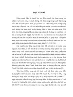
Chart 3.10. General results
Comments: patients achieved very good and good results accounted for
96.83% (very good: 85.72%; good: 11.11%), average: 3.17%, no poor results.
of accompanying fibula fractures at the level of low-tibial fractures and low tibial
fractures, only one peripheral pin is screwed. Conduct bone fixation marks with
screw braces before nailing the pelvic bone marrow, (while correcting fibula axis,
combining correction of tibial deformity).
* Treatment after surgery:
- Use antibiotics, analgesic, anti-edema after surgery.
- X-ray examination after 48 hours of surgery.
- Instruct patients to exercise.
- Appoint patients to follow up after 3, 6, 9 and after 12 months. Record the
rehabilitation process of the limb and X-ray examination to evaluate the fracture
after 6 months, after 12 months.
2.2.2.4. Method of evaluating results
* Evaluating close results
Evaluation of the results is based on Larson and Bostman standards: The
changes in the incision and the results of recovery of postoperative bone surgery
(based on the tibial X-ray film in 2 straight and inclined postures). Evaluate bone
Chapter 4. DISCUSSION
4.1. Characteristics of tibial anatomy in Vietnamese adults
4.1.1. Characteristics of age, gender, height of the research group
114 Viet people (67 males, 47 females) of adult age, average age of 41,31 ±
15,59 male group is 38.64 ± 13.66, less than the female group of 45.11 ± 17.35
with p <0.05. Average height is 161.77 ± 7.85cm, male group: 167.39 ± 3.62cm is
higher than female group: 153.77 ± 4.64cm with p <0.05.
4.1.2. Characteristics of tibia anatomy in some Vietnamese adults
4.1.2.1. The tibia length is related to age, gender, height.
* The tibia length is related to age group and gender
The average absolute tibial length in the male group: Youth: 37.35 ± 2.13cm;
Middle-aged: 36.91 ± 1.75cm; Elderly: 36.41 ± 2.35. Female group: youth: 35.19 ±
fixation results: Bone length, tibia. The lower end of the medullary nail compared
to the joint surface. The position of the pin holes and screws compared to the break.
1.55 cm; Middle-aged: 35.41 ± 2.16cm; Elderly: 34.08 ± 2.32. Average length of
Evaluate rotation, angular displacement.
Elderly: 23.22 ± 2.38. Female group: Youth: 23.32 ± 1.57cm: Middle-aged: 23.34 ±
* Evaluating near and far results (over 12 months)
Status of surgical scarring, bone healing, rehabilitation (muscle atrophy, short
limb, movement amplitude of the knee, movement of the ankles and the degree of
pain during compression).
tibia bone in male group: Youth: 24.76 ± 1.7cm; Middle-aged: 23.8 ± 1.28cm:
1.81cm; Elderly: 21.75 ± 1.76.
4.1.2.2. Size of tibia marrow canal
9
16
Time to evaluate results is over 12 months. Results of functional rehabilitation
3.2.7. Distant results after 12 months
evaluation of Ter. Schiphort consists of 4 levels: Very good, good, medium and poor.
3.2.7.1. Results of bone healing
35
KX mác
Results of bone healing and bone healing time
* Evaluating the bone healing based on the clinical results and straight and
tilted X-ray films. The result of bone healing is divided into 3 levels: Solid bone,
30
Gãy loại A1
unstable bone, false joint.
25
Gãy loại A2
20
Gãy loại A3
15
Gãy loại B1
10
Gãy loại B2
according to false test method theory of differences between the two averages in
Independent Sample T test, X2 value, standard deviation (X ± SD), One - Way
5
Gãy loại B3
ANOVA.
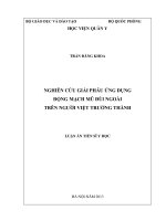
Chart 3.9. The result of tibial bone healing (n = 63)
0
Rất tốt
Tốt
Trung bình
Kém
Gãy loại C1
Comments: 100% bone healing, very good 57 patients (90.48%), good 4
patients (6.35%), average 2 patients (3.17%).
* Result of fibula healing: Twenty-three fibular fixation patients were in
good bone, not displaced. 34 fibula fracture patients without bone fixation are also
seamless but still deviate.
3.2.7.2. Status of surgical scar: 100% of patients have soft scar.
3.2.7.3. Results of rehabilitation
3.2.7.3.1. Restoring knee joint movement:
100% of patients with normal knee joints have knee folds of> 120 - 1400.
3.2.7.3.2. Results of recovery of ankle joint movement
Table 3.37. Results of recovery of ankle joint movement (n = 63)
Movement level
Number of patients
Rate %
Normal
61
96,82
Less restrictions
2
3,18
Total
63
100
Comments: Mobilizing normal ankle joint 61 patients (96.82%), limiting at
least 2 patients (3.18%) (restriction of pubis fold).
3.2.7.3.3. Short limbs level: No patient with short limbs.
2.2.3. Data processing methods
Data processing by SPSS statistical software 20.0: Comparison of absolute length of
tibia, length of tibial body, root canal size in positions, by gender, age and height,
10
15
Chapter 3. RESEARCH RESULTS
3.2.4.2.2. Screwing results: 100% of the screws into the right holes.
3.2.5. Results of rehabilitation
3.1. Characteristics of tibial anatomy in Vietnamese adults
3.2.5.3. Time to travel normally
3.1.1. Study group characteristics
Table 3.1. Characteristics of age, gender and height
Age
Height
(Min - Max)
Gender
X ± SD
X ± SD (cm)
Male (67)
38,64 ± 13,66
(20 - 80)
167,39 ± 3,62
Female (47)
45,11 ± 17,35
(20 - 83)
153,77 ± 4,64
Male+Female(114)
41,31 ± 15,59
(20 - 83)
161,77 ± 7,85
p < 0,05
p < 0,05
p
Chart 3.7. Time to travel normally (n = 63)
(Min - Max)
25
(158 - 176)
(146 - 165)
(146 - 176)
20
Comments: The male group is higher than the female group (with p <0.05).
KX mác
Gãy loại A1
Gãy loại A2
15
Gãy loại A3
10
Gãy loại B1
3.1.2. The absolute length and length of the tibia bone body are related to age,
Gãy loại B2
gender and height
5
3.1.2.2. Absolute length and length of tibial body are related to age group and gender
0
Table 3.3. Absolute tibial length associated with age and gender group (n = 228)
Absolute tibia bone length (X ± SD) - cm
Age
group
Male (67)
(Min - Max)
Female (47)
(Min - Max)
18 - 44
37,35 ± 2,13
(33,4 - 46)
35,19 ± 1,55
(32,6 - 38,9)
45 - 59
36,91 ± 1,75
(34,2 - 42)
35,41 ± 2,16
(31,2 - 39,2)
≥ 60
36,41 ± 2,35
(33,3 - 38,8)
34,08 ± 2,32
(32,1 - 41)
p < 0,05
p < 0,05
p
Gãy loại B3
Gãy loại C1
< 2 tháng
2 - 3 tháng
3 - 4 tháng
> 4 tháng
Comments: Normal travel time of 2 months: 41.27%.
3.2.6. Follow up after 6 months of surgery
3.2.6.1. Evaluation of rehabilitation after 6 months of surgery
Chart 3.8. Assessing the rehabilitation after 6 months (n = 63)
Comments: The group of children with absolute length of tibia is longer than the
elderly group with p <0.05.
3.1.6. Size of tibia marrow canal in the narrowest position associated with age
Table 3.13. Size of tibia marrow canal in the narrowest position associated with
age (n = 228)
Age
Size of tibia marrow canal bed in the narrowest position
(X ± SD) - (cm) (8 to 10cm from the end of the tibia body)
group
p
Inner-Outer
Min - Max
Front-Behind
Min - Max
18 - 44
1,13 ± 0,12
(0,8 - 1,4)
0,98 ± 0,1
(0,7 - 1,3)
p < 0,05
45 - 59
1,16 ± 0,14
(0,8 - 1,4)
1 ± 0,12
(0,7 - 1,3)
p < 0,05
≥ 60
1,22 ± 0,14
(1 - 1,4)
1,05 ± 0,12
(0,9 - 1,3)
p < 0,05
p < 0,05
p < 0,05
p
35
KX mác
30
Gãy loại A1
25
Gãy loại A2
20
Gãy loại A3
15
Gãy loại B1
10
Gãy loại B2
5
Gãy loại B3
0
Rất tốt
Tốt
Trung bình
Kém
Gãy loại C1
Comments: After 6 months very good 54 patients (85.72%), good 7 patients
(11.11%), average 2 patients (3.17%).
14
11
Comments: Two screws were installed under the parallel fracture area for
Comments: Medium size internal marrow canal - outside and before - after in
patients with a distance from fracture area to tibiotalar joint> 60mm.
the older group wider in the younger group with p <0.05. Select the diameter of
3.2.3.8. Surgery of bone fixation fracture area fibula
nails based on the size of the narrowest root canal.
Table 3.28. Surgery of bone fixation fracture area fibula (n = 57)
Fibular fixation
Number of patients
Rate %
Không bone fixation
34
59,65
bone fixation bằng nẹp vít
23
40,35
Total
57
100
3.1.8. Inner ankle length
Comments: Twenty-three patients with bone fixation by screwing (40.35%)
are fractures of fibula at the same level as fracture area of the tibia and capturing 1
pin.
Table 3.17. The inner ankle length related to gender (n = 228)
The inner ankle length
(Min - Max)
Gender
(X ± SD) - (cm)
Male (67 people)
1,45 ± 0,13
(1,30 - 1,70)
Female (47 people)
1,40 ± 0,11
(1,30 - 1,70)
Male+Female (114 people)
1,43 ± 0,13
(1,30 - 1,70)
p
p < 0,05
Comments: Ankle length among men than among women with long p <0.05.
3.2.4. Treatment results
3.1.8.2. The inner ankle length related to height
3.2.4.1. Near results
* Status of incision surgery: 100% of patients will have a first-term wound without
infection.
* Early complications: No bleeding complications, pinched cavity, fat embolism ...
3.2.4.2. Results of bone fixation
3.2.4.2.1. Results of correcting fracture area under AO
Table 3.29. Correction results of fracture area according to classification of AO
fracture (n = 63)
Classification
of fractures
Type A1
Type A2
Type A3
Type B1
Type B2
Type B3
Type C1
Total
Rate %
3.1.8.1. The inner ankle length related to gender
Results of rectification of fracture area
Very good
Good
Average
Weak
4
32
3
11
1
2
1
6
2
1
57
4
2
0
90,48
6,35
3,17
0
Total
4
35
14
1
6
2
1
63
100
Comments: Very good 90.48%, good 6.35%, 3.17% on average, all break
into A3 type. For 2 patients with large deviations: 1 patient was from fracture area
to tibiotalar joint belonging to fracture area from 31 - 40mm.
Height (cm)
< 152
153 - 154
155 - 156
157 - 159
160 - 162
> 163
Table 3.18. Ankle length in relation to height (n = 228)
The inner ankle length (X ± SD) - (cm) (Min - Max)
1,41 ± 0,1
(1,3 - 1,7)
1,45 ± 0,16
(1,3 - 1,7)
1,37 ± 0,07
(1,3 - 1,5)
1,35 ± 0,07
(1,3 - 1,5)
1,45 ± 0,16
(1,3 - 1,7)
1,44 ± 0,12
(1,3 - 1,7)
p
p<
0,05
Comments: The higher the height group, the longer the ankle length with p <0.05.
The ankle length along with the distance from the end screw hole to the end of the
nail is the limit to avoid tibiotalar joint violations.
3.2. Results of clinical treatment application
3.2.1. Characteristics of researched patients
3.2.1.1. Age and gender
Age
18 - 44
45 - 59
≥ 60
Total
Rate %
(X ± SD)
Gender
Male
26
8
5
39
61,90
Total
Female
11
37
12
20
1
6
24
63
38,10
100
42,75 ± 13,97 age (18 - 78 age)
Rate %
58,73
31,75
9,52
100
12
13
Comments: Number of patients with bone fixation surgery during <24h is 47
Table 3.19. Age and gender characteristics (n = 63)
Comments: Male/female ratio: 1.6/1. Average age: 42.75 ± 13.97 years
patients (74.60%), from 2-4 days 14 patients (22.22%), from 5 to 7 days 1 patient
(18-78 years). Youth with the highest rate: 58.73%.
(1.59 %),> 7 days 1 patient (1.59%).
3.2.2. Classification of injury
3.2.3.2. Anesthesia method: 63 patients who underwent tibial fixation had spinal
3.2.2.4. Distance from fracture area to tibiotalar joint
anesthesia.
Table 3.22. Distance from fracture area to tibiotalar joint (n = 63)
Distance from fracture area to tibiotalar joint
Number of patients Rate %
31 - 40mm
14
22,22
41 - 60mm
14
22,22
61 - 80mm
19
30,16
> 80mm
16
25,40
Total
63
100
3.2.3.3. Bone fixation technique: 63 patients are bone fixation sealed with
Comments: Distance from fracture area to tibiotalar joint from 61 - 80mm at
Rate %
most 19 patients (30.16%).
3.2.2.5. Tibial fractures classified according to AO
Table 3.23. Classifying tibia fractures according to AO (n = 63)
Classification of
Number of
Rate %
Total
Total rate %
fractures
patients
A1
4
6,35
53
Type A
A2
35
55,55
84,12
A3
14
22,22
B1
1
1,59
9
B2
6
9,52
14,29
Type B
B3
2
3,18
Type C
C1
1
1,59
1
1,59
Total
63
100
Comments: Classification by AO, fracture type A: 84.12%, type B: 14.29%, type C: 1.59%.
3.2.3. Treatment method
interlocking intramedullary nail.
3.2.3.4. Medullary canal drilling: 63 patients drilled their canals when nailed.
3.2.3.5. Nail size
Table 3.25. Nail diameter and nail length (n = 63)
Nail diameter
8 mm
9 mm
10 mm
Total
Nail length
280mm
0
2
0
2
300mm
4
22
2
28
320mm
1
19
1
21
340mm
0
12
0
12
5
55
3
63
Total
7,94
87,30
4,76
Rate %
100
Comments: Nail 9mmx300mm most used
3.2.3.7. Distance from fracture area bone to tibiotalar joint related to Screwing
technique
Table 3.27. Distance from fracture area to tibiotalar joint related to screwing
Distance from
fracture area to
tibiotalar joint
3.2.3.1. Time from fracture to surgery
Table 3.24. Time from fracture to surgery (n = 63)
Time
Number of patients
< 24h
47
From 2 - 4 days
14
From 5 - 7 days
1
> 7 days
1
Total
63
Rate (%)
74.60
22.22
1.59
1.59
100
3,17
44,44
33,33
19,05
100
31 - 40mm
41 - 60 mm
61 - 80mm
> 80mm
Total
2 screws
under
parallel
fracture
area
0
0
12
14
26
technique (n = 63)
Screwing technique
2 screws
under
1 screw
perpendi
under
cular
fracture
fracture
area
area
0
12
8
6
4
3
0
2
12
23
1 screw
above +
1 screw
under
fracture
area
2
0
0
0
2
Total
Rate
%
14
14
19
16
63
22,22
22,22
30,16
25,40
100