2015 annual update in intensive care and emergency medicine 2015newmedicalbooks
Bạn đang xem bản rút gọn của tài liệu. Xem và tải ngay bản đầy đủ của tài liệu tại đây (6.65 MB, 582 trang )
2015
Annual Update
in Intensive Care
and Emergency
Medicine 2015
Edited by J.-L.Vincent
123
Annual Update in Intensive Care and
Emergency Medicine 2015
The series Annual Update in Intensive Care and Emergency Medicine is the continuation of the series entitled Yearbook of Intensive Care Medicine in Europe and
Intensive Care Medicine: Annual Update in the United States.
Jean-Louis Vincent
Editor
Annual Update in
Intensive Care and
Emergency Medicine 2015
Editor
Prof. Jean-Louis Vincent
Université libre de Bruxelles
Dept. of Intensive Care
Erasme Hospital
Brussels, Belgium
ISSN 2191-5709
ISBN 978-3-319-13760-5
DOI 10.1007/978-3-319-13761-2
ISBN 978-3-319-13761-2 (eBook)
Springer Cham Heidelberg New York Dordrecht London
© Springer International Publishing Switzerland 2015
This work is subject to copyright. All rights are reserved, whether the whole or part of the material is
concerned, specifically the rights of translation, reprinting, reuse of illustrations, recitation, broadcasting,
reproduction on microfilm or in any other way, and storage in data banks. Duplication of this publication
or parts thereof is permitted only under the provisions of the German Copyright Law of September 9,
1965, in its current version, and permission for use must always be obtained from Springer. Violations
are liable to prosecution under the German Copyright Law.
The use of general descriptive names, registered names, trademarks, etc. in this publication does not imply, even in the absence of a specific statement, that such names are exempt from the relevant protective
laws and regulations and therefore free for general use.
The publisher, the authors and the editors are safe to assume that the advice and information in this book
are believed to be true and accurate at the date of publication. Neither the publisher nor the authors or
the editors give a warranty, express or implied, with respect to the material contained herein or for any
errors or omissions that may have been made.
Cover design: WMXDesign GmbH, Heidelberg
Printed on acid-free paper
Springer International Publishing AG Switzerland is part of Springer Science+Business Media
(www.springer.com)
Contents
Common Abbreviations . . . . . . . . . . . . . . . . . . . . . . . . . . . . . . . .
Part I
xi
Infections
Early Identification of Ventilator-associated Pneumonia Causative
Pathogens: Focus on the Value of Gram-stain Examination . . . . .
C. Chiurazzi, A. Motos-Galera, and A. Torres
3
Central Line-associated Bloodstream Infections: A Critical Look
at the Role and Research of Quality Improvement Interventions
and Strategies . . . . . . . . . . . . . . . . . . . . . . . . . . . . . . . . . . . 15
K. Blot, D. Vogelaers, and S. Blot
Clostridium difficile Infection . . . . . . . . . . . . . . . . . . . . . . . . . . . . . 25
M. H. Wilcox, M. J. G. T. Vehreschild, and C. E. Nord
Viral Sepsis . . . . . . . . . . . . . . . . . . . . . . . . . . . . . . . . . . . . . . . . 37
P. Amin and V. Amin
Part II
Antimicrobials and Resistance
Light and Shade of New Antibiotics . . . . . . . . . . . . . . . . . . . . . . . . 63
M. Bassetti, P. Della Siega, and D. Pecori
Optimizing Antimicrobial Efficacy at Minimal Toxicity: A Novel
Indication for Continuous Renal Replacement Therapy? . . . . . . . 85
P. M. Honoré, R. Jacobs, and H. D. Spapen
v
vi
Contents
Combatting Resistance in Intensive Care: The Multimodal Approach
of the Spanish ICU “Zero Resistance” Program . . . . . . . . . . . . . 91
The Scientific Expert Committee for the “Zero Resistance” Project
Immune System Dysfunction and Multidrug-resistant Bacteria in
Critically Ill Patients: Inflammasones and Future Perspectives . . . 105
M. Girardis, S. Busani, and S. De Biasi
Part III
Sepsis
Tachycardia in Septic Shock: Pathophysiological Implications
and Pharmacological Treatment . . . . . . . . . . . . . . . . . . . . . . . 115
A. Morelli, A. D’Egidio, and M. Passariello
Angiotensin II in Septic Shock . . . . . . . . . . . . . . . . . . . . . . . . . . . . 129
T. D. Corrêa, J. Takala, and S. M. Jakob
ˇ-Blockers in Critically Ill Patients: From Physiology to Clinical Evidence 139
S. Coppola, S. Froio, and D. Chiumello
Part IV
Oxygenation and Respiratory Failure
Prehospital Endotracheal Intubation: Elemental or Detrimental? . . . . . 155
P. E. Pepe, L. P. Roppolo, and R. L. Fowler
Hyperoxia in Intensive Care and Emergency Medicine:
Dr. Jekyll or Mr. Hyde? An Update . . . . . . . . . . . . . . . . . . . . . 167
S. Hafner, P. Radermacher, and P. Asfar
Extracorporeal Gas Exchange for Acute Respiratory Failure
in Adult Patients: A Systematic Review . . . . . . . . . . . . . . . . . . 179
M. Schmidt, C. Hodgson, and A. Combes
Update on the Role of Extracorporeal CO2 Removal as an Adjunct
to Mechanical Ventilation in ARDS . . . . . . . . . . . . . . . . . . . . . 207
P. Morimont, A. Batchinsky, and B. Lambermont
Fundamentals and Timing of Tracheostomy:
ICU Team and Patient Perspectives . . . . . . . . . . . . . . . . . . . . . 219
V. Pandian and M. Mirski
Shared Decision-making to Pursue, Withhold or Withdraw Invasive
Mechanical Ventilation in Acute Respiratory Failure . . . . . . . . . . 233
M. E. Wilson, P. R. Bauer, and O. Gajic
Contents
Part V
vii
Monitoring
New Fully Non-invasive Hemodynamic Monitoring Technologies:
Groovy or Paltry Tools . . . . . . . . . . . . . . . . . . . . . . . . . . . . . 249
J. Benes and E. Kasal
Assessing Global Perfusion During Sepsis: SvO2 , Venoarterial PCO2
Gap or Both? . . . . . . . . . . . . . . . . . . . . . . . . . . . . . . . . . . . 259
J.-L. Teboul and X. Monnet
An Update on Cerebral Oxygenation Monitoring, an Innovative
Application in Cardiac Arrest and Neurological Emergencies . . . . 273
B. Schneider, T. J. Abramo, and G. Albert
Part VI
Cardiac Arrest
Out-of-hospital Cardiac Arrest and Survival to Hospital Discharge:
A Series of Systemic Reviews and Meta-analyses . . . . . . . . . . . . 289
M. Vargas, Y. Sutherasan, and P. Pelosi
Cooling Techniques for Targeted Temperature Management
Post-cardiac Arrest . . . . . . . . . . . . . . . . . . . . . . . . . . . . . . . . 315
C. Vaity, N. Al-Subaie, and M. Cecconi
Part VII
Fluids
How Does Volume Make the Blood Go Around? . . . . . . . . . . . . . . . . 327
S. Magder
Clinical Implications from Dynamic Modeling of Crystalloid Fluids . . . 339
R. G. Hahn
Part VIII
Renal Injury
Urinary Electrolyte Monitoring in the Critically Ill:
Revisiting Renal Physiology . . . . . . . . . . . . . . . . . . . . . . . . . . 351
P. Caironi, T. Langer, and M. Ferrari
Management of AKI: The Role of Biomarkers . . . . . . . . . . . . . . . . . 365
Z. Ricci, G. Villa, and C. Ronco
Bone Morphogenetic Protein 7: An Emerging Therapeutic Target
for Sepsis-associated Acute Kidney Injury . . . . . . . . . . . . . . . . . 379
X. Chen, X. Wen, and J. A. Kellum
viii
Contents
Long-term Sequelae from Acute Kidney Injury: Potential Mechanisms
for the Observed Poor Renal Outcomes . . . . . . . . . . . . . . . . . . 391
M. Varrier, L. G. Forni, and M. Ostermann
Part IX
Hepatic and Abdominal Issues
Application of the Acute Kidney Injury Network Criteria in Patients with
Cirrhosis and Ascites: Benefits and Limitations . . . . . . . . . . . . . 405
P. Angeli, M. Tonon, and S. Piano
Intensive Care Management of Severe Acute Liver Failure . . . . . . . . . 415
S. Warrillow and R. Bellomo
Human Albumin: An Important Bullet Against Bacterial Infection
in Patients with Liver Cirrhosis? . . . . . . . . . . . . . . . . . . . . . . . 431
M. Bernardi, M. Domenicali, and P. Caraceni
Open Abdomen Management: Challenges and Solutions
for the ICU Team . . . . . . . . . . . . . . . . . . . . . . . . . . . . . . . . . 447
J. J. De Waele and M. L. N. G. Malbrain
Part X
Nutrition
Protein Intake in Critical Illness . . . . . . . . . . . . . . . . . . . . . . . . . . . 459
O. Rooyackers and J. Wernerman
Part XI
Trauma and Massive Bleeding
Rational and Timely Use of Coagulation Factor Concentrates
in Massive Bleeding Without Point-of-Care
Coagulation Monitoring . . . . . . . . . . . . . . . . . . . . . . . . . . . . 471
O. Grottke, D. R. Spahn, and R. Rossaint
Optimal Temperature Management in Trauma:
Warm, Cool or In-between? . . . . . . . . . . . . . . . . . . . . . . . . . . 481
M. C. Reade and M. Lumsden-Steel
Detection of Consciousness in the Severely Injured Brain . . . . . . . . . . 495
J. Stender, A. Gjedde, and S. Laureys
Contents
Part XII
ix
Neuromuscular Considerations
The Role of Local and Systemic Inflammation in the Pathogenesis
of Intensive Care Unit-acquired Weakness . . . . . . . . . . . . . . . . 509
E. Witteveen, M. J. Schultz, and J. Horn
Critical Illness is Top Sport . . . . . . . . . . . . . . . . . . . . . . . . . . . . . . 519
M. Suker, C. Ince, and C. van Eijck
Part XIII
Rapid Response Teams
Vital Signs: From Monitoring to Prevention of Deterioration
in General Wards . . . . . . . . . . . . . . . . . . . . . . . . . . . . . . . . . 533
M. Cardona-Morrell, M. Nicholson, and K. Hillman
Rapid Response Systems: Are they Really Effective? . . . . . . . . . . . . . 547
C. Sandroni, S. D’Arrigo, and M. Antonelli
Severe Sepsis Beyond the Emergency Department and ICU:
Targeting Early Identification and Treatment
on the Hospital Floor . . . . . . . . . . . . . . . . . . . . . . . . . . . . . . 557
C. A. Schorr, J. Sebastien, and R. P. Dellinger
Part XIV
Data Management
State of the Art Review: The Data Revolution in Critical Care . . . . . . . 573
M. Ghassemi, L. A. Celi, and D. J. Stone
Creating a Learning Healthcare System in the ICU . . . . . . . . . . . . . . 587
J. Yu and J. M. Kahn
Index . . . . . . . . . . . . . . . . . . . . . . . . . . . . . . . . . . . . . . . . . . . . 597
Common Abbreviations
AKI
ARDS
BAL
COPD
CPR
CT
CVP
DO2
EKG
EMR
FiO2
GCS
GFR
ICP
ICU
IL
INR
LV
MAP
MRI
NF-ÄB
NO
OR
PAC
PEEP
RBC
RCT
ROS
RRT
RV
SAPS
ScvO2
SOFA
TBI
TNF
VAP
Acute kidney injury
Acute respiratory distress syndrome
Bronchoalveolar lavage
Chronic obstructive pulmonary disease
Cardiopulmonary resuscitation
Computed tomography
Central venous pressure
Oxygen delivery
Electrocardiogram
Electronic medical record
Inspired fraction of oxygen
Glasgow Coma Scale
Glomerular filtration rate
Intracranial pressure
Intensive care unit
Interleukin
International normalized ratio
Left ventricular
Mean arterial pressure
Magnetic resonance imaging
Nuclear factor kappa-B
Nitric oxide
Odds ratio
Pulmonary artery cather
Positive end-expiratory pressure
Red blood cell
Randomized controlled trial
Reactive oxygen species
Renal replacement therapy
Right ventricular
Simplified acute physiology score
Central venous oxygen saturation
Sequential organ failure assessment
Traumatic brain injury
Tumor necrosis factor
Ventilator-associated pneumonia
xi
Part I
Infections
Early Identification of Ventilator-associated
Pneumonia Causative Pathogens: Focus on the
Value of Gram-stain Examination
C. Chiurazzi, A. Motos-Galera, and A. Torres
Introduction
Ventilator-associated pneumonia (VAP) is a common nosocomial infection in critically ill patients, associated with increased morbidity and healthcare costs. Early
identification of causative pathogens plays a critical role in the administration of
appropriate antibiotic therapy and patient outcomes. In particular, in patients with
clinical suspicion of VAP, respiratory samples should be obtained promptly to corroborate the provision of effective antibiotic treatment, while avoiding unnecessary
antibiotic use that would promote the development of resistance. In this context, the
value of the Gram-stain examination, and its potential impact on adequate empiric
antibiotic treatment and major outcomes, is still under debate. In this manuscript,
we review the most recent evidence on methods for early identification of VAP
causative pathogens, with specific focus on Gram-stain examination of respiratory
samples, and we highlight potential methodological limitations and future areas of
investigation.
Incidence, Etiology and Diagnosis of Ventilator-associated
Pneumonia
VAP is the second most common nosocomial infection in patients admitted to intensive care units (ICUs) [1]. VAP occurs in 9–27% of all ventilated patients [2,
3]. However, the incidences of VAP vary considerably among patient populations,
e. g., trauma patients or those undergoing cardiac and neurological surgery are at
greater risk. Additionally, various comorbidities and co-factors, such as prolonged
mechanical ventilation, chronic pulmonary disease, prior use of antibiotics, acute
C. Chiurazzi A. Motos-Galera A. Torres
Department of Pulmonary and Critical Care Medicine, Thorax Institute, Hospital Clinic,
Barcelona, Spain
e-mail:
© Springer International Publishing Switzerland 2015
J.-L. Vincent (ed.), Annual Update in Intensive Care and Emergency Medicine 2015,
DOI 10.1007/978-3-319-13761-2_1
3
4
C. Chiurazzi et al.
respiratory distress syndrome (ARDS) may increase the risk of VAP. Patients who
develop VAP present longer ICU- and hospital stays [4]. As a result, VAP is associated with increased healthcare costs, estimated at around US$ 40,000 per patient
who develops VAP [5, 6]. A recent report in VAP patients indicated that the overall attributable mortality was 13%. Nevertheless, mortality rates are inconsistent
among studies. Indeed, in a study by Bekaert and collaborators [7], a relatively
limited attributable VAP-associated mortality was reported. Importantly, late-onset
VAP is often caused by multidrug resistant (MDR) pathogens and is associated with
worse outcomes in comparison with VAP that develops early during the course of
mechanical ventilation.
VAP is frequently caused by aerobic, Gram-negative pathogens (Pseudomonas
aeruginosa, Acinetobacter baumannii, Klebsiella pneumoniae, Escherichia coli);
the most frequent Gram-positive pathogen is Staphylococcus aureus. Underlying
diseases may predispose patients to infection with specific organisms. For example,
patients with chronic obstructive pulmonary disease (COPD) are often colonized
and develop VAP caused by Haemophilus influenzae, Moraxella catarrhalis and P.
aeruginosa, whereas, Haemophilus spp. and Streptococcus pneumoniae are frequent causative pathogens in trauma patients. A. baumannii, S. aureus, and P.
aeruginosa are the most common causative pathogens in ARDS patients. Importantly, VAP is often caused by potentially MDR pathogens, i. e., P. aeruginosa, S.
aureus, Acinetobacter spp., Stenotrophomonas maltophilia, Burkholderia cepacia
and extended-spectrum ˇ-lactamase (ESBL) K. pneumoniae. Patients at risk of
being colonized by MDR pathogens are extremely varied, commonly present comorbid conditions, are ventilated for longer periods of time and receive antibiotics
during the course of their hospitalization. A recent study [8] demonstrated that
severity of illness did not affect etiology and risk factors for MDR pathogens. The
incidence of MDR pathogens is also closely linked to local factors and varies widely
from one institution to another [9].
VAP is commonly suspected in a patient receiving mechanical ventilation for at
least 48 hours, who develops new or progressive radiographic infiltrates, and at least
two clinical signs of infection, such as fever/hypothermia, leukocytosis/leukopenia
and purulent secretions. Other clinical signs may be of some value on a specific
case-by-case basis, e. g., worsening gas exchange, increased inflammatory markers.
Unfortunately, in critically ill patients, clinical signs of infection have marginal
diagnostic specificity/sensitivity. Thus, the Clinical Pulmonary Infection Score
(CPIS) is often calculated [10]. The CPIS is based on six clinical assessments
(temperature, blood leukocyte count, volume and purulence of tracheal secretions,
oxygenation, pulmonary radiographic findings, and semiquantitative culture of tracheal aspirate), each worth between 0 and 2 points. The CPIS showed a good
correlation (r = 0.84, p < 0.0001) with quantitative bacteriology of bronchoalveolar
lavage (BAL) samples. Moreover, a value 6 was the threshold to accurately identify patients with pneumonia.
Importantly, in patients with VAP, the diagnostic strategy should be sensitive
enough to identify the greatest number of infected patients and enable early initiation of adequate empiric antibiotic treatment. On the other hand, patients without
Early Identification of Ventilator-associated Pneumonia Causative Pathogens
5
Clinical Diagnostic Strategy
No
Clinical suspicion of VAP?
No further investigation
Yes
LRT and blood cultures
(before starting or changing ATBs)
Gram-stain examination of LRT sample
Start empiric ATBs
Yes
No
Positive cultures
Samples were
obtained before
administering
antibiotics?
Stop antibiotics
No
Yes
Consider short
course of
antibiotics
Adjust antibiotics
(based on culture results, clinical response)
Microbiological Diagnostic Strategy
Clinical suspicion of VAP?
Observe
(consider
other loci of
infection)
No
Yes
No further
investigation
Bronchoscopy (BAL/PSB), or
Blinded BAS/TBAS
Gram-stain examination
No
Positive
quantitative
cultures
Observe
(consider
other loci of
infection)
Yes
Start ATBs
(based on
culture results)
Continue/adjust ATBs
(another infection?)
No
Severe sepsis
Yes
No
Bacteria present
Yes
Start ATBs
Start ATBs
(based on guidelines,
(based on microscopic
local prevalence of examination, local prevalence
of pathogens)
pathogens)
No
Positive
quantitative
cultures
Positive
quantitative
cultures
Yes
Yes
No Continue/adjust ATBs
(consider other loci of
infection)
Adjust ATBs
Adjust ATBs
(based on culture results) (based on culture results)
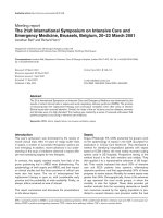
Fig. 1 On the top, a proposed clinical strategy for the diagnosis and treatment of ventilatorassociated pneumonia (VAP). Gram-stain examination of tracheal secretions can be performed.
The main drawback of this strategy is the potential overuse of antibiotics. On the bottom, the
microbiological strategy for the diagnosis and treatment of VAP. Lower respiratory tract (LRT)
samples are obtained through invasive (bronchoalveolar lavage [BAL], protected specimen brush
[PSB]) or non-invasive (tracheal aspiration) techniques. Of note, this strategy has high specificity
for the diagnosis of VAP, but lower sensitivity compared to the clinical strategy. BAS: bronchial
aspirate; ATB: antibiotic; TBAS: tracheobronchial aspirate
6
C. Chiurazzi et al.
infection should be discriminated to avoid overtreatment with antimicrobial drugs,
and selection of MDR microorganisms.
In a patient with clinical suspicion of VAP, two diagnostic algorithms can be
used following clinical suspicion of nosocomial pneumonia (Fig. 1). The clinical
approach recommends treating every patient with suspicion of having a pulmonary
infection with new antibiotics. Samples of respiratory secretions, such as endotracheal aspirate (ETA), should be obtained before the initiation of antibiotic treatment. In this strategy, the selection of appropriate empirical therapy is based on risk
factors and local resistance patterns. The etiology of pneumonia is defined by semiquantitative cultures of ETA or sputum, with potential Gram-stain examination of
the sample. Antimicrobial therapy is adjusted according to culture results or clinical response. This clinical strategy provides antimicrobial treatment to the majority
of the patients. The main drawback is that the high sensitivity of semi-quantitative
cultures of tracheal aspirates may lead to antibiotic overtreatment.
The bacteriological strategy is based on the results of quantitative cultures of
lower respiratory tract secretions. Samples can be obtained using ETA, BAL or
protected specimen brush (PSB). Specific threshold cut-offs for each test (105 –
106 CFU/mL for ETA, 104 CFU/mL for BAL, and 103 CFU/mL for PSB) are applied to discriminate between colonizing microorganisms and those producing infection. Ideally, Gram-stain examination of these samples can be performed to
improve early adequacy of antibiotic treatment. The bacteriological strategy attempts to identify patients with true VAP, reduce overuse of antibiotics and improve
outcomes. Yet, false negative results may be obtained using this strategy, which
leads to delayed antibiotic treatment and worse outcomes.
The Importance of Rapid Diagnostic Techniques
for Ventilator-associated Pneumonia
Early diagnosis and initiation of appropriate antibiotic therapy for VAP is associated with improved outcomes; conversely, delayed or inappropriate administration
of targeted antibiotic therapy is associated with increased mortality. In particular,
inadequate therapy during the first 48 hours following clinical suspicion of VAP is
associated with a 3-fold increase in mortality (91%), in comparison with patients
appropriately treated (38%) [11]. The importance of a prompt microbiological diagnosis of VAP is aimed not only at optimizing antimicrobial treatment, but also
at narrowing or de-escalating the initial empiric treatment, as soon as antimicrobial
susceptibility data are available.
The main limitation in the use of standard microbiology cultures for the diagnosis of VAP and guiding empiric therapy is that the results are not available for
48 hours. Thus, several alternative techniques to microbial cultures have been developed to achieve a more rapid and accurate diagnosis of VAP (Table 1).
In this context, the Gram-stain examination of respiratory samples, described in
the following paragraphs, can promptly provide information regarding the type of
microorganisms and the purulency of the biomaterial (defined as 25 neutrophils
Early Identification of Ventilator-associated Pneumonia Causative Pathogens
7
Table 1 Diagnostic methods for the identification of ventilator-associated pneumonia causative
pathogens
Method
Bacterial culture
Required time
to generate
results
48–72 h
Gram-stain
1h
Nucleic
acid-based
amplification
method
(i. e., multiplex
real-time PCR)
Mass spectrometry (MS)
(e. g., matrixassisted laser
desorption
ionization
time-of-fly
(MALDI-TOF)
Electrospray
ionization (ESI
MS)
1h
1–2 minutes,
after standard
bacterial
culture
4–6 h
Advantages
Drawbacks
Diagnostic gold standard
Quantitative analysis
Assessment of antibiotic
susceptibility
Identification of bacterial
species
Rapid test
Inexpensive test
Direct analysis of clinical
samples
Time to identify causative
pathogen of an infection is
overly long
Direct analysis of clinical
samples
Multiple causative
pathogens are tested
Assessment of antibiotic
susceptibility
Identification of bacterial
species
Identification of bacterial
toxins Assessment of
antibiotic susceptibility
Direct analysis of clinical
samples
Semi-quantitative
analysis
Expertise required
Considerable colonization is
needed to identify causative
pathogens
Qualitative analysis
No information on antibiotic
susceptibility
No identification of bacterial
species
Expensive
Lack of clinical validation
Reduced reliability during
poly-microbial colonization
Analysis performed only after
standard culture
High risk of contamination
(open work platform)
Expensive test
Reduced reliability during
poly-microbial colonization
PCR: polymerase chain reaction.
and Ä 10 squamous epithelial cells per low power field) [2]. As an alternative, new
molecular-based methods for early identification of respiratory pathogens have been
developed. Similar to the Gram-stain examination, molecular methods are aimed at
identifying the causative agent of infection in a timely manner [12]; yet, these novel
techniques can also determine antimicrobial susceptibility profiles. Molecular diagnostic techniques simultaneously target a wide range of bacterial species and
resistance genes through polymerase chain reaction (PCR) amplification of nucleic
acid. The technique most frequently applied is multiplex real-time PCR and de-
8
C. Chiurazzi et al.
tection through arrays, such as two dimensional micro-chips or three-dimensional
beads and dye-labeled probes. More recently, rapid detection and identification
of pathogens directly from clinical specimens can be performed with the use of
matrix-assisted laser desorption ionization time-of-fly (MALDI-TOF) and PCRelectroSpray ionization mass spectrometry (PCR/ESI-MS) systems, which rely,
however, on the use of expensive operating systems [13].
Some of the main advantages with use of molecular diagnostic techniques are
the rapid results and the possibility to detect very low quantities of target sequences
irrespective of pathogen viability or concomitant use of antibiotics. Additionally,
these techniques also target specific sequences related to antimicrobial resistance
and improve detection of microorganisms that are difficult to culture using conventional methods [14]. The main limitations are potential contamination, overlap
among genetic sequences of different pathogens, lack of validation of some assays,
complex interpretation of the results, and increased costs [12]. Finally, the majority
of these systems only provide qualitative results, and it is difficult to distinguish
between colonizers and invasive pathogens [13].
Gram-stain Examination of Respiratory Samples: Methodological
Notes
Gram-stain examination is a technique applied to cluster bacterial species into two
groups – Gram-positive and Gram-negative – based on specific features of their cell
wall. The Gram-stain procedure begins by placing a very thin layer of respiratory
sample onto a glass slide. The sample should be air-dried rather than heated, because the heat distorts bacterial and cell morphology. The sample is then stained
with crystal violet and iodine. The length of time that crystal violet and iodine are
left on the smear is not critical. A minimal 10-second stain with these reagents
is sufficient. A decolorizing agent, such as ethanol or acetone, is then applied
briefly, and the solution is rinsed across the smear. Gram-positive bacteria retain
the crystal violet and iodine, because their thick cell wall comprises peptidoglycan.
Conversely, a thinner cell-wall layer characterizes Gram-negative pathogens; thus,
the stains are diffused from the bacteria with the use of ethanol. Finally, a counterstain, such as a red dye, safranin or fuchsin, is applied for at least 30 seconds to
allow staining of Gram-negative bacteria and a clear distinction from Gram-positive
microorganisms.
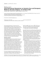
Upon microscopic examination, Gram-positive bacteria appear purple-blue;
whereas, Gram-negative microorganisms are reddish (Fig. 2). Several other bacterial features may help in the correct identification of pathogens. In particular,
the bacterial shape, e. g. cocci, rods, fusiform, narrows the range of potential
causative pathogens. In addition, the presence and quantification of inflammatory
cells increases the likelihood of an ongoing infection. Finally, the presence of
oropharyngeal squamous epithelial cells corroborates contamination of the sample
with saliva. Ideally, squamous epithelial cells should be less than 1% of all cells
present in the field of view [15].
Early Identification of Ventilator-associated Pneumonia Causative Pathogens
Fig. 2 Gram-stain images.
a Gram-stain appearance
of bronchoalveolar aspirate
showing Streptococcus pneumoniae and Haemophilus
influenzae. b Gram-stain appearance of bronchoalveolar
aspirate showing Gramnegative bacilli and some
intracellular bacteria. c Gram
stain appearance of tracheal
aspirate showing Nocardia.
(1000 × magnification, Nikon
Eclipse 50i Microscopy,
Nikon digital sight- NIS Elements). Micrographs were
kindly provided by Dr. Puig,
Microbiology Department,
Hospital Clinic, Barcelona,
Spain
a
b
c
9
10
C. Chiurazzi et al.
Gram-stain is a very rapid tool in the diagnosis of VAP and provides useful information on etiology; indeed, results may be ready within an hour. Additionally, the
test is inexpensive to perform in comparison with newer molecular tests. A recent
meta-analysis [16] found no difference in Gram-stain results in patients undergoing
antibiotic therapy and those without therapy. Thus, in comparison with standard microbiology cultures, Gram-stain is not significantly influenced by ongoing antibiotic
therapy.
Nevertheless, several limitations should be highlighted. First of all, the Gramstain technique requires considerable experience to adequately assess the samples
and provide reliable results. Additionally, considerable colonization of the sample is
needed – at least 105 organisms per milliliter – to identify pathogens on microscopy
[17]. Finally, the technique does not quantify pathogens and does not provide any
information on bacterial viability.
The Value of Gram-stain in Ventilator-associated Pneumonia
Given the rapid results and the valuable interpretation of respiratory samples using
Gram-stain, there has been considerable interest in recent years on the role of this
technique in the diagnosis of VAP, as detailed in Table 2.
In a recent meta-analysis, O’Horo and colleagues pooled data from 24 studies published from 1994 to 2008; the primary aim was to determine the value
of Gram-stain examination in the diagnosis of patients with clinical suspicion of
VAP [16]. Additionally, the possible role of Gram-stain examination in guiding
empiric therapy was assessed. The meta-analysis included a total of 3,148 respiratory samples obtained through BAL, mini-BAL, ETA and PSB. Gram-stain
examination was associated with a sensitivity of 0.79 and specificity of 0.74. Additionally, there was fair agreement (Ä 0.54) between bacteria identified through
microscopy and those identified by culture. However, it is important to emphasize
that among the studies included in the analysis, several did not report antibiotic
use; furthermore, the studied populations, the methods used to obtain respiratory
specimens and the Gram-stain examination were highly heterogeneous. Based on
these limitations, the authors concluded that Gram-stain examination should not be
recommended to guide early antimicrobial therapy; nevertheless Gram-stain examination was slightly more sensitive in the diagnosis of VAP caused by Gram-positive
bacteria; finally, Gram-stain results had a very high negative predictive value.
In the last two decades, several key studies assessed the role of Gram-stain examination in the diagnosis of VAP. In a study published by Blot et al. in 2000 [18], ETA
and PSB samples were concomitantly obtained from 91 suspected cases of VAP to
evaluate concordance between Gram-stain and microbiology results. The sensitivity
and specificity of Gram-stain examination in the diagnosis of microbiologicallyconfirmed pneumonia were, respectively, 91% and 64% for ETA and 70% and 96%
for samples obtained through PSB. Thus, the authors proposed a diagnostic algorithm based on three possible combinations: 1) When Gram-stain examination of
ETA samples is negative, VAP is highly improbable and therapy should be delayed
Early Identification of Ventilator-associated Pneumonia Causative Pathogens
11
Table 2 Studies assessing the value of Gram-stain examination in the diagnosis of
microbiologically-confirmed ventilator-associated pneumonia
Study
Year
Allaouchiche et al.
[23]
Allaouchiche et al.
[24]
Blot et al.
[18]
1996
Number Collection Study Design
of
Methods
samples
163
BAL
Prospective
cohort study
1999
146
BAL
2000
91
BAL/ETA Prospective
cohort study
Duflo et al. 2001
[25]
116
MiniBAL
Prospective
cohort study
Davis et al. 2005
[26]
155
BAL
Retrospective
chart review
Kopelman
[27]
2006
227
BAL
Retrospective
chart review
Veinstein
et al. [19]
2006
78
PTC/ETA
Albert
et al. [21]
2008
705
Goldberg
2008
et al. [28]
O’Horo
2012
et al. [16]
Gottesman 2014
et al. [22]
309
3141
115
Prospective
cohort study
Multicenter
prospective
trial
BAL/ETA Retrospective
analysis of
multicenter
randomized
control trial
BAL
Prospective
trial
BAL/PTC/ Meta-analysis
ETA
ETA
Prospective
cohort study
Main results vs. bacterial
identification through standard
cultures
Se 92, Sp 76.5,
PPV 69, NPV 91,
Ä 0.44
Se 90.2, Sp 73.7,
PPV 64.8, NPV 93.3,
Ä 0.586
ETA: Se 89, Sp 56, PPV 53,
NPV 90
PTC: Se 74, Sp 97, PPV 93,
NPV 87
Se 76.2, Sp 100,
PPV 100, NPV 75.4,
Ä 0.73
GP: Se 87, Sp 59, PPV 68,
NPV 83
GN: Se 73, Sp 49, PPV 78,
NPV 42
GP: Se 79.7, Sp 65.6 %,
PPV 47.7 %, NPP 89.2 %
GN: Se 67.0%, Sp 73.6 %,
PPV 68.9 %, NPV 71.8 %
Se 83, Sp 74,
PPV 79, NPV 79 (combining the
two techniques)
Se 74, Sp 72,
PPV 75, NPV 70,
Ä 0.36
Se 90, Sp 67,
PPV 45, NPV 96
Se 79, Sp 74,
PPV 40, NPV 90
GP: Se 90.47, Sp 82, PPV 57,
NPV 97
GN: Se 69.6, Sp 77, PPV 97,
NPV 20
Sterile culture: Se 50, Sp 79,
PPV 13, NPV 96
Ä 0.54
Se: sensitivity (%); Sp: specificity (%); PPV: positive predictive value (%); NPV: negative predictive value (%); BAL: bronchoalveolar lavage; ETA: endotracheal aspirate; GP: Gram-positive;
GN: Gram-negative; PTC: plugged telescoping catheter; Ä: kappa statistic.
12
C. Chiurazzi et al.
until microbiology results become available; 2) when Gram-stain examination of
PSB samples is positive, VAP is probable and antibiotic therapy should be promptly
administered and later readjusted based on microbiology results; finally, 3) when
Gram-stain examination of PSB samples is negative, but Gram-stain examination
of ETA is positive, diagnosis of VAP should be confirmed from standard microbiology results; antibiotic therapy should be initiated only in patients with severe
signs of infection. In a later report by the same group [19], the value of concomitant
Gram-stain evaluation of PSB and ETA samples was reassessed and the aforementioned diagnostic algorithm validated. Seventy-six patients with clinical suspicion
of VAP were enrolled into the trial. The diagnostic algorithm allowed early appropriate antibiotic therapy in 83% of the patients with microbiologically confirmed
pneumonia, and 74% of those without confirmed infection. The rate of appropriate
diagnosis and therapy using this algorithm was significantly higher compared with
a strategy based on the CPIS (80 vs. 50%, p < 0.001). Thus, it seems that combining Gram-stain examination of the distal airways (PSB) with microbiological
confirmation of VAP could help guide initial antibiotic therapy, particularly when
severe signs of infection are also taken into account. Nevertheless, further larger
studies are needed to confirm these findings, particularly, in patients with greater
VAP severity.
In 2006, the Canadian Critical Care Trials group published a study on 740 patients included in a randomized trial to compare two diagnostic strategies of VAP
(BAL with quantitative culture of the BAL fluid or ETA with non-quantitative
culture of the aspirate) [20]. In a subsequent analysis of these patients [21], investigators retrospectively examined the correlation between Gram-stain examination of
respiratory samples and microbiology results. They found a very poor association,
both in the analysis of ETA and BAL samples, and warned about the risks associated with withholding antibiotic therapy based on Gram-stain results. Nevertheless,
similar to the results by O’Horo et al. [16], they found a high negative predictive
value associated with Gram-positive microorganisms (93%). Thus, it would be reasonable to stop empiric therapy against Gram-positive bacteria when Gram-stain
examination yields negative results and no previous history of methicillin-resistant
S. aureus is confirmed.
The most recent prospective clinical trial [22] that assessed the diagnostic efficacy of Gram-stain examination, specifically focused on the negative predictive
value of this technique in the context of S. aureus VAP. Gottesman et al. [22] enrolled 114 patients with clinical suspicion of VAP, excluding patients with a recent
change in antibiotic therapy in the previous 48 hours. Interestingly, Gram-stain sensitivity was 90.5% for Gram-positive cocci, 69.6% for Gram-negative rods and 50%
for negative cultures; whereas, specificity was 82.5, 77.8 and 79%, respectively. In
agreement with previous publications, these authors reported a high negative predictive values for Gram-positive cocci (97%) as well as for negative culture (96%),
but a low negative predictive value for Gram-negative rods (20%). Finally, the
positive predictive values for Gram-positive pathogens, negative results and Gramnegative microorganisms were 57, 97 and 13%, respectively. Although this study
had a few limitations – single center study, lack of power due to only 21 cases of S.
Early Identification of Ventilator-associated Pneumonia Causative Pathogens
13
aureus VAP – it was confirmed that the absence of Gram-positive bacteria on early
microscopic examination has a high negative predictive value and could help avoid
unnecessary antibiotics against these pathogens.
Conclusion
In conclusion, the use and validity of Gram-stain examination in the diagnosis of
VAP is still highly debated. A few studies in particular support its use, specifically
in patients at risk of Gram-positive colonization, when samples from distal airways
are obtained and concomitant standard microbiology techniques are applied. Nevertheless, further studies are needed to corroborate the value of this “old” technique,
particularly now that several alternative molecular methods for early diagnosis of
VAP are being developed. Importantly, identifying the causative agent of infection
in a timely manner and determining its antimicrobial susceptibility profile is pivotal
in the management of VAP patients. Conventional microbiology methods are overly
long for optimal patient care and potentially increase risks for development of MDR
pathogens. Development and validation of molecular diagnostic techniques and a
reappraisal of Gram-stain examination within a multi-tiered diagnostic approach
should be a primary focus to improve patient care.
References
1. Hunter JD (2012) Ventilator associated pneumonia. BMJ 344:e3325
2. American Thoracic Society and Infectious Diseases Society of America (2005) Guidelines
for the management of adults with hospital-acquired, ventilator-associated, and healthcareassociated pneumonia. Am J Respir Crit Care Med 171:388–416
3. Fagon JY, Chastre J (2003) Diagnosis and treatment of nosocomial pneumonia in ALI/ARDS
patients. Eur Respir J Suppl 42:83
4. Rello J, Ollendorf DA, Oster G et al (2002) Epidemiology and outcomes of ventilatorassociated pneumonia in a large US database. Chest 122:2115–2121
5. Warren DK, Shukla SJ, Olsen MA et al (2003) Outcome and attributable cost of ventilatorassociated pneumonia among intensive care unit patients in a suburban medical center. Crit
Care Med 31:1312–1317
6. Zimlichman E, Henderson D, Tamir O et al (2013) Health care-associated infections: a metaanalysis of costs and financial impact on the US health care system. JAMA Intern Med
173:2039–2046
7. Bekaert M, Timsit JF, Vansteelandt S et al (2011) Attributable mortality of ventilatorassociated pneumonia: a reappraisal using causal analysis. Am J Respir Crit Care Med
184:1133–1139
8. Di Pasquale M, Ferrer M, Esperatti M et al (2014) Assessment of severity of ICU-acquired
pneumonia and association with etiology. Crit Care Med 42:303–312
9. Rello J, Sa-Borges M, Correa H, Leal SR, Baraibar J (1999) Variations in etiology of ventilatorassociated pneumonia across four treatment sites: implications for antimicrobial prescribing
practices. Am J Respir Crit Care Med 160:608–613
10. Pugin J, Auckenthaler R, Mili N, Janssens JP, Lew PD, Suter PM (1991) Diagnosis of
ventilator-associated pneumonia by bacteriologic analysis of bronchoscopic and nonbronchoscopic “blind” bronchoalveolar lavage fluid. Am Rev Respir Dis 143:1121–1129
14
C. Chiurazzi et al.
11. Luna CM, Vujacich P, Niederman MS et al (1997) Impact of BAL data on the therapy and
outcome of ventilator-associated pneumonia. Chest 111:676–685
12. Mothershed EA, Whitney AM (2006) Nucleic acid-based methods for the detection of bacterial pathogens: present and future considerations for the clinical laboratory. Clin Chim Acta
363:206–220
13. Lung M, Codina G (2012) Molecular diagnosis in HAP/VAP. Curr Opin Crit Care 18:487–494
14. Bhat N, O’Brien KL, Karron RA, Driscoll AJ, Murdoch DR (2012) Use and evaluation of
molecular diagnostics for pneumonia etiology studies. Clin Infect Dis 54(Suppl 2):S153–S158
15. Bouza E, Torres MV, Burillo A (2005) The contribution of the microbiology laboratory to the
diagnosis of ventilator-associated pneumonia. Enferm Infecc Microbiol Clin 23(Suppl 3):2–9
16. O’Horo JC, Thompson D, Safdar N (2012) Is the gram stain useful in the microbiologic diagnosis of VAP? A meta-analysis. Clin Infect Dis 55:551–561
17. Brooks GF, Carroll KC, Butel JS, Morse SA, Mietzner TA (2007) Jawetz, Melnick, & Adelberg’s Medical Microbiology. McGraw Hill, New York
18. Blot F, Raynard B, Chachaty E, Tancrede C, Antoun S, Nitenberg G (2000) Value of gram stain
examination of lower respiratory tract secretions for early diagnosis of nosocomial pneumonia.
Am J Respir Crit Care Med 162:1731–1737
19. Veinstein A, Brun-Buisson C, Derrode N et al (2006) Validation of an algorithm based on
direct examination of specimens in suspected ventilator-associated pneumonia. Intensive Care
Med 32:676–683
20. Canadian Critical Care Trials Group (2006) A randomized trial of diagnostic techniques for
ventilator-associated pneumonia. N Engl J Med 355:2619–2630
21. Albert M, Friedrich JO, Adhikari NK, Day AG, Verdant C, Heyland DK (2008) Utility of
Gram-stain in the clinical management of suspected ventilator-associated pneumonia. Secondary analysis of a multicenter randomized trial. J Crit Care 23:74–81
22. Gottesman T, Yossepowitch O, Lerner E et al (2014) The accuracy of Gram-stain of respiratory
specimens in excluding Staphylococcus aureus in ventilator-associated pneumonia. J Crit Care
29:739–742
23. Allaouchiche B, Jaumain H, Dumontet C, Motin J (1996) Early diagnosis of ventilatorassociated pneumonia. Is it possible to define a cutoff value of infected cells in BAL fluid?
Chest 110:1558–1565
24. Allaouchiche B, Jaumain H, Chassard D, Bouletreau P (1999) Gram-stain of bronchoalveolar
lavage fluid in the early diagnosis of ventilator-associated pneumonia. Br J Anaesth 83:845–
849
25. Duflo F, Allaouchiche B, Debon R, Bordet F, Chassard D (2001) An evaluation of the Gramstain in protected bronchoalveolar lavage fluid for the early diagnosis of ventilator-associated
pneumonia. Anesth Analg 92:442–447
26. Davis KA, Eckert MJ, Reed RL et al (2005) Ventilator-associated pneumonia in injured patients: do you trust your Gram’s stain? J Trauma 58:462–466
27. Kopelman TR (2006) Can empiric broad-spectrum antibiotics for ventilator-associated pneumonia be narrowed based on Gram’s stain results of bronchoalveolar lavage fluid. Am J Surg
192:812–816
28. Goldberg AE, Malhotra AK, Riaz OJ et al (2008) Predictive value of broncho-alveolar lavage
fluid Gram’s stain in the diagnosis of ventilator-associated pneumonia: a prospective study. J
Trauma 65:871–876
Central Line-associated Bloodstream
Infections: A Critical Look at the Role and
Research of Quality Improvement
Interventions and Strategies
K. Blot, D. Vogelaers, and S. Blot
Introduction
Central venous catheters (CVC) are ubiquitous in the intensive care unit (ICU).
Central lines are necessary for infusion, withdrawal of blood, or hemodynamic
monitoring. Unfortunately, use of these devices predisposes to the development
of central line-associated bloodstream infections (CLABSI). Approximately half of
the patients admitted to the ICU require a CVC [1], and these catheters account for
the majority of CLABSIs [2]. In the USA, up to 5 million CVCs are inserted each
year and approximately 200,000 patients reportedly develop a CLABSI; the number of deaths attributable to these infections has been estimated at 25,000 (12.5%),
equating to 0.5% of CVC insertions [3]. The 2009 Extended Prevalence of Infection in Intensive Care (EPIC II) study reported that, of 13,796 adult patients, 7,087
(51%) were classified as infected on the day of the study; BSIs accounted for 15%
of these infections, however, this percentage includes BSIs of unknown origin (not
related to an infection at another site, including intravascular-access devices) and
secondary BSIs (related to an infection with the same organism at another site).
CLABSIs were responsible for 4.7% of all ICU infections [4]. A 2011 systematic review calculated that CLABSIs were associated with the highest number of
preventable deaths and associated costs compared to other healthcare-associated
infections [5].
CLABSIs have been shown to cause additional patient morbidity, leading to
longer ICU length of stay (LOS) and increased hospital costs [6]. These infections
can lead to metastatic infection, severe sepsis and multiple organ failure (MOF).
Published estimates of extra hospital costs attributable to CLABSI vary: $6,005–
9,738 [7], C13,585 [6], $25,849–$29,156 [8], and $34,508–$56,000 [9]. Total
yearly costs to the US healthcare system range between $300 million and $2 billion
[10]. Reported attributable catheter-related BSI mortality ranges from 0–35% [9]
K. Blot
D. Vogelaers S. Blot
Faculty of Medicine and Health Sciences, Ghent University, Ghent, Belgium
e-mail:
© Springer International Publishing Switzerland 2015
J.-L. Vincent (ed.), Annual Update in Intensive Care and Emergency Medicine 2015,
DOI 10.1007/978-3-319-13761-2_2
15