2018 annual update in intensive care and emergency medicine
Bạn đang xem bản rút gọn của tài liệu. Xem và tải ngay bản đầy đủ của tài liệu tại đây (16.94 MB, 675 trang )
2018
Annual Update
in Intensive Care
and Emergency
Medicine 2018
Edited by J.-L.Vincent
123
Annual Update in Intensive Care and
Emergency Medicine 2018
The series Annual Update in Intensive Care and Emergency Medicine is the continuation of the series entitled Yearbook of Intensive Care Medicine in Europe and
Intensive Care Medicine: Annual Update in the United States.
Jean-Louis Vincent
Editor
Annual Update in
Intensive Care and
Emergency Medicine 2018
Editor
Prof. Jean-Louis Vincent
Dept. of Intensive Care
Erasme Hospital
Université libre de Bruxelles
Brussels, Belgium
The first printed copies of the book were unfortunately printed with an incorrect version of
Fig. 1 in Chapter Assessment of Fluid Responsiveness in Patients with Intraabdominal Hypertension (page 410). An erratum sheet with the correct version was placed in the affected
copies. This copy has been printed with the correct version.
ISSN 2191-5709
ISSN 2191-5717 (electronic)
Annual Update in Intensive Care and Emergency Medicine
ISBN 978-3-319-73669-3
ISBN 978-3-319-73670-9 (eBook)
/>© Springer International Publishing AG 2018
This work is subject to copyright. All rights are reserved by the Publisher, whether the whole or part
of the material is concerned, specifically the rights of translation, reprinting, reuse of illustrations,
recitation, broadcasting, reproduction on microfilms or in any other physical way, and transmission or
information storage and retrieval, electronic adaptation, computer software, or by similar or dissimilar
methodology now known or hereafter developed.
The use of general descriptive names, registered names, trademarks, service marks, etc. in this publication does not imply, even in the absence of a specific statement, that such names are exempt from the
relevant protective laws and regulations and therefore free for general use.
The publisher, the authors and the editors are safe to assume that the advice and information in this book
are believed to be true and accurate at the date of publication. Neither the publisher nor the authors or
the editors give a warranty, express or implied, with respect to the material contained herein or for any
errors or omissions that may have been made. The publisher remains neutral with regard to jurisdictional
claims in published maps and institutional affiliations.
Cover design: WMXDesign GmbH, Heidelberg
Printed on acid-free paper
This Springer imprint is published by Springer Nature
The registered company is Springer International Publishing AG
The registred company adress is: Gewerbestrasse 11, 6330 Cham, Switzerland
Contents
Common Abbreviations . . . . . . . . . . . . . . . . . . . . . . . . . . . . . . . . .
Part I
xi
Sepsis: Underlying Mechanisms
Lipid Mediators in the Pathogenesis and Resolution of Sepsis and ARDS
B. Hamilton, L. B. Ware, and M. A. Matthay
3
Immune Paralysis in Sepsis: Recent Insights and Future Development . .
B. M. Tang, V. Herwanto, and A. S. McLean
13
Persistent Inflammation, Immunosuppression and Catabolism
after Severe Injury or Infection . . . . . . . . . . . . . . . . . . . . . . . . . . . .
P. A. Efron, F. A. Moore, and S. C. Brakenridge
Part II
25
Infections and Antimicrobial Issues
Current Trends in Epidemiology and Antimicrobial Resistance
in Neonatal Sepsis . . . . . . . . . . . . . . . . . . . . . . . . . . . . . . . . . . . .
S. Chavez-Bueno and R. J. McCulloh
39
Prolonged Infusion of Beta-lactam Antibiotics in Critically Ill Patients:
Revisiting the Evidence . . . . . . . . . . . . . . . . . . . . . . . . . . . . . . . . .
S. A. M. Dhaese, V. Stove, and J. J. De Waele
53
Colistin Dosing in Continuous Renal Replacement Therapy . . . . . . . . .
P. M. Honore, M. L. N. G. Malbrain, and H. D. Spapen
71
v
vi
Part III
Contents
Cardiovascular Concerns
Left Ventricular Diastolic Dysfunction in the Critically Ill . . . . . . . . . .
F. Guarracino, P. Bertini, and M. R. Pinsky
Management of Intraoperative Hypotension: Prediction, Prevention
and Personalization . . . . . . . . . . . . . . . . . . . . . . . . . . . . . . . . . . .
T. W. L. Scheeren and B. Saugel
Vasodilatory Shock in the ICU: Perils, Pitfalls and Therapeutic Options .
S. Vallabhajosyula, J. C. Jentzer, and A. K. Khanna
79
89
99
Angiotensin in Critical Care . . . . . . . . . . . . . . . . . . . . . . . . . . . . . . 113
A. Hall, L. W. Busse, and M. Ostermann
Part IV
Cardiovascular Resuscitation
Making Sense of Early High-dose Intravenous Vitamin C
in Ischemia/Reperfusion Injury . . . . . . . . . . . . . . . . . . . . . . . . . . . 125
A. M. E. Spoelstra-de Man, P. W. G. Elbers, and H. M. Oudemans-van Straaten
Optimal Oxygen and Carbon Dioxide Targets During
and after Resuscitated Cardiac Arrest . . . . . . . . . . . . . . . . . . . . . . . 141
M. B. Skrifvars, G. M. Eastwood, and R. Bellomo
Outcome after Cardiopulmonary Resuscitation . . . . . . . . . . . . . . . . . 155
C. J. R. Gough and J. P. Nolan
Medico-economic Evaluation of Out-of-hospital Cardiac Arrest Patient
Management . . . . . . . . . . . . . . . . . . . . . . . . . . . . . . . . . . . . . . . . 165
G. Geri
Part V
Respiratory Support
A Systematic Review of the High-flow Nasal Cannula for Adult Patients . 177
Y. Helviz and S. Einav
Role of Tissue Viscoelasticity in the Pathogenesis of Ventilator-induced
Lung Injury . . . . . . . . . . . . . . . . . . . . . . . . . . . . . . . . . . . . . . . . 193
A. Protti and E. Votta
Alveolar Recruitment in Patients with Assisted Ventilation:
Open Up the Lung in Spontaneous Breathing . . . . . . . . . . . . . . . . . . 205
A. Lovas and Z. Molnár
Contents
vii
Close Down the Lungs and Keep them Resting to Minimize Ventilator-induced Lung Injury . . . . . . . . . . . . . . . . . . . . . . . . . . . . . . . . . . . . 217
P. Pelosi, P. R. M. Rocco, and M. Gama de Abreu
Diaphragm Dysfunction during Weaning from Mechanical Ventilation:
An Underestimated Phenomenon with Clinical Implications . . . . . . . . . 231
M. Dres and A. Demoule
Part VI
Monitoring: New Aspects
Emerging Technology Platforms for Optical Molecular Imaging
and Sensing at the Alveolar Level in the Critically ill . . . . . . . . . . . . . . 247
T. H. Craven, T. S. Walsh, and K. Dhaliwal
Contributors to Differences between Mixed and Central Venous Oxygen
Saturation . . . . . . . . . . . . . . . . . . . . . . . . . . . . . . . . . . . . . . . . . 263
T. D. Corrêa, J. Takala, and S. M. Jakob
Bioelectrical Impedance Analysis in Critical Care . . . . . . . . . . . . . . . 275
P. Formenti, L. Bolgiaghi, and D. Chiumello
Part VII
Acute Renal Failure
Acute Kidney Injury and Microcirculatory Shock . . . . . . . . . . . . . . . 293
P. Guerci, B. Ergin, and C. Ince
Critical Care Ultrasonography and Acute Kidney Injury . . . . . . . . . . . 309
R. Wiersema, J. Koeze, and I. C. C. van der Horst
Acute Kidney Injury Risk Prediction . . . . . . . . . . . . . . . . . . . . . . . . 321
K. Kashani
Early Detection of Acute Kidney Injury after Cardiac Surgery:
A Problem Solved? . . . . . . . . . . . . . . . . . . . . . . . . . . . . . . . . . . . . 333
M. Heringlake, C. Schmidt, and A. E. Berggreen
Biomarker-guided Care Bundles for Acute Kidney Injury: The Time has
Come . . . . . . . . . . . . . . . . . . . . . . . . . . . . . . . . . . . . . . . . . . . . . 345
J. A. Kellum, A. Zarbock, and I. Göcze
viii
Contents
Part VIII
Renal Replacement Therapy
High Cut-off Membranes for Continuous Renal Replacement Therapy . . 357
Z. Ricci, S. Romagnoli, and C. Ronco
The Role of Intraoperative Renal Replacement Therapy
in Liver Transplantation . . . . . . . . . . . . . . . . . . . . . . . . . . . . . . . . 371
C. J. Karvellas and S. M. Bagshaw
Part IX
Fluid Administration
Effects of Fluids on the Macro- and Microcirculations . . . . . . . . . . . . . 383
V. A. Bennett, A. Vidouris, and M. Cecconi
Regulation of Cardiac Output and Manipulation with Fluids . . . . . . . . 395
H. D. Aya, M. Cecconi, and M. I. Monge García
Assessment of Fluid Responsiveness in Patients
with Intraabdominal Hypertension . . . . . . . . . . . . . . . . . . . . . . . . . 407
A. Beurton, X. Monnet, and J.-L. Teboul
Assessment of Fluid Overload in Critically Ill Patients:
Role of Bioelectrical Impedance Analysis . . . . . . . . . . . . . . . . . . . . . 417
M. L. N. G. Malbrain, E. De Waele, and P. M. Honoré
Part X
Coagulopathy and Blood Products
Prothrombin Complex Concentrate: Anticoagulation Reversal
and Beyond . . . . . . . . . . . . . . . . . . . . . . . . . . . . . . . . . . . . . . . . . 439
O. Grottke and H. Schöchl
Advances in Mechanisms, Diagnosis and Treatment of Coagulopathy
and Progression of Hemorrhage After Traumatic Brain Injury . . . . . . . 451
M. Maegele
Blood Transfusion in Critically Ill Patients with Traumatic Brain Injury 473
A. F. Turgeon, F. Lauzier, and D. A. Fergusson
Part XI
Acute Cerebral Concerns
Systemic Inflammation and Cerebral Dysfunction . . . . . . . . . . . . . . . 487
A. M. Peters van Ton, P. Pickkers, and W. F. Abdo
Contents
ix
Opening a Window to the Injured Brain: Non-invasive Neuromonitoring
with Quantitative Pupillometry . . . . . . . . . . . . . . . . . . . . . . . . . . . . 503
D. Solari, J.-P. Miroz, and M. Oddo
Brain Ultrasound: How, Why, When and Where? . . . . . . . . . . . . . . . . 519
C. Robba and G. Citerio
Continuous Electroencephalography Monitoring in Adults
in the Intensive Care Unit . . . . . . . . . . . . . . . . . . . . . . . . . . . . . . . 535
A. Caricato, I. Melchionda, and M. Antonelli
Respiratory Management in Patients with Severe Brain Injury . . . . . . . 549
K. Asehnoune, A. Roquilly, and R. Cinotti
Part XII
Therapeutic Issues
Central ˛2-adrenoreceptor Agonists in Intensive Care . . . . . . . . . . . . 561
D. Liu and M. C. Reade
Rituximab-related Severe Toxicity . . . . . . . . . . . . . . . . . . . . . . . . . . 579
E. Ghrenassia, E. Mariotte, and E. Azoulay
Between Dream and Reality in Nutritional Therapy: How to Fill the Gap 597
E. De Waele, P. M. Honoré, and M. L. N. G. Malbrain
Part XIII
Moving the Patient
Inter-hospital Transport on Extracorporeal Membrane Oxygenation . . . 609
R. S. Stephens, D. Abrams, and D. Brodie
Early Mobilization of Patients in Intensive Care: Organization,
Communication and Safety Factors that Influence Translation
into Clinical Practice . . . . . . . . . . . . . . . . . . . . . . . . . . . . . . . . . . 621
C. L. Hodgson, E. Capell, and C. J. Tipping
Part XIV
The Future
The Emerging Role of the Microbiota in the ICU . . . . . . . . . . . . . . . . 635
N. S. Wolff, F. Hugenholtz, and W. J. Wiersinga
In Pursuit of Precision Medicine in the Critically Ill . . . . . . . . . . . . . . 649
M. Shankar-Hari, C. Summers, and K. Baillie
Future Roles for Xenon in Emergency Medicine and Critical Care . . . . 659
T. Laitio and M. Maze
x
Contents
Electronic Health Record Research in Critical Care:
The End of the Randomized Controlled Trial? . . . . . . . . . . . . . . . . . . 673
S. Harris, N. MacCallum, and D. Brealey
Using Telemedicine in the ICU Setting . . . . . . . . . . . . . . . . . . . . . . . 691
P. R. Menon, T. D. Rabinowitz, and R. D. Stapleton
Index . . . . . . . . . . . . . . . . . . . . . . . . . . . . . . . . . . . . . . . . . . . . . 701
Common Abbreviations
AKI
ARDS
BMI
CBF
COPD
CPB
CPR
CRRT
CT
CVP
DO2
ECMO
EEG
GFR
ICP
ICU
IL
IVC
LPS
MAP
NO
OHCA
OR
PEEP
PPV
RAP
RCT
ROS
RV
SOFA
SVV
TBI
TNF
Acute kidney injury
Acute respiratory distress syndrome
Body mass index
Cerebral blood flow
Chronic obstructive pulmonary disease
Cardiopulmonary bypass
Cardiopulmonary resuscitation
Continuous renal replacement therapy
Computed tomography
Central venous pressure
Oxygen delivery
Extracorporeal membrane oxygenation
Electroencephalogram
Glomerular filtration rate
Intracranial pressure
Intensive care unit
Interleukin
Inferior vena cava
Lipopolysaccharide
Mean arterial pressure
Nitric oxide
Out-of-hospital cardiac arrest
Odds ratio
Positive end-expiratory pressure
Pulse pressure variation
Right atrial pressure
Randomized controlled trial
Reactive oxygen species
Right ventricular
Sequential organ failure assessment
Stroke volume variation
Traumatic brain injury
Tumor necrosis factor
xi
Part I
Sepsis: Underlying Mechanisms
Lipid Mediators in the Pathogenesis
and Resolution of Sepsis and ARDS
B. Hamilton, L. B. Ware, and M. A. Matthay
Introduction
Recent research has demonstrated the likely importance of lipid mediators in both
the pathogenesis and the resolution of sepsis and the acute respiratory distress
syndrome (ARDS) [1–3]. Compared to cytokines, lipid mediators have been little studied. However, newer methods using mass spectrometry and comprehensive
lipidomic analysis have facilitated more detailed investigations into lipid mediator
profiles [4]. Use of broad lipid mediator profiling may uncover previously unidentified patterns in a variety of disease processes [3], including sepsis and ARDS.
The first section of this review will describe a relatively new class of lipid
molecules that plays a major role in the resolution of acute inflammation and infection, termed specialized pro-resolving mediators (SPMs). The second section
will review evidence that supports an important role for these endogenous lipid
mediators in the resolution of localized infections as illustrated in experimental
models, including viral and bacterial infections. The last section will consider the
contribution of pro-inflammatory and pro-resolving lipid mediators in the resolution phase of sepsis and ARDS, including prostaglandins, leukotrienes, lipoxins,
protectins and resolvins, with a focus on clinical and biological data from patients
with sepsis or ARDS.
B. Hamilton
Department of Surgery, University of California
San Francisco, CA, USA
L. B. Ware
Department of Medicine and Division of Allergy, Critical Care and Pulmonary Medicine,
Vanderbilt University
Nashville, TN, USA
M. A. Matthay ( )
Cardiovascular Research Institute, University of California
San Francisco, CA, USA
e-mail:
© Springer International Publishing AG 2018
J.-L. Vincent (ed.), Annual Update in Intensive Care and Emergency Medicine 2018,
Annual Update in Intensive Care and Emergency Medicine,
/>
3
4
B. Hamilton et al.
Specialized Pro-Resolving Mediators and Resolution
of Inflammation
The initial inflammatory responses to tissue infection have been recognized and
studied for more than three decades, specifically the production of arachidonic acid
metabolites, including thromboxane, prostaglandins and cysteinyl leukotrienes [1].
In the presence of infection, prostaglandin E2 (PGE2) increases local blood flow
and leukotrienes C and D increase vascular permeability to augment delivery of
host defense factors to the site of infection. Pro-inflammatory cytokines, such as
interleukin (IL)-8, the pro-inflammatory lipid leukotriene B4 (LTB4) and activated
complement factors (C3a and C5a), are key chemoattractants for neutrophils and
M1-like pro-inflammatory monocytes to the site of infection [2]. Plasma factors
including immunoglobulins accumulate in the extravascular site of infection.
Once the invading pathogen has been neutralized by these initial pro-inflammatory innate immune responses, the process of lipid-mediated resolution begins.
This process has been termed class switching in which arachidonic acid metabolism
changes from production of leukotrienes to the generation of SPMs. This new class
of pro-resolving lipid mediators was initially described in studies from the laboratory of Charles Serhan [1]. These SPMs are primarily generated from essential
fatty acids that include arachidonic acid, eicosapentaenoic acid (EHA) and docosahexaenoic acid (DHA). A major class of the SPMs is the lipoxins. In the circulation, lipoxins can be synthesized from leukocyte-derived 5-lipoxygenase and
platelet-derived 12-lipoxygenase. In the extravascular compartments, lipoxins are
produced by conversion of arachidonic acid by epithelial cell- or monocyte-derived
15-lipoxygenase and leukocyte-derived 5-lipoxygenase. In addition to the pro-resolving lipoxins, acute inflammatory and infectious exudates also include other
SPMs, specifically resolvins, protectins and maresins. The receptors for some of
the SPMs have been identified. The lipoxin A4 (LXA4) receptor is termed ALX in
humans (and FPR2 in mice) and is a G-protein coupled receptor with high affinity.
The receptor for resolvin D1 is also a G-protein receptor, termed GPR18, although
resolvin D1 can also bind to ALX with high affinity [2]. Receptors for the other
SPMs have not been comprehensively identified.
Several reviews have described the major features of how these SPMs function
to resolve the different components of the acute inflammatory response [1, 2]. Initially, SPMs inhibit transendothelial and transepithelial migration of neutrophils. At
the same time, SPMs enhance the capacity of macrophages to clear tissue debris,
pathogens, and apoptotic neutrophils by a process termed efferocytosis. SPMs also
induce production of the anti-inflammatory cytokine IL-10 and inhibit pro-inflammatory cytokine production in macrophages and in epithelial cells. In pulmonary
studies, LXA4 has several effects that favor resolution of acute lung injury. LXA4
increases transepithelial electrical resistance by enhancing tight junctions through
increased expression of zona occludens-1 and claudin-1 [5]. LXA4 also reverses the
endotoxin-induced production of extracellular matrix and perivascular lung stiffening as measured by atomic force microscopy [6]. In addition, LXA4 increases NaK-ATPase dependent alveolar fluid clearance across lung epithelium in rats in the
Lipid Mediators in the Pathogenesis and Resolution of Sepsis and ARDS
5
presence of oleic acid-induced lung injury [7]. SPMs can also shift the balance to
resolution by enhancing natural killer cells to accelerate neutrophil apoptosis. There
is also some evidence that SPMs may activate lymphocytes to enhance resolution of
acute lung injury. Resolvin E1 can decrease the production of IL-17 from T helper
17 cells, an effect that would dampen pro-inflammatory responses [2].
Specialized Pro-Resolving Mediators and Resolution of Infection
The role of pro-resolving lipid mediators in the resolution of infection needs to be
assessed in the context of the contribution of both the pro-inflammatory and the
pro-resolving lipids, without focusing exclusively on the SPMs. Modern methods
for lipidomic profiling have made possible a more comprehensive understanding of
the lipid mediators that induce and resolve inflammation in the presence of infection
[4].
In the case of influenza infection, lipid chromatography and mass spectrometry
were used to study 141 lipid species in mouse models of influenza (X31/H3N2 and
PR8/H1N1) and also in nasopharyngeal samples from patients with influenza infection from the 2009–2011 seasons [8]. In the mouse studies, the protein levels of
cytokines and chemokines indicated a straightforward positive relationship between
the influenza pathogenicity and the immune response. However, the lipidomic patterns showed overlap between the pro- and anti-inflammatory pathways and more
complex dynamics. On balance, the pro-resolving lipids predominated in the resolving phase of the viral infections. In the human samples, there was a general
increase in both the pro-inflammatory lipids and the pro-resolving lipids in the more
severely ill patients. Thus, determining the specific contributions of the endogenous
pro-resolving lipids will require more complex experiments with blockade of key
receptors. In one mouse study of X31/H3N2 influenza infection, supplemental therapy with substrate to enhance production of the pro-resolving lipid protectin D1
improved survival and lung pathology [9].
In a mouse model of bacterial pneumonia due to Klebsiella pneumoniae, early
treatment with LXA4 at 1 h decreased the inflammatory response and in fact worsened the infection and decreased survival. However, treatment with LXA4 at 24 h
increased survival. The results are difficult to interpret, in part, because antibiotictreated arms were not included [10]. In another mouse study that combined hydrochloric acid-induced injury with live Escherichia coli instilled into one lung,
resolvin E1 was administered as a pre-treatment. The treated mice had less lung injury, reduced tissue levels of pro-inflammatory cytokines, improved bacterial clearance and better survival [11]. However, resolvin E1 was not tested as a therapy after
the development of acid-induced lung injury with Gram-negative pneumonia. In
a cecal-ligation model of bacterial peritonitis in mice, LXA4 was given as a therapy
5 h after the initial surgery. The treated mice had enhanced 8-day survival in the
absence of antibiotic therapy. The LXA4 treated mice had a reduced bacterial load,
an increase in peritoneal macrophages and less systemic inflammation as reflected
by lower plasma levels of IL-6 and monocyte chemotactic protein-1 [12].
6
B. Hamilton et al.
Some studies have identified an important role for SPMs in promoting protection
against bacterial periodontitis [2]. For example, resolvin E1 has therapeutic benefits in experimental models of aggressive periodontitis. In tuberculosis, the balance
between pro-inflammatory and pro-resolving lipids is a determinant of survival. In
mouse models of tuberculosis, excess production of either LTB4 or LXA4 had deleterious results with dysregulated production of tumor necrosis factor (TNF) [13].
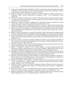
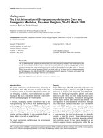
Finally, in a recent experimental study from our research group, the beneficial
effects on survival of bone marrow-derived mesenchymal stromal cells (MSCs) in
endotoxin-induced lung injury in mice depended in part on the secretion of LXA4
by the MSCs [14]. In these studies, pretreatment with the LXA4 receptor inhibitor,
WRW4, prevented the beneficial effects of MSCs on severity of lung injury and survival. In addition, administration of LXA4 alone increased survival from endotoxininduced lung injury (Fig. 1).
a
100
48 h survival rate (%)
80
#
60
*
40
No injury
LPS
20
LPS +MSC
0
b
0
10
20
30
hours
40
50
100
80
48 h survival rate (%)
Fig. 1 The effects of mesenchymal stromal cells
(MSCs), ALX/FPR2 agonists
(lipotoxin A4 [LXA4]) and
antagonist (WRW4) on 48hour survival of lipopolysaccharide (LPS)-injured mice
(a and b). Four hours after
LPS injury (5 mg/kg, intra-tracheal), mice received
MSCs (500,000 cells), LXA4
(10 ug/kg), WRW4 (1 mg/kg)
or vehicle intra-tracheally.
Statistical analysis was performed using a log-rank test.
Results are expressed as percentage survival (n = 25–35
per group). * p < 0.05 versus
no injury, # p < 0.05 versus
LPS group. Reproduced from
[14] with permission
#
#
60
40
*
LPS
LPS +MSC
20
LPS +MSC+WRW4
LPS +LXA4
0
0
10
20
30
hours
40
50
Lipid Mediators in the Pathogenesis and Resolution of Sepsis and ARDS
7
Contribution of Arachidonic Acid Metabolites in Sepsis and ARDS
In a recent clinical study of 22 patients, plasma was collected within 48 h after
the onset of sepsis and follow up samples on days 3 and 7 [3]. More than 30
bioactive compounds were measured by mass spectrometry and lipid profiling.
Patients were divided into survivors and non-survivors. Some interesting patterns
emerged from this study. In the patients who did not survive, there were significantly higher levels of the inflammation-initiating prostaglandin F2˛ (PGF2˛) and
the pro-inflammatory LTB4, but there were also elevated levels of the pro-resolving mediators, resolvin E1, resolvin D5 and 17r-protectin D1. This pattern persisted
through day 7. Thus, the higher pro-resolving lipids in the non-survivors could be
interpreted as a failed endogenous attempt to resolve the infection and inflammation. However, the multiplicity of factors, including comorbidities, that determine
mortality in sepsis patients makes interpretation of these results challenging. This
study did not include measurements of biomarkers such as IL-6 and IL-8, or other
biomarkers that have been used to profile biological responses in sepsis.
Before the availability of more comprehensive lipidomic assays, our research
group used radioimmunoassay and high pressure liquid chromatography to measure selected products of arachidonic acid metabolism in the pulmonary edema fluid
in the early phase of patients with ARDS, including several patients with sepsis
[15]. There were 10 patients with ARDS based on bilateral chest radiographic infiltrates and severe arterial hypoxemia, a normal pulmonary arterial wedge pressure
in seven patients and a normal central venous pressure in three patients. The 10 patients with ARDS had an edema fluid to plasma total protein ratio of 0.80 ˙ 0.16,
consistent with increased protein permeability edema. There were five control patients with hydrostatic pulmonary edema, three of whom had an elevated pulmonary
arterial wedge pressure (28, 30 and 33 mmHg) and the other two patients had decreased left ventricular function on echocardiography. In these five patients with
hydrostatic pulmonary edema, the mean edema fluid-to-plasma total protein ratio was 0.46 ˙ 0.14, consistent with hydrostatic edema. Radioimmunoassay and
high pressure liquid chromatography measured several products of arachidonic acid
metabolism in the pulmonary edema fluid of these patients, including PGE2, thromboxane A2 (TXA2), LTB4, LTC4 and LTD4. LTD4 was significantly elevated in the
edema fluid from the 10 patients with ARDS compared to in the five patients with
hydrostatic edema (mean ˙ SD 19 ˙ 7 versus 4 ˙ 1 pmol/ml, p < 0.001). LTB4 levels were numerically elevated in the ARDS edema fluid samples compared to the
hydrostatic edema fluid samples (11 ˙ 8 versus 4 ˙ 3 pmol/ml), although this difference did not reach statistical significance. Of the 10 patients with ARDS, five
had sepsis as the primary cause of ARDS. Prior studies had focused on cyclooxygenase products of arachidonic metabolism, which had been recognized for their
vasoconstrictor properties [16]. This clinical study was focused on the leukotrienes,
especially LTB4 and LTD4. The elevated LTD4 was thought to be a likely contributor to the increase in lung vascular permeability. LTB4 was recognized at the
time to be an important neutrophil chemoattractant that allowed large numbers of
neutrophils to cross the normally tight alveolar epithelial barrier in humans without
8
B. Hamilton et al.
inducing a significant increase in protein permeability [17]. A follow-up study documented the presence of both LTD4 and LTE4 in the edema fluid of patients with
ARDS at significantly higher concentrations than in patients with hydrostatic edema
[18]. Biologically, LTE4 has similar properties to LTD4 for increasing vascular permeability. These studies were done prior to the recognition of the pro-resolution
lipid pathways.
In more recent work, our research group studied 20 mechanically ventilated
patients with acute pulmonary edema, 14 with ARDS and six with hydrostatic pulmonary edema [19]. The patients were categorized as ARDS or hydrostatic edema
based on clinical data and the edema fluid-to-plasma protein ratio, as in prior studies. Undiluted pulmonary edema fluid was collected, centrifuged and frozen within
24 h of intensive care unit (ICU) admission from ventilated patients with pulmonary
edema. The etiology of ARDS was infectious in nine of the 14 patients (pneumonia or sepsis) and is provided in Table 1. The baseline clinical data and patient
characteristics are provided in Table 2. The clinical characteristics were comparable between patients with hydrostatic edema and those with ARDS except that
oxygenation was significantly worse in the patients with ARDS.
To take advantage of the comprehensive lipidomic analysis using more advanced
liquid chromatography and mass spectrometry and multiple reaction monitoring
methods [20], seven pro-inflammatory or pro-resolving lipid mediators were measured including arachidonic acid, PGE2, PGF2˛, TXB2, LTB4, LTE4 and LXA4.
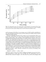
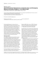
Levels of three of the lipid mediators were significantly higher in the ARDS edema
fluid, specifically LTB4, LTE4 and LXA4 (p < 0.05) (Fig. 2). These findings provide
evidence for the likely contribution of the two pro-inflammatory leukotrienes, LTB4
Table 1 Etiology and underlying medical disorders in the patients with hydrostatic pulmonary
edema (HPE) and those with acute respiratory distress syndrome (ARDS)
Etiology
Pneumonia
HPE
0
ARDS
5
Myocardial infarct
Sepsis
1
0
0
4
TACO/TRALI
1
1
Idiopathic
Volume overload
Drug overdose
Reperfusion injury
Neurogenic
Heart failure
Hypertension
0
1
0
0
1
1
1
2
0
1
1
0
0
0
Underlying disorder
Community-acquired; myasthenia gravis;
metastatic cancer; perioperative; fungal
Peri-catheterization
S/p small bowel resection; gastroparesis &
end-stage liver disease; sepsis vs. aspiration
with cardiac arrest
End-stage liver disease with TACO; transfusion s/p spinal fusion with TRALI
Intracranial tumor; acute hepatic failure
Mitral stenosis/congestive heart failure
Fulminant hepatic failure
S/p lung transplant
Subarachnoid hemorrhage
Hypoxic respiratory failure
ESRD
TACO: transfusion-associated circulatory overload; TRALI: transfusion-related acute lung injury;
s/p: status post; ESRD: end-stage renal disease
Lipid Mediators in the Pathogenesis and Resolution of Sepsis and ARDS
9
Table 2 Baseline clinical characteristics in the patients with hydrostatic pulmonary edema (HPE)
and those with acute respiratory distress syndrome (ARDS)
Characteristic
Male, n (%)
Age, years, median (IQR)
PaO2 /FiO2 ratio, median (IQR)
Lung injury score, median (IQR)
Tidal volume per kg, median (IQR)
Use of vasopressors, n (%)
Alveolar fluid clearance, median (IQR) (%/hour)
Days ventilated, median (IQR)
Death, n (%)
HPE
3 (50%)
63 (51, 71)
115 (106, 137)
3.0 (2.4, 3.0)
6.3 (5.9, 7.4)
2 (50%)
4.2 (2.4, 7.8)
4.5 (2.5, 5.8)
0 (0%)
ARDS
8 (57%)
46 (37, 55)
53 (47, 76)
3.0 (2.7, 3.5)
6.6 (4.8, 8.4)
10 (91%)
0.6 (0.0, 3.3)
3.0 (2.0, 6.0)
7 (43%)
p value
1.00
0.12
0.03
0.19
1.00
0.15
0.17
0.79
0.06
Continuous data are shown as median with interquartile ranges (IQR; 25th to 75th percentile) and
compared using Wilcoxon rank-sum tests because of the non-normal distribution of the data. Categorical data are shown as number and percent and compared using Fisher’s exact test
and LTE4, in the pathogenesis of the increased protein permeability in ARDS. The
statistically higher level of LXA4 is particularly interesting given the growing data
that pro-resolving lipids play an important role in tissue repair. Elevation of LXA4
early in ARDS may indicate that the process of resolving injury has been initiated
8
p = 0.149
p = 0.134
p = 0.773
p = 0.023
p = 0.035
p = 0.019
p = 0.076
Level Ln (pg/μ)
4
Condition
ARDS
0
HPE
–4
AA
PGE2
PGF2α
LXA4
LTB4
Lipid Mediator
LTE4
TXB2
Fig. 2 Lipid mediator levels in the undiluted pulmonary edema fluid of the patients with hydrostatic pulmonary edema (HPE) and acute respiratory distress syndrome (ARDS). The levels are
displayed on the y-axis in Ln (natural log transformed) as pg/µl and the data are shown as median
with confidence intervals (25th to 75th intervals). The seven measured lipid mediators were arachidonic acid (AA), prostaglandin E2 (PGE2), prostaglandin F2˛ (PGF2˛), lipoxin A4 (LXA4),
leukotriene B4 (LTB4), leukotriene E4 (LTE4) and thromboxane B2 (TXB2). p < 0.05 for LTB4,
LTE4 and LXA4
10
B. Hamilton et al.
at an early stage, similar to some of the experimental studies cited earlier in this
review. Thus, the lipid mediator levels measured in the alveolar fluid compartment
demonstrate distinct patterns in patients with ARDS versus hydrostatic edema. Further studies are needed to determine the association and function of lipid mediators
in the pathogenesis of ARDS.
Conclusion
The availability of comprehensive lipidomic and mass spectrometry assays has
made it possible to study both pro-inflammatory and pro-resolving lipids in experimental and clinical studies of sepsis and acute lung injury. The important contribution of SPMs in the resolution of tissue injury has now been established in
several clinically relevant experimental models of infection, sepsis and acute lung
injury. More clinical studies are needed to characterize the pro-inflammatory and
pro-resolving lipid patterns in patients with sepsis and ARDS, potentially making it
possible to endotype these patients into sub-populations that have different clinical
outcomes, as our group has done by combining protein biomarkers and clinical data
using latent class analysis [21, 22]. Given developments in lipid mediator pharmacology, identification of specific targets could lead to novel therapeutic strategies
for sepsis and ARDS.
References
1.
Serhan CN (2014) Pro-resolving lipid mediators are leads for resolution physiology. Nature
510:92–101
2. Basil MC, Levy BD (2016) Specialized pro-resolving mediators: endogenous regulators of
infection and inflammation. Nat Rev Immunol 16:51–67
3. Dalli J, Colas RA, Quintana C et al (2017) Human sepsis eicosanoid and proresolving lipid
mediator temporal profiles: correlations with survival and clinical outcomes. Crit Care Med
45:58–68
4. Cajka T, Fiehn O (2014) Comprehensive analysis of lipids in biological systems by liquid
chromatography-mass spectrometry. Trends Anal Chem 61:192–206
5. Grumbach Y, Quynh NVT, Chiron R, Urbach V (2009) LXA4 stimulates ZO-1 expression
and transepithelial resistance in human airway epithelial cells. Am J Physiol Lung Cell Mol
Physiol 296:L101–L108
6. Meng F, Mambetsariev I, Tian Y et al (2015) Attenuation of lipopolysaccharide-induced lung
vascular stiffening by lipoxin reduces lung inflammation. Am J Respir Cell Mol Biol 52:152–
161
7. Wang Q, Lian QQ, Li B et al (2013) Lipoxin A4 activates alveolar epithelial sodium channel,
Na,K-ATPase, and increases alveolar fluld clearance. Am J Respir Cell Mol Biol 48:610–618
8. Tam VC, Quehenberger O, Oshansky C et al (2013) Lipidomic profiling of influenza infection
identifies mediators that induce and resolve inflammation. Cell 154:213–227
9. Morita M, Kuba K, Ichikawa A et al (2013) The lipid mediator protectin D1 inhibits influenza
viral replication and improves severe influenza. Cell 153:112–125
10. Sordi R, Menez-de-Lima O Jr, Horewicz V et al (2013) Dual role of lipoxin A4 in pneumosepsis pathogenesis. Int Immunopharm 17:283–292
Lipid Mediators in the Pathogenesis and Resolution of Sepsis and ARDS
11
11. Seki H, Fukunaga K, Artia M et al (2009) The anti-inflammatory and proresolving mediator resolving E1 protects mice from bacterial pneumonia and acute lung injury. J Immunol
184:836–843
12. Walker J, Dichter E, Lacorte G et al (2011) Lipoxin A4 increases survival by decreasing
systemic inflammation and bacterial load in sepsis. Shock 36:410–416
13. Tobin D, Roca JF, Oh SF et al (2012) Host genotype-specific therapies can optimize the inflammatory response to mycobacterial infections. Cell 148:434–446
14. Fang X, Abbott J, Cheng L, Lee JW, Levy BD, Matthay MA (2015) Human mesenchymal
stem (stromal) cells promote the resolution of acute lung injury in part through lipoxin A4.
J Immunol 195:875–881
15. Matthay M, Eschenbacher WL, Goetzl EJ (1984) Elevated concentrations of leukotriene D4
in pulmonary edema fluid of patients with the adult respiratory distress syndrome. J Clin
Immunol 4:479–483
16. Snapper JR, Hutchinson AA, Ogletree ML, Brigham KL (1983) Effects of cyclooxygenase
inhibitors on the alterations in lung mechanics caused by endotoxemia in the unanesthetized
sheep. J Clin Invest 72:63–76
17. Martin TR, Pistoresse BP, Chi EY, Goodman RB, Matthay MA (1989) Effects of leukotriene
B4 in the human lung. J Clin Invest 84:1609–1619
18. Ratnoff WD, Matthay MA, Wong MY et al (1988) Sulfidopeptide-leukotriene peptidases in
pulmonary edema fluid from patients with the adult respiratory distress syndrome. J Clin Immunol 8:250–258
19. Hamilton B, Gronert K, Gotts JE, Calfee CS, Ware LB, Matthay MA (2017) Integrated analysis method of soluble lipid mediators in alveolar fluid discriminates ARDS from hydrostatic
pulmonary edema. Am J Respir Crit Care Med 195:A4356 (abst)
20. von Moltke J, Trinidad NJ, Moayeri M et al (2012) Rapid induction of inflammatory lipid
mediators by the inflammasome in vivo. Nature 490:107–111
21. Calfee CS, Delucchi K, Parsons PE, Thompson BT, Ware LB, Matthay MA (2014) Subphenotypes in acute respiratory distress syndrome: latent class analysis of data from two randomized
controlled trials. Lancet Respir Med 2:611–620
22. Famous K, Delucchi K, Ware LB et al (2017) Acute respiratory distress syndrome subphenotypes respond differently to randomized fluid management strategy. Am J Respir Crit Care
Med 195:331–338
Immune Paralysis in Sepsis: Recent Insights
and Future Development
B. M. Tang, V. Herwanto, and A. S. McLean
Introduction
Immune paralysis, or the inability of the immune response to recover despite clearance of pathogens by antimicrobials, is a major cause of death in patients with
sepsis. Persistent immune paralysis leads to failure to eradicate the primary infection and increased susceptibility to secondary infection [1, 2]. The clinical relevance
of this immunosuppressed state in sepsis patients is evidenced by the frequent occurrence of infection with opportunistic and multidrug-resistant bacterial pathogens
and the reactivation of latent viruses (cytomegalovirus, Epstein-Barr virus and herpes simplex virus-1) [3–8]. Here, we review recent insights related to the cellular
mechanisms of sepsis-induced immune paralysis and the development of novel therapies for treating immune paralysis.
How Does Immune Paralysis Occur?
We begin with a brief review of the established literature on the mechanisms of
immune paralysis. These mechanisms have been well studied in animal models and
human studies. They fall into three main categories as follows:
B. M. Tang
Department of Intensive Care Medicine, Nepean Hospital
Kingswood, NSW 274, Australia
Centre for Immunology and Allergy Research, Westmead Institute for Medical Research
Westmead, NSW 2145, Australia
V. Herwanto A. S. McLean ( )
Department of Intensive Care Medicine, Nepean Hospital
Kingswood, NSW 274, Australia
e-mail:
© Springer International Publishing AG 2018
J.-L. Vincent (ed.), Annual Update in Intensive Care and Emergency Medicine 2018,
Annual Update in Intensive Care and Emergency Medicine,
/>
13
14
B. M. Tang et al.
Death of Immune Cells
Sepsis causes progressive, apoptosis-induced loss of cells of the immune system.
Apoptosis is prominent in CD4 T+ -cells, CD8+ T-cells, B-cells, natural killer (NK)
cells and follicular dendritic cells in sepsis patients. Two pathways for apoptosis
have been identified: (1) the death-receptor pathway; and (2) the mitochondrialmediated pathway [9].
The detrimental effects of apoptosis are not only related to the severe loss of
immune cells but also to the impact that apoptotic cell uptake has on the surviving
immune cells. Uptake of apoptotic cells by monocytes, macrophages and dendritic
cells either leads to increased anti-inflammatory cytokine production (e.g., interleukin [IL]-10) or results in an anergy state (see below) that further exacerbates the
immune suppressive state [10, 11].
Immune Cell Exhaustion or ‘Anergy’
A robust cytokine response, after stimulation by pathogens or bacterial antigens
(e.g., lipopolysaccharide [LPS]), is a common characteristic of healthy, well-functioning immune cells. The progressive loss of such a response is a well-recognized
condition in sepsis. This condition has been named as “immune cell exhaustion”,
“anergy” or “endotoxin tolerance” [12]. T-cell anergy, or an impaired response to
an antigen with decreased release of cytokines in the T cells, can lead to immune
dysfunction in sepsis patients. Immune cell anergy also occurs in macrophages
and monocytes. Loss of their expression of surface receptor, major histocompatibility complex (MHC) class II, contributes to macrophage and monocyte dysfunction [13]. Furthermore, the decrease in monocyte CD14/human leukocyte antigen
(HLA)-DR co-expression correlates with the degree of immune dysfunction and
results in a poorer outcome in severe sepsis [14].
Anti-Inflammatory State
During sepsis, the anti-inflammatory cytokine, IL-10, is produced by T regulatory
(Treg) and T helper (Th)2 cells and suppresses the Th1 response. This suppressive
environment results in a marked decrease in monocyte production of pro-inflammatory cytokines tumor necrosis factor (TNF)-˛, IL-1ˇ, and IL-6 [13, 14].
What Are the New Insights from Recent Studies?
The above three processes, although well supported by many studies, are unlikely to
be the only mechanisms that underpin sepsis-induced immune paralysis. Additional
mechanisms have been discovered in more recent studies.
Immune Paralysis in Sepsis: Recent Insights and Future Development
15
Immune-Metabolic Dysfunction
Immune cells rely on oxidative phosphorylation as their main energy source. However, during sepsis, immune cells shift their metabolism towards aerobic glycolysis
[15, 16]. This shift is an important adaptive mechanism that helps maintain host
defense. The failure of this shift may explain immune paralysis during sepsis. In
a recent landmark study, investigators found that in immune cells during sepsis both
oxidative phosphorylation and aerobic glycolysis were greatly diminished. The investigators also observed that the expected metabolic shift did not occur [17]. The
cellular consequence of this metabolic failure is significant, as immune cells require
an adequate supply of adenosine triphosphate and other metabolic intermediates
(e.g., NAD+ ) to maintain critical cellular functions during host defense, including
activation, differentiation and proliferation [18].
Transcriptomics Changes
Changes in cellular function are controlled, in part, at a gene-expression level.
Therefore, studies on gene-expression changes (i.e., transcriptomics) have revealed
considerable insight into the host response in sepsis. The findings from these studies demonstrated increased gene-expressions in pro-inflammatory, anti-inflammatory, and mitochondrial dysfunction and decreased gene-expression in translational
initiation, mTOR signaling, adaptive immunity and antigen presentation [19–21].
A recent landmark gene-expression study explored the correlation between geneexpression changes and patient level outcomes (e.g., mortality). The authors discovered a subgroup of sepsis patients who displayed gene-expression changes that
corresponded to an immunosuppressive phenotype and termed these gene-expression changes the “sepsis response signature” 1. Genes included in this gene-expression signature indicate changes in T cell exhaustion, endotoxin tolerance, and
downregulation of HLA class II. The authors showed that the presence of this immunosuppressive signature predicted poor prognosis [22].
Epigenetic Modifications
Gene-expression can be modulated at an epigenetic level. Epigenetic modification
could retain unfavorable changes in gene-expression and maintain these changes
beyond the acute phase of infection. This ‘imprinting’ process may contribute to
the persistence of the immune suppressive state during the post-resuscitation period
of sepsis. For example, epigenetic imprinting might occur in progenitor cells in the
bone marrow and in other immune tissues, such as spleen and thymus. This effect
may explain why the immune system is not completely recovered by the generation
of new immune cells from the bone marrow. Similarly, epigenetic reprogramming
may be retained in the progenitor cells of patients who survive sepsis, allowing
them to perpetuate the epigenetic marks into well differentiated cells, which fur-