Pocket atlas of sectional anatomy vol III, spine, extremeties, joints t moeller, e reif (thieme, 2007) 1
Bạn đang xem bản rút gọn của tài liệu. Xem và tải ngay bản đầy đủ của tài liệu tại đây (23.7 MB, 80 trang )
Moeller / Reif, Sectional Anatomy © 2007 Thieme
All rights reserved. Usage subject to terms and conditions of license.
Moeller / Reif, Sectional Anatomy © 2007 Thieme
All rights reserved. Usage subject to terms and conditions of license.
Moeller / Reif, Sectional Anatomy © 2007 Thieme
All rights reserved. Usage subject to terms and conditions of license.
Pocket Atlas of
Sectional Anatomy
Computer Tomography
and Magnetic Resonance Imaging
Volume 3
Spine, Extremeties, Joints
Torsten B. Moeller, MD
Department of Radiology
Caritas Hospital
Dillingen, Germany
Emil Reif, MD
Department of Radiology
Caritas Hospital
Dillingen, Germany
485 Illustrations
Thieme
Stuttgart · New York
Moeller / Reif, Sectional Anatomy © 2007 Thieme
All rights reserved. Usage subject to terms and conditions of license.
IV
Inhalt
Library of Congress Cataloging-in-Publication
Data is available from the publisher.
Translator: Barbara Herzberger, MD,
Munich, Germany
Illustrator: Barbara Gay, Stuttgart, Germany
© 2007 Georg Thieme Verlag,
Rüdigerstrasse 14, 70469 Stuttgart, Germany
Thieme New York, 333 Seventh Avenue,
New York, NY 10001, USA
Cover design: Thieme Marketing
Typesetting by Primustype Hurler,
Notzingen
Printed in Germany by Appl, Wemding
10-ISBN 3–13–143171–7 (GTV)
13-ISBN 978–3-13–143171–4 (GTV)
10-ISBN 1–58890–566–7 (TNY)
13-ISBN 978–1-58890–566–6 (TNY)
123456
Important note: Medicine is an everchanging science undergoing continual
development. Research and clinical experience are continually expanding our
knowledge, in particular our knowledge
of proper treatment and drug therapy.
Insofar as this book mentions any dosage
or application, readers may rest assured
that the authors, editors, and publishers
have made every effort to ensure that
such references are in accordance with
the state of knowledge at the time of
production of the book.
Nevertheless, this does not involve, imply, or express any guarantee or responsibility on the part of the publishers
in respect to any dosage instructions and
forms of applications stated in the book.
Every user is requested to examine
carefully the manufacturers’ leaflets
accompanying each drug and to check, if
necessary in consultation with a physician or specialist, whether the dosage
schedules mentioned therein or the
contraindications stated by the manufacturers differ from the statements
made in the present book. Such examination is particularly important with
drugs that are either rarely used or have
been newly released on the market.
Every dosage schedule or every form of
application used is entirely at the user’s
own risk and responsibility. The authors
and publishers request every user to
report to the publishers any discrepancies or inaccuracies noticed. If errors in
this work are found after publication,
errata will be posted at www.thieme.
com on the product description page.
Some of the product names, patents, and
registered designs referred to in this
book are in fact registered trademarks or
proprietary names even though specific
reference to this fact is not always made
in the text. Therefore, the appearance of
a name without designation as proprietary is not to be construed as a representation by the publisher that it is in
the public domain.
This book, including all parts thereof, is
legally protected by copyright. Any use,
exploitation, or commercialization outside the narrow limits set by copyright
legislation, without the publisher’s consent, is illegal and liable to prosecution.
This applies in particular to photostat
reproduction, copying, mimeographing,
preparation of microfilms, and electronic data processing and storage.
Moeller / Reif, Sectional Anatomy © 2007 Thieme
All rights reserved. Usage subject to terms and conditions of license.
V
For my American relatives
Bernie and Arlene, Bryan, Nancy,
Rick and Bill, Shirley, Mike, Michael,
Austin and Amanda, Audrey, Mike,
Kristin and Katelyn, Claudia, Dale,
Bryan, Jamie, and Meghan
Moeller / Reif, Sectional Anatomy © 2007 Thieme
All rights reserved. Usage subject to terms and conditions of license.
Moeller / Reif, Sectional Anatomy © 2007 Thieme
All rights reserved. Usage subject to terms and conditions of license.
VII
Preface
Magnetic resonance imaging (MRI) of the musculoskeletal system is
an established and important component in the diagnosis of diseases
of the joints, soft tissues, bones, and bone marrow. We are therefore
pleased to collect together images of the joints and the spinal column
in a separate volume on the musculoskeletal system. Demonstrating
the growing importance of new developments in MRI in recent years,
with ever-increasing resolution, many images were acquired with 3tesla units. We are deeply grateful to the manufacturers, Siemens and
Philips, for making this possible.
We believe that colored atlases are the ideal medium to represent the
highly detailed images achieved nowadays with improved resolution
techniques. Volume 3 of the Pocket Atlas of Sectional Anatomy provides a color illustration facing each magnetic resonance image, as in
the preceding volumes on the skull, thorax, and abdomen. To ensure
the greatest possible precision in details, we still produce these
illustrations ourselves. Each is accompanied by a sectional image
and an orientation aid. Uniform color schemes ensure optimal clarity,
as similar structures, such as arteries, veins, nerves, tendons, etc., are
consistently represented in the same color. Individual muscle groups
are represented uniformly, but differentiated from other muscle
groups, so that classification is possible even when numerous groups
of muscles are shown in the same image. Maximal lucidity prevails
even in highly detailed representations. This is made possible by the
high quality of the production and printing process that are characteristic of Thieme International.
Our special thanks go to our radiology assistants, Silke Köhl, Sabine
Mattil, Stephanie Müller, Heike Philippi, Brigitte Schild, Petra Weber,
and Tanja Breunig, and also to Birgit Reuter and Marion Hellinger
from the Siemens Manufacturing Center, for providing these images.
We are deeply grateful to our medical colleagues Sigrid Roth and,
especially, Simone Zenner, for their intensive discussions and helpful
suggestions.
Torsten B. Moeller
Emil Reif
Moeller / Reif, Sectional Anatomy © 2007 Thieme
All rights reserved. Usage subject to terms and conditions of license.
Moeller / Reif, Sectional Anatomy © 2007 Thieme
All rights reserved. Usage subject to terms and conditions of license.
IX
Contents
Upper Extremity . . . . . . . . . . . . . . . . . . . . . . . . . . . . . . . . . .
1
Arm, Axial . . . . . . . . . . . . . . . . . . . . . . . . . . . . . . . . . . . . . . . . . . . . . . . . . . . . . .
Shoulder, Coronal. . . . . . . . . . . . . . . . . . . . . . . . . . . . . . . . . . . . . . . . . . . . . . .
Shoulder, Sagittal . . . . . . . . . . . . . . . . . . . . . . . . . . . . . . . . . . . . . . . . . . . . . . .
Elbow, Coronal . . . . . . . . . . . . . . . . . . . . . . . . . . . . . . . . . . . . . . . . . . . . . . . . .
Elbow, Sagittal. . . . . . . . . . . . . . . . . . . . . . . . . . . . . . . . . . . . . . . . . . . . . . . . . .
Hand, Coronal . . . . . . . . . . . . . . . . . . . . . . . . . . . . . . . . . . . . . . . . . . . . . . . . . .
Hand, Sagittal . . . . . . . . . . . . . . . . . . . . . . . . . . . . . . . . . . . . . . . . . . . . . . . . . .
2
62
74
84
90
98
106
Lower Extremity . . . . . . . . . . . . . . . . . . . . . . . . . . . . . . . . . . 115
Leg, Axial . . . . . . . . . . . . . . . . . . . . . . . . . . . . . . . . . . . . . . . . . . . . . . . . . . . . . . .
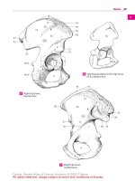
Hip, Coronal . . . . . . . . . . . . . . . . . . . . . . . . . . . . . . . . . . . . . . . . . . . . . . . . . . . .
Hip, Sagittal . . . . . . . . . . . . . . . . . . . . . . . . . . . . . . . . . . . . . . . . . . . . . . . . . . . .
Knee, Coronal . . . . . . . . . . . . . . . . . . . . . . . . . . . . . . . . . . . . . . . . . . . . . . . . . .
Knee, Sagittal. . . . . . . . . . . . . . . . . . . . . . . . . . . . . . . . . . . . . . . . . . . . . . . . . . .
Foot, Coronal . . . . . . . . . . . . . . . . . . . . . . . . . . . . . . . . . . . . . . . . . . . . . . . . . . .
Foot, Sagittal . . . . . . . . . . . . . . . . . . . . . . . . . . . . . . . . . . . . . . . . . . . . . . . . . . .
116
180
190
200
212
232
254
Spine . . . . . . . . . . . . . . . . . . . . . . . . . . . . . . . . . . . . . . . . . . . . . . . . . 263
Spine, Sagittal . . . . . . . . . . . . . . . . . . . . . . . . . . . . . . . . . . . . . . . . . . . . . . . . . .
Cerivcal Spine, Sagittal. . . . . . . . . . . . . . . . . . . . . . . . . . . . . . . . . . . . . . . . . .
Cervical Spine, Coronal . . . . . . . . . . . . . . . . . . . . . . . . . . . . . . . . . . . . . . . . .
Cervical Spine, Axial . . . . . . . . . . . . . . . . . . . . . . . . . . . . . . . . . . . . . . . . . . . .
Thoracic Spine, Sagittal . . . . . . . . . . . . . . . . . . . . . . . . . . . . . . . . . . . . . . . . .
Thoracid Spine, Axial . . . . . . . . . . . . . . . . . . . . . . . . . . . . . . . . . . . . . . . . . . .
Lumbar Spine, Sagittal . . . . . . . . . . . . . . . . . . . . . . . . . . . . . . . . . . . . . . . . . .
Lumbar Spine, Coronal. . . . . . . . . . . . . . . . . . . . . . . . . . . . . . . . . . . . . . . . . .
Lumbar Spine, Axial. . . . . . . . . . . . . . . . . . . . . . . . . . . . . . . . . . . . . . . . . . . . .
264
266
272
278
288
294
296
304
310
Bibliography . . . . . . . . . . . . . . . . . . . . . . . . . . . . . . . . . . . . . . . . . . . . . . . . . . . . 316
Index . . . . . . . . . . . . . . . . . . . . . . . . . . . . . . . . . . . . . . . . . . . . . . . . . . . . . . . . . . . 318
Moeller / Reif, Sectional Anatomy © 2007 Thieme
All rights reserved. Usage subject to terms and conditions of license.
Moeller / Reif, Sectional Anatomy © 2007 Thieme
All rights reserved. Usage subject to terms and conditions of license.
Color Code: Upper Extremity
Arteries
Nerves
Veins
Bones
Fatty tissue
Cartilage
Tendon
Disk, labrum etc.
Fluid
Muscles of Trunk:
Serratus anterior
Omohyoid
Trapezius
Subclavius
Intercostal
Muscles of Shoulder:
Deltoid
Infraspinatus
Pectoralis major and pectoralis minor
Subscapularis
Coracobrachialis
Latissimus dorsi
Dorsal Muscles of Lower Arm:
Supinator
Extensor pollicis longus and brevis
Extensor indicis
Muscles of Hand:
Dorsal and palmar interosseous
Lumbrical
1
Radial Muscles of Lower Arm:
Brachioradialis
Extensor carpi radialis longus
Extensor carpi radialis brevis
Volar Muscles of Lower Arm
(superficial layer):
Pronator teres
Flexor digitorum superficialis
Flexor carpi radialis and flexor carpi ulnaris
Palmaris longus and palmaris brevis
Volar Muscles of Lower Arm
(deep layer):
Flexor digitorum profundus
Flexor pollicis longus
Pronator quadratus
Muscles of Little (Fifth) Finger:
Abductor digiti minimi
Flexor digiti minimi brevis
Opponens digiti minimi
Muscles of Thumb:
Abductor pollicis longus
and abductor pollicis brevis
Opponens pollicis
Flexor pollicis brevis
Adductor pollicis
Volar Muscles of Upper Arm:
Biceps brachii
Brachialis
Dorsal Muscles of Upper Arm:
Triceps brachii
Anconeus
Dorsal Muscles of Lower Arm
(superficial layer):
Extensor digitorum
Extensor digiti minimi
Extensor carpi ulnaris
Moeller / Reif, Sectional Anatomy © 2007 Thieme
All rights reserved. Usage subject to terms and conditions of license.
2
Upper Extremity
Ventral
Lateral
Medial
Dorsal
Moeller / Reif, Sectional Anatomy © 2007 Thieme
All rights reserved. Usage subject to terms and conditions of license.
Arm, Axial
3
2
1
3
3
4
5
6
7
9
8
11
10
13
12
15
14
17
16
1
1
2
3
4
5
6
7
8
9
10
Trapezius muscle
Deltoid muscle (clavicular part)
Clavicle
Coracoclavicular ligament
Acromioclavicular joint
Suprascapular artery and vein
Acromion
Subclavius muscle
Deltoid muscle (acromial part)
Omohyoid muscle
11 Supraspinatus muscle (central
tendon)
12 Rib
13 Deltoid muscle (spinal part)
14 Serratus anterior muscle
15 Supraspinatus muscle (dorsal
ligament)
16 Supraspinatus muscle (ventral
ligament)
17 Spine of scapula
Moeller / Reif, Sectional Anatomy © 2007 Thieme
All rights reserved. Usage subject to terms and conditions of license.
4
Upper Extremity
Ventral
Lateral
Medial
Dorsal
Moeller / Reif, Sectional Anatomy © 2007 Thieme
All rights reserved. Usage subject to terms and conditions of license.
Arm, Axial
5
2
1
4
6
3
5
8
7
10
9
12
11
14
13
16
15
18
17
19
20
21
1
2
3
4
5
6
7
8
9
10
11
Coracohumeral ligament
Deltoid muscle (clavicular part)
Middle glenohumeral ligament
Coracoid process
Supraspinatus muscle (tendon)
Clavicle
Humerus (greater tubercle)
Subclavius muscle
Deltoid muscle (acromial part)
Coracoclavicular ligament
Head of humerus
12
13
14
15
16
17
18
19
20
21
Serratus anterior muscle
Superior glenoid labrum
Rib
Glenoid
Internal intercostal muscle
Deltoid muscle (spinal part)
External intercostal muscle
Infraspinatus muscle
Supraspinatus muscle
Spine of scapula
Moeller / Reif, Sectional Anatomy © 2007 Thieme
All rights reserved. Usage subject to terms and conditions of license.
6
Upper Extremity
Ventral
Lateral
Medial
Dorsal
Moeller / Reif, Sectional Anatomy © 2007 Thieme
All rights reserved. Usage subject to terms and conditions of license.
Arm, Axial
7
2
1
5
4
6
8
7
10
3
12
9
14
11
13
16
18
15
20
7
22
17
24
19
26
28
21
23
25
27
1
2
3
4
5
6
7
8
9
10
11
12
13
14
Coracohumeral ligament
Deltoid muscle (clavicular part)
Middle glenohumeral ligament
Coracoid process
Humerus (lesser tubercle)
Pectoralis major muscle
Biceps brachii muscle (long
head, tendon)
Clavicle
Intertubercular sulcus (bicipital
groove)
Pectoralis minor muscle
(tendon)
Humerus (greater tubercle)
Subclavius muscle
Deltoid muscle (acromial part)
Brachial plexus
15
16
17
18
19
20
21
22
23
24
25
26
27
28
Head of humerus
Glenoid
Superior glenoid labrum
Rib
Intraspinatus muscle
(tendon attachment)
Coracoclavicular ligament
Spine of scapula
Lung
Deltoid muscle (spinal part)
Internal and external intercostal
muscles
Supraspinatus muscle
Suprascapular artery and vein
Infraspinatus muscle
Serratus anterior muscle
Moeller / Reif, Sectional Anatomy © 2007 Thieme
All rights reserved. Usage subject to terms and conditions of license.
8
Upper Extremity
Ventral
Lateral
Medial
Dorsal
Moeller / Reif, Sectional Anatomy © 2007 Thieme
All rights reserved. Usage subject to terms and conditions of license.
Arm, Axial
1
9
2
3
4
5
6
7
8
9
10
11
13
12
15
14
17
16
19
18
21
20
23
22
24
25
26
28
27
29
1 Deltoid muscle (clavicular part)
2 Pectoralis major muscle
3 Coracobrachialis muscle
(+ tendon)
4 Cephalic vein
5 Biceps brachii muscle (short
head, tendon)
6 Subclavius muscle
7 Humerus (lesser tubercle)
8 Pectoralis minor muscle
9 Biceps brachii muscle (long
head, tendon)
10 Axillary artery and vein
11 Humerus (greater tubercle)
12 Brachial plexus and
subscapular nerve
13 Middle glenohumeral ligament
14
15
16
17
18
19
20
21
22
23
24
25
26
27
28
29
Subscapularis muscle
Deltoid muscle (acromial part)
Internal intercostal muscle
Anterior glenoid labrum
Serratus anterior muscle
Head of humerus
Rib
Humeroscapular joint
Intercostal artery, vein, and nerve
Posterior glenoid labrum
Glenoid
Infraspinatus muscle
Suprascapular artery, vein, and nerve
Scapula
External intercostal muscle
Deltoid muscle (spinal part)
Moeller / Reif, Sectional Anatomy © 2007 Thieme
All rights reserved. Usage subject to terms and conditions of license.
10
Upper Extremity
Ventral
Lateral
Medial
Dorsal
Moeller / Reif, Sectional Anatomy © 2007 Thieme
All rights reserved. Usage subject to terms and conditions of license.
Arm, Axial
11
1
3
2
5
7
4
9
6
11
8
13
10
15
12
17
14
16
19
18
20
21
22
23
24
25
1 Cephalic vein
2 Pectoralis major muscle
3 Coracobrachialis muscle
(+ tendon)
4 Pectoralis minor muscle
5 Biceps brachii muscle (short
head, tendon)
6 Axillary artery and vein
7 Humerus (lesser tubercle)
8 Brachial plexus
9 Biceps brachii muscle (long
head, tendon)
10 Rib
11 Humerus
12
13
14
15
16
17
18
19
20
21
22
23
24
25
Serratus anterior muscle
Inferior glenoid labrum
Lung
Glenoid
Intercostal artery, vein, and nerve
Joint capsule
External intercostal muscle
Suprascapular artery, vein, and nerve
Internal intercostal muscle
Deltoid muscle
Scapula
Teres minor muscle
Serratus posterior muscle
Infraspinatus muscle
Moeller / Reif, Sectional Anatomy © 2007 Thieme
All rights reserved. Usage subject to terms and conditions of license.
12
Upper Extremity
Ventral
Lateral
Medial
Dorsal
Moeller / Reif, Sectional Anatomy © 2007 Thieme
All rights reserved. Usage subject to terms and conditions of license.
Arm, Axial
1
13
2
3
5
4
7
6
8
9
11
10
13
12
15
14
17
16
19
18
20
22
24
26
21
23
25
1 Cephalic vein
2 Pectoralis major muscle
3 Biceps brachii muscle (short
head, tendon)
4 Pectoralis minor muscle
5 Biceps brachii muscle (long
head, tendon)
6 Coracobrachialis muscle
7 Humerus
8 Long thoracic nerve
9 Latissimus dorsi muscle and
teres major muscle
10 Axillary artery and vein and
brachial plexus
11 Deltoid muscle
12 Rib
13 Triceps brachii muscle (lateral
head)
14 Anterior circumflex humeral artery
and vein
15 Axillary nerve
16 Subscapularis muscle
17 Posterior circumflex humeral artery
and vein
18 Lung
19 Triceps brachii muscle (long head)
20 Internal intercostal muscle and
innermost intercostal muscle
21 Circumflex scapular artery
22 External intercostal muscle
23 Teres minor muscle
24 Serratus anterior muscle
25 Infraspinatus muscle
26 Scapula
Moeller / Reif, Sectional Anatomy © 2007 Thieme
All rights reserved. Usage subject to terms and conditions of license.