Lation, characterization and molecular identification of culturable gut bacteria in diamondback moth, Plutella Xylostella (Linnaeus)
Bạn đang xem bản rút gọn của tài liệu. Xem và tải ngay bản đầy đủ của tài liệu tại đây (364.32 KB, 8 trang )
Int.J.Curr.Microbiol.App.Sci (2019) 8(2): 3291-3298
International Journal of Current Microbiology and Applied Sciences
ISSN: 2319-7706 Volume 8 Number 02 (2019)
Journal homepage:
Original Research Article
/>
Isolation, Characterization and Molecular Identification of Culturable Gut
Bacteria in Diamondback Moth, Plutella xylostella (Linnaeus)
W. Vijaykumar1*, R. Muthuraju1, B. Shivanna2, K. Archana1 and B.S. Nalini1
1
Department of Agricultural Microbiology, 2Department of Agricultural Entomology,
University of Agricultural Sciences, Bengaluru-560065, India
*Corresponding author
ABSTRACT
Keywords
Diamondback moth
(DBM),
Endosymbionts,
Catalase and
IMVIC tests, 16S
rRNA, Serratia spp
Article Info
Accepted:
22 January 2019
Available Online:
10 February 2019
Diamondback moth, Plutella xylostella is a major pest of cruciferous crops worldwide and
it has developed resistance to almost all synthetic insecticides. It was known to harbour
microorganisms which play important role in growth and development of the host. In the
present study bacterial strains were isolated from third instar larvae of P. xylostella
collected from Sugatur, Kolar District of the state Karnataka. Morphological and
Biochemical characterization were done, among them most of the bacterial isolates were
gram negative and negative for some biochemical tests. Further, total bacterial genomic
DNA was extracted from the bacterial isolates and amplified using PCR with 16S rRNA
primers (expected size 1000 bp). Ten different bacterial isolates were isolated and five
were identified at genus level such as Serratia marcescens (DBM1 and DBM9), Serratia
nematodiphila (DBM2), Serratiasp. (DBM3) and Myroidesodoratus (DBM4). The
Serratia spp. is the most predominant bacterial isolate in this region. These studies
suggested that a combination of molecular and traditional culturing methods can be
effectively used to analyze and determine the diversity of gut microflora. These bacterial
strains may play important roles in growth and development of P. xylostella.
Introduction
Diamondback moth, Plutella xylostella (L.)
(Lepidoptera: Plutellidae) is the most
important destructive pest of the cruciferous
vegetables like brassica, cabbage, cauliflower,
radish, knol-khol, turnip, mustard and
amaranthus in many parts of the world (Saeed
et al., 2010). The developing resistance and
decline of insecticide efficiency against DBM
become a limiting factor in cultivation of
commercial crops like cabbage and
cauliflower in India. Insect system harbors a
wide range of microbial community (Hunt
and Charnley, 1981). Microorganisms play an
important role in the growth and development
of insects. Microbial symbionts provide an
diverse range of benefits in insect nutrition,
e.g. by providing essential amino acids,
digestive enzymes or vitamins (Brune, 2014).
3291
Int.J.Curr.Microbiol.App.Sci (2019) 8(2): 3291-3298
The insects created opportunities for bacteria,
and these bacteria occupy right niches in host
bodies. The interactive relationship between
microbiota and their host exist, and these coevolution of microorganisms and their insect
hosts led to a stable mutualistic relationship
(Genta et al., 2006). The diversity of insects is
reflected in large and varied microbial
communities inhabiting the gut (Dillan and
Dillan, 2004). The most important beneficial
function of the indigenous intestinal
microbiota is their ability to withstand the
colonization of the gut by non-indigenous
species including pathogens and therefore
prevent enteric infections (Berg, 1996), such
as gut bacteria could mediate disease
resistance and fight against damage from bad
factors in host insects (Dillon and Charnley,
1996). Besides, the insect gut bacteria can
cause insects population changes and
phenotype manipulation (Rajagopal, 2009).
Lin et al., (2014) collected different life
stages (fourth instar larvae, pupae and adults)
of the Diamondback moth, P.xylostella, to
find out different microbial abundance and
diversity of gut bacteria. A large quantity of
bacteria was found in all life stages, out of
which higher quantity of bacteria was found
in larval gut. Firmicutes bacteria, Bacillus sp.,
were the most dominant species in every life
stage. Phylogenetic analysis showed the
sequences of the bacteria belonged to the
Actinobacteria,
Proteobacteria
and
Firmicutes. Serratia sp. in proteobacteria was
abundant in the larval gut. Their study also
suggested that a combination of molecular
and traditional culturing methods can be
effectively used to analyze and to determine
the diversity of gut microflora.
Materials and Methods
Collection and mass rearing of DBM
The field caught population of DBM larvae
were collected from the cabbage field of
Sugatur village, Kolar District of Karnataka.
This region is predominantly cabbage
growing region of South Karnataka. The
collected populations were brought to the
laboratory and reared on mustard seedlings
raised in plastic ice cream cups (8× 4 cm) by
adopting the method described (Liu and
Tzeng, 1984) with suitable modifications. The
rearing procedure was continued for at least
one generation till sufficient number of larvae
was available for further studies.
Isolation
bacteria
and
characterization
of
gut
The third instar larvae of DBM were selected,
starved for 24 hours and surface sterilized
with 70% ethanol for 1 minute followed by
0.1% sodium hypochlorite for 1 minute, then
rinsed with sterile distilled water for 2 to 3
times to remove the external microflora. Gut
homogenate (100 µl) were plated on Nutrient
Agar (NA) and Luria Bertani (LB) media in
three replicates and incubated at 300C for 48 h
and surveyed every 24 h for new colonies.
The colonies were differentiated based on
size, shape, color, margin and morphology
and a single representative isolate of each
morphotype transferred to new plates and
made pure culture.
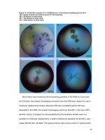
Biochemical characterization of isolated
bacteria
The isolates were gram stained and subjected
to basic biochemical characterization,
including catalase and IMVIC reaction.
IMVIC reactions consist of Indole production
test in tryptone broth, after adding kovac’s
reagent, cherry red ring on the top layer of
broth indicates the production of indole
(positive). Methyl Red and Voges Proskauer
tests in an MR-VP broth, for methyl red test,
after adding methyl red, the production of red
colour indicates the positive result and having
ability to oxidize glucose. For Voges
3292
Int.J.Curr.Microbiol.App.Sci (2019) 8(2): 3291-3298
Proskauer, VP reagent 1 and 2 were added,
and then pinkish red color appeared which
indicates the positive result. Citrate utilization
test in Simmons Citrate Agar, changes in
color as an indicator in the media, which is
from green to blue, indicates positive for this
test and Catalase test in trypticase soy agar
media, formation of bubbles after adding
hydrogen peroxide indicates positive result
for this test (Benson, 2002).
Molecular identification
DNA extraction and PCR amplification
The isolates were multiplied in LBbroth and
the genomic DNA isolated by CTAB
(CetylTrimethyl Ammonium Bromide) lysis
method. Isolated bacteria were multiplied in
LB broth. Pellet was obtained by
centrifugation at 10000 rpm for 1 minute and
was resuspended in TE buffer, SDS, RNase,
Proteinase K and lysozyme was added. Tubes
were kept in hot water bath for 30 minutes at
65°C. The5M NaCl and CTAB buffer was
added, then incubated in hot water bath for 30
minutes at 65°C.Equal volume of Chloroform:
Isoamyl alcohol (24:1) was added and were
centrifuged for 5 minutes at 10000 rpm.
Supernatant was transferred to a new tube and
added equal volume of Phenol: Chloroform:
Isoamyl alcohol (25:24:1), then centrifuged
for 5 minutes in 10000 rpm. The DNA was
precipitated by adding 600 µl of chilled
isopropanol and centrifuged after overnight
incubation. The pellet was washed with of 70
% chilled ethanol, air dried and dissolved in
80 µl of TE buffer.
The isolated DNA was checked for quantity
with 1% agarose gel. Then, the DNA will be
amplified in PCR with 16S rRNA gene with
having primers (Fp1: GAGTTTGATC
CTGGTCA and Rp2: ACGGCTACCTTGTT
ACGACTT). PCR conditions were as
follows: Initial denaturation at 94 °C for 4
mins, 35 cycles of denaturation at 94 °C for 1
min, annealing at 60 °C for 30 sec, extension
at 72 °C for 1 min and final extension at 72
°C for 10 mins. Initial denaturation at 94 °C
for 3 mins, 35 cycles of denaturation at 94 °C
for 1 min, annealing at 60 °C for 1 min,
extension at 72 °C for 1 min and final
extension at 72 °C for 2.5 mins. The
amplified unpurified PCR products were
verified with agarose gel (1%) electrophoresis
and purified. The obtained nucleotide
sequences were submitted to the National
Centre for Biotechnology Information (NCBI)
( />database.
Phylogenetic analysis
The phylogenetic analysis was performed
with nucleotide sequences using molecular
evolutionary genetic analysis (MEGA 7),
after multiple alignment of the data by
CLUSTAL W. the tree was constructed using
closely related sequences using neighbor
joining algorithm. Based on maximum query
coverage the bacterial species was identified.
Results and Discussion
Isolation and characterization gut bacterial
isolates
The totally ten bacterial isolates were
obtained based on their morphology, six
bacteria in the nutrient agar media and four
bacteria in LB media showed in Table 1. The
bacterial isolates were predominantly slightly
dry texture, raised, pasty looking, white in
color. Some colonies were irregular, concave,
yellow color and others were smooth,
circular, creamy white color. Most of the
isolates were rod shaped, gram negative
bacteria. Among ten isolates, three isolates
were positive and seven were negative for
gram reactions. Six isolates were rod shaped
and four were cocci shaped bacteria.
3293
Int.J.Curr.Microbiol.App.Sci (2019) 8(2): 3291-3298
Biochemical characterization of isolated
bacteria
isolates, molecular
performed (Fig. 1).
The almost same type of bacterial colonies
were analysed through morphological
character. Therefore, all the bacterial isolates
were
subjected
for
biochemical
characterization. Most of the isolates
predominantly showed positive results for
IMVIC test except catalase test which get
positive result for only one isolate. Among
ten isolates, five isolates had positive result
and remaining five had negative results for
indole production test. Methyl red testing was
positive for six isolates and negative for four
isolates. Three isolates were positive and
seven
isolates
were
negative
for
Vogesproskauer. Five Isolates had positive
and five isolates had negative results on
citrate test. Only one isolate showed positive
result and remaining nine isolates showed
negative results for catalase test (Table 2). For
further confirmation and identification of
Molecular
isolates
identification
identification
of
bacterial
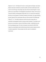
In total, five bacterial isolates from larvae
were identified and sequenced. The genomic
DNA was isolated from six bacterial isolates.
The thick DNA bands were visualized on
agarose gel under gel documentation
photograph represents the presence of DNA
and which was subjected to PCR
amplification in thermocycler with 16S rRNA
primers. The amplified product was expected
1000 bp in size. The molecular identification
indicated that the genus Serratia was
invariably associated in third instar larvae of
DBM. The bacterial isolates were identified
as Serratia marcescens (DBM1 and DBM5),
Serratia nematodiphila (DBM2), Serratiasp.
(DBM3) and Myroides odoratus (DBM4)
(Table 3 and Fig. 2 and 3).
Table.1 Morphological features of bacterial isolates from larvae of DBM
SI. No.
Isolates
-3
Colony morphology
Cell shape
Gram reaction
DBM 1
10 ,R3, I1
Round, regular,
dark yellow
Straight Rod
Negative
DBM 2
10-3, R3, I3
Yellow
Cocci
Positive
DBM 3
10-5, R1, I2
DBM 4
DBM 5
White
Cocci
Positive
-3
Light yellow
Cocci
Negative
-3
Opaque, irregular, White
Rod
Negative
-3
10 , R3, I2
10 , R3, I4
DBM 6
10 , R2, I5
Large, irregular,
convex, White
Rod
Positive
DBM 7
10-3, R3, I1
Large, concave, White
Rod
Negative
-3
was
DBM 8
10 , R3, I2
Filamentous, Creamy
dark yellow
Rod
Negative
DBM 9
10-3, R2, I1
Small, round,
mucoidWhite
Rod
Negative
DBM 10
10-3, R2, I2
Creamy yellow
Cocci
Negative
3294
Int.J.Curr.Microbiol.App.Sci (2019) 8(2): 3291-3298
Table.2 Biochemical features of bacterial strains isolated from larvae
1.
SI. No.
1
2
3
4
5
DBM 1
+
+
DBM 2
+
+
DBM 3
+
+
DBM 4
DBM 5
+
+
+
DBM 6
+
+
DBM 7
+
+
DBM 8
+
DBM 9
+
+
+
+
DBM 10
+
+
Indole production test, 2. Methyl red test, 3. Voges proskauer test, 4. Citrate utilization test, 5. Catalase
test. + - Positive, - - Negative
Table.3 Identified bacterial isolatesin larvae
Isolate
DBM1
DBM5
DBM7
DBM8
DBM9
Identified bacterial endosymbionts
Serratiamarcescens
Serratia nematodiphila
Serratiasp.
Myroidesodoratus
Serratiamarcescens
Gene bank accession number(s)
MK044840
MK044841
MK044842
MK044843
MK044844
Fig.1 Biochemical characterization of isolated bacteria of Diamondback moth (Plutella
xylostella)
MR-VP TEST
INDOLE TEST
CITRATETEST
UTILIZATION TEST
CATALASE TEST
3295
Int.J.Curr.Microbiol.App.Sci (2019) 8(2): 3291-3298
Fig.2Agarose gel showing amplification of 1000 bp gene corresponding to 16S rRNA, MMarker DNA-1000bp: First population
M
1
2
3
4
(1) DBM1, (2) DBM5,(3) DBM7, (5) DBM8, (6) DBM9
Fig.3 Phylogenetic tree of bacterial strains isolated from P. xylostella
Only less than one percent of bacteria could
be cultured, even though, feed process such as
feeding on different host plants, medium
component and culture time affected
culturable bacteria species, a few strains
detected in the larval gut might be just existed
in the environment even they were fasted for
2 h, and some bacteria even need few days to
grow on the medium.
All the bacterial isolates were subjected for
Catalase and IMVIC test. Changes in color of
the media or broth and bubble formation after
adding reagent for particular test which
indicated positive results for that particular
test. Anand et al.(2009) obtained eleven
isolates from digestive tract of Bombyxmori
and labelled as Isolate 1 to 11. They
characterized them morphologically and
3296
5
Int.J.Curr.Microbiol.App.Sci (2019) 8(2): 3291-3298
biochemically. Totally nine isolates for
catalase test, nine isolates for citrate
utilization test, six Isolates for Indole
production test and Six isolates for methyl red
test and five isolates for Voges-proskauer test
shown positive result.
Totally ten bacteria strains were cultured from
larval gut and six isolates were identified
through molecular approach. The bacteria
isolates identified after sequencing from the
larvae of DBM were Serratia marcescens,
Serratia nematodiphila, Serratia sp., and
Myroidesodoratus. Among these Serratia sp
was most predominant (Table 3). The cultureindependent bacteria, Enterococcus sp., were
the main component of P. xylostella gut
microbiota in the laboratory (Raymond et al.,
2008). In the larval gut of other lepidopterous
insects, such as the small white butterfly
(Pierisrapae L.), Proteobacteria was the most
highly represented phylum (Robinson et al.,
2010).
Acknowledgement
I am greatful to Division of Agricultural
Microbiology for giving opportunity to does
this research work and also thankfull to
Division of Agricultural Entomology,
University
of
Agricultural
Sciences,
Bengaluru, India, for providing me with all
the required facilities to complete my research
programme.
References
Anand, A. A. P., Vennison, S. J., Sankar, S.
G., Prabhu, D. I. G., Vasan, P. T.,
Raghuraman, T., Geoffrey, C. J. AND
Vendan, S. E., 2009, Isolation and
characterization of bacteria from the
gut of Bombyxmori that degrade
cellulose, xylan, pectin and starch and
their impact on digestion. J. Insect
Sci., 10(1): 107.
Benson,
H.J.,
2002,
Microbiological
applications. pp. 170–200. Boston,
MA: McGraw Hill.
Berg,
R.D.,
1996,
The
indigenous
gastrointestinal microflora. Trends
Microbiol., 4: 430–435.
Brune, A., 2014, Symbiotic digestion of
lignocellulose in termite guts. Nat.
Rev. Microbiol., 12: 168–180.
Dillon, R.J. and Charnley, A.K., 1996,
Colonization of the guts of germ-free
desert locusts, Schistocerca gregaria,
by
the
bacterium
Panto
eaagglomerans. J. Invertebr. Path.,
67: 11–14.
Dillon, R. J. and Dillon, V. M., 2004, The gut
bacteria of insects: Nonpathogenic
interactions. Annu. Rev. Entomol., 49:
71-92.
Genta, F.A., Dillon, R.J., Terra, W.R. and
Ferreira, C., 2006, Potential role for
gut microbiota in cell wall digestion
and glucoside detoxification in
Tenebriomolitor larvae. J. Invertebr.
Path., 52: 593–601.
Hunt, J. and Charnley, A. K., 1981,
Abundance and distribution of the gut
flora of the desert locust. Schistocerca
gregaria. J. Invertebr. Pathol., 38:
378–385.
Lin, Xiao-Li., Pan, Qin-Jian., Tian, HongGang., Douglas, E. Angela and Liu,
Tong-Xian., 2014, Bacteria abundance
and diversity of different life stages of
Plutella
xylostella
(Lepidoptera:
Plutellidae), revealed by bacteria
culture-dependent and PCR-DGGE
methods. Insect Sci., 00: 1–11.
Liu, M. Y. and Tzeng, Y. J., 1984, Rearing
diamondback moth (Lepidoptera:
Yponomuetidae) on rape seedling by
modification of the Koshihara and
yamada method. J. Econ. Entomol., 77
(6): 1608-1609.
Rajagopal, R., 2009, Beneficial interactions
between insects and gut bacteria.
3297
Int.J.Curr.Microbiol.App.Sci (2019) 8(2): 3291-3298
Indian J. Microbiol., 49: 114–119.
Raymond, B., West, S.A., Griffin, A.S. and
Bonsall, M.B., 2008, The dynamics of
cooperative bacterial virulence in the
field. Sci., 337: 85–88.
Robinson, C.J., Schloss, P., Ramos, Y., Raffa,
K. and Handelsman, J., 2010,
Robustness
of
the
bacterial
community in the cabbage white
butterfly larvalmidgut. Microbial
Ecology, 59:199–211.
Saeed, R., Ali, H.S., Sarfraz, A. S. and Syed,
M.Z., 2010, Effect of different host
plants on the fitness of diamondback
moth, Plutella xylostella (Lepidoptera:
Plutellidae). Crop Protection, 29:
178–182.
How to cite this article:
Vijaykumar, W., R. Muthuraju, B. Shivanna, K. Archana and Nalini, B.S. 2019. Isolation,
Characterization and Molecular Identification of Culturable Gut Bacteria in Diamondback
Moth, Plutella xylostella (Linnaeus). Int.J.Curr.Microbiol.App.Sci. 8(02): 3291-3298.
doi: />
3298