In vitro expression of enolase from Streptococcus suis serotype 2 and it’s antigenicity
Bạn đang xem bản rút gọn của tài liệu. Xem và tải ngay bản đầy đủ của tài liệu tại đây (1.08 MB, 6 trang )
Life Sciences | Agriculture, Biotechnology
In vitro expression of enolase from Streptococcus
suis serotype 2 and it’s antigenicity
Hoang Bach Nguyen1,2*, Thi Dang Khoa Nguyen1,2, Gessica Tore3, Van An Le1,2, Alberto Alberti3
Carlo Urbani Centre, Hue University of Medicine and Pharmacy
Department of Microbiology, Hue University of Medicine and Pharmacy
3
Department of Veterinary Medicine, University of Sassari, Italy
1
2
Received 20 November 2017; accepted 6 April 2018
Abstract:
Introduction
Streptococcus suis serotype 2 (SS2) is one of the most
important pathogens in the porcine industry and an
important zoonotic agent. The absence of suitable
vaccine or virulence markers makes SS2 infections
more difficult to control. An immunoproteomics
approach is used for identifying antigenic proteins in
SS2 recognized enolase, which may represent strainspecific antigenic proteins and potential protective
antigens. This study aims to clone, express enolase
gene from SS2 and use western blotting to evaluate
the antigenicity. Enolase gene from the SS2 strain was
amplified with specific primers. The obtained PCR
product was inserted into an expression vector, pGEX4T1. The recombinant vector was then transformed
into BL21 cells for protein expression. Subsequently,
the immunological activity of the recombinant enolase
was tested by western blotting with human sera in
the convalescent phase. The glutathione S-transferase
(GST)-tag fusion enolase was purified by glutathione
sepharose affinity chromatography for further
studies. The SS2 recombinant enolase was successfully
expressed in E. coli. The western blotting analysis
demonstrated that enolase has an antigenic property,
which is recognized by patients naturally infected with
SS2.
Streptococcus suis, commensal and opportunistic
pathogens of swinea, α-hemolytic gram-positive cocci with
35 different serotypes. Streptococcus suis is an important
pathogen in pigs and an emerging zoonotic agent in humans
who are in contact with pigs or with their products [1]. S.
suis infections in human are most often reported in countries
where there is a high population of pigs. By the end of 2012,
a total of 1584 cases had been reported in the literature
(including 189 probable cases identified in 3 outbreaks),
mainly from Thailand (36%), Vietnam (30%) and China
(22%) [2]. Human infections with S. suis most frequently
manifest as purulent meningitis, but septic shock with
multiple organ failure, endocarditis, pneumonia, arthritis
and peritonitis have also been reported [1]. In Thailand,
China (including Hong Kong) and Vietnam, S. suis is an
important cause of adult endocarditis, sepsis, and especially,
meningitis [3]. Thirty-five serotypes (types 1-34 and 1/2)
have been described based on capsular polysaccharides;
serotype 2 is considered to be the most common pathogen
in both humans and pigs [1, 4].
Keywords: antigenicity, enolase, protein expression,
Streptococcus suis serotype 2.
Classification numbers: 3.1, 3.5
The lack of thorough knowledge about virulence
markers and protective antigens can hinder the control of
SS2 infections. Several approaches have been adopted to
develop vaccines for S. suis. However, little success was
achieved because the protection was either a serotype or
strain dependent. More recently, interest has shifted towards
the protein antigens of S. suis as vaccine candidates.
Recently, immunoproteomics has become an effective
approach for identifying immunoreactive proteins, which
are essential antigens in the development of vaccine; they
are also biomarkers for diagnostic and molecular therapy.
*Corresponding author: Email:
24
Vietnam Journal of Science,
Technology and Engineering
JUne 2018 • Vol.60 Number 2
Life Sciences | Agriculture, Biotechnology
Enolase, which is one of the remarkable antigenic proteins
of SS2, was identified through immunoproteomics.
Enolase is a potential virulence factor of SS2; it
was originally identified as a key glycolytic enzyme
that catalyses the dehydration of 2-phosphoglycerate to
phospho-enolpyruvate in the last steps of the catabolic
glycolytic pathway [5, 6]. Recent studies have found that
enolase plays an important role in many physiological
and pathological processes, such as facilitating pathogen
invasion into the host [7-9], cancer metastasis [10-14] and
apoptosis [15, 16]. Recently, it was also recognized as an
immunodominant antigen involved in the virulence of
the Streptococcus species [6, 8, 17, 18]. It is noteworthy
that enolase is an antigenic protein recognised by patient
sera with SS2 infection based on the immunoproteomics
approach [19]. In this study, the enolase gene from SS2 was
cloned and expressed to confirm its antigenic property.
Methods and materials
Bacterial strain and patient sera collection
SS2 strains were isolated and identified in patients
infected with Streptococcus suis serotype 2, as described in
previous publication [19]. The patient sera were collected
from the SS2 infected patients in convalescent phases who
were hospitalized for treatment. Both the bacterial isolates
and patient sera were stored in a deep freezer at -80oC [20].
One Shot™ TOP10 Chemically Competent E. coli (Thermo
Fisher Scientific, Massachusetts, USA) and One Shot BL21
Star (DE3) Chemically Competent E. Coli (Thermo Fisher
Scientific, Massachusetts, USA) were used as recipients
of recombinant enolase, containing the vector pGEX- 4T1
(Amersham).
Amplification of enolase gene and cloning enolase
gene into expression vector
A PCR assay was performed by using forward primer
(5’-GGC GGA TCC ATG TCA ATT ATT ACT GAT G-3’)
and reverse primer (5’-ACG CTC GAG TTA TTT TTT
CAA GTT GTA GAA TGA G-3’), which specifically targets
1305-bp of the enolase gene of locus taq SSUBM407_1397
of Streptococcus suis BM407 complete genome data
(NC_012926.1). The primes were design in the Geneious
v8.1 software, which contained BamHI and XhoI sites at
5’and 3’ ends, respectively [21]. The PCR amplification
was profiled as follows: initial denaturation at 94°C for 2
minutes, followed by 36 cycles of 95oC for 15s, 55oC for
30s and 68oC for 30s in Veriti® Thermal Cycler (Applied
Biosystems, CA, USA).
The PCR products were purified using the QIA quick
PCR Purification Kit (Qiagen, Hilden, Germany), following
the manufacturer’s instructions. Ten of the purified enolase
PCR products and 0.32 µM of the primer were used for direct
sequencing. To sequence both strands, two specific PCR
primers were run for each enolase PCR product samples.
The chromatograms were analysed by the Geneious software
v8.1 and compared with the enolase sequence data available
in the GenBank ( />using the BLASTn plugin of the Geneious software [21].
All sequences were aligned using ClustalX [22].
The 1305 bp PCR product, which corresponded to the
obtained enolase, was restricted, gel eluted and inserted into
the expression vector pGEX-4T1 in frame with the GST-tag
sequence. The recombinant plasmid was introduced into E.
coli TOP10 by heat shock transformation, and the clones
were selected by growing the cultures on Luria-Bertani
(LB) agar in the presence of 100 µg/ml ampicillin. For
identifying the positives clones, miniprep was performed,
and then, the putative clones were checked by restriction
digestion.
Expression of recombinant S. suis enolase
The recombinant vectors were transformed into E. coli
BL21 for expression. The transformation procedure followed
the instructions of TransformAid Bacterial Transformation
Kit (Thermo Fisher Scientific, USA) [23]. A single
recombinant colony of BL21 was grown on a SOB (super
optimal broth) medium at 37°C overnight with shaking. The
protein expression was induced by the addition of 0.1 mM
isopropyl b-D-1-thiogalactopyranoside (IPTG). One hour
later, the cells were collected by centrigugation, and the
cell pellet at were stored at -20°C; this collection process
continued for a period of 4 hours with 1 hour intervals.
SDS-PAGE and Western Blotting
The recombinant enolase protein was resolved in 2
separate acrylamide gels. After electrophoresis, one gel was
alternatively stained with SimplyBlue™ SafeStain (Novex,
Life Technologies), and then digitalized, with the Gel Doc™
XR+ System (Bio-rad, USA).
JUne 2018 • Vol.60 Number 2
Vietnam Journal of Science,
Technology and Engineering
25
Life Sciences | Agriculture, Biotechnology
The second gel was subjected to western blot assay. The
proteins were transferred onto a nitrocellulose membrane
(Hybond-C Extra, Amersham, GE) with a Mini-TransBlot Cell (Bio-Rad, CA, USA) at 250 mA (100 V) for
one hour at 4°C. Then, the membranes were blocked with
PBS-T containing 5% (w/v) skim milk. Membranes were
incubated for one hour with a patient serum in 1:1500
dilutions in PBS-T, containing 2% (w/v) skim milk in
a Mini-PROTEAN II Multiscreen Apparatus (Bio-Rad,
CA, USA). The incubated membranes with the Goat AntiHuman IgG-HRP conjugated (Southern Biotech, Alabama,
USA) as secondary antibodies diluted in the ration 1:50000
in PBS-T, containing 5% (w/v) skim milk, in one hour.
The membranes were developed with Luminata™ Forte
Western HRP substrate (Merck Millipore Corp., Darmstadt,
Germany), and images were acquired with a ChemiDoc™
XRS+ System and were analysed by Image Lab Software
(Bio-Rad, CA, USA).
Affinity purification of GST fusion enolase
The GST-tag added to the enolase allowed its isolation
by affinity chromatography on glutathione column.
Overnight cultures of E. coli BL21 cells harbouring
recombinant enolase were sub-cultured in an LB medium
with ampicillin 100 µg/ml and chloramphenicol 35 µg/
ml and incubated at 37oC until an optical density of 0.6 at
600 nm (OD600) was reached. Then, the E. coli were grown
with 0.1 mM IPTG for 3 hours. The cells were centrifuged,
and then, the pellet was re-suspended in ice-cold 1X PBS
buffer with protease inhibitors. After sonication on ice,
the lysates were centrifuged at 10,000×g for 10 minutes,
and the recombinant protein was purified from the filtered
supernatants by affinity chromatography. The procedure
was performed as described in the Recombinant Protein
Purification Handbook-GE. Healthcare [24].
Results and discussion
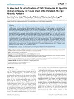
Enolase sequences analysis
Amplification of the full length coding sequences (CDS)
of the enolase gene was confirmed by gel electrophoresis of
the amplified fragments. The expected size of about 1305
bp was obtained, which corresponded to the SS2 enolase
gene (Fig. 1).
26
Vietnam Journal of Science,
Technology and Engineering
Fig. 1. PCR amplification of enolase gene from S. suis
serotype 2. The PCR products were separated on 1.0%
agarose gel. SM: 1 kb DNA Ladder (D0428, SIGMA DNA);
lane 1: PCR product; lane 2: Negative control.
The raw data of enolase sequences were analysed with
a multiple alignment algorithm in Geneious v8.1 with
the enolase sequence from the reference genome of S.
suis BM407 (NC_012926) in the Genbank (https://www.
ncbi.nlm.nih.gov/nuccore/NC_012926.1). The sequence
assembly data showed that our sequences matched with the
eno CDS at locus_tag SSUBM407_1397 of the reference
genome sequence (Fig. 2).
Cloning and expression of enolase in pGEX-4T1
The PCR products and pGEX-4T1 vector were digested
by the BamHI/XhoI and purified by the ZymoClean™ Gel
DNA Recovery Kit (Zymo Research Corp., USA). The
enolase was cloned into the TOP10 E. coli. Double digestion
of the enolase recombinant vector by BamHI/XhoI showed
that the expected size of 1300 bp fragment on agarose gel
electrophoresis corresponded to the enolase gene (Fig. 3).
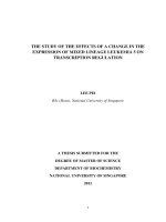
Expression of recombinant enolase
The enolase gene was 1305 bp in size and encoded into
a 435 amino acid protein with a predicted molecular mass
of 52 kDa. The result of expression from the pGEX-4T1
is a GST-tagged fusion enolase in which the functional
GST protein (26 kDa) is fused with the N-terminus of the
recombinant enolase. By comparing the SDS-PAGE results
of the non-induced and IPTG-induced BL21 strains (Fig. 4),
it can be seen that induced BL21- pGEX 4T1-enolase lands
JUne 2018 • Vol.60 Number 2
Life Sciences | Agriculture, Biotechnology
recombinant
vector by BamHI/XhoI
showed that theofexpected
of 1300sequences
bp fragment by Align/Assembly plugin of Geneious v8.1.
Fig.2. Analyse
the chromatograms
S. suis size
enolase
on agarose gel electrophoresis corresponded to the enolase gene (Fig. 3).
4969 bp
1305 bp
1500 bp
SM
1
2
3
4
5
6
7
Fig.
3. gelAgarose
gel
electrophoresis
of digested
pGEX-4T1
Fig. 4. SDS-PAGE analysis of recombinant enolase. Lane
Fig. 3.
Agarose
electrophoresis
of pGEX-4T1
recombinant vector
BamHI/XhoI.
recombinant vector digested BamHI/XhoI. SM: 1 kb DNA 1: PageRuler™ Prestained Protein Ladder Plus (Thermo
SM: ladder;
1kb DNA ladder;
lane lane
1: 1:pGEX-4T1-enolase undigested; lane 3-7: Fisher Scientific Inc.); Lane 2: Non-induced BL21; Lane
pGEX-4T1-enolase digested with BamHI/XhoI.
3-5: Induced BL21-pGEX 4T1 empty; Lane 6-10: Induced
BL21-pGEX 4T1-enolase at time zero, 1h, 2h, 3h and 4h,
have a strong band with an approximate size of 78 kDA. The respectively.
other lanes (2-5) also have a light band with an approximate
size of 78 kDA. The antigenic property of the recombinant
enolase in the total protein of the induced BL21-pGEX 4T1enolase was evaluated in the western blot assay.
of the recombinant enolase with sera from naturally infected
Testing antigenic property of recombinant enolase
The protein reacted to the convalescent sera with a strong
Western blotting was used to test the antigenic property
patients. The primary antibody was diluted in a 1:1000 ratio,
and the secondary antibody was diluted in a 1:50000 ratio.
band with a predicted size of 78 kDa (Fig. 5).
JUne 2018 • Vol.60 Number 2
Vietnam Journal of Science,
Technology and Engineering
27
Life Sciences | Agriculture, Biotechnology
which is present at the surface of all 35 S. suis serotypes. It
contributes to S. suis adhesion to and invasion of host cells
[8], and it is a highly conserved protein [25].
Conclusions
We have successfully cloned the SS2 enolase gene into
the pGEX 4T1 vector and the recombinant enolase into
BL21 cells. The recombinant enolase has antigenicity and
can be recognized by patient sera infected SS2 in western
blot assay.
REFERENCES
[1] Z.R. Lun, Q.P. Wang, X.G. Chen, A.X. Li, X.Q. Zhu (2007),
“Streptococcus suis: an emerging zoonotic pathogen”, Lancet Infect. Dis.,
7(3), pp.201-209.
Fig. 5. Western blotting analysis of recombinant enolase.
Convalescent serum (A) and negative serum (B). MW:
PageRuler™ Prestained Protein Ladder Plus (Thermo
Fisher Scientific Inc.).
Purification of recombinant enolase
Protein purification was confirmed through a SDSPAGE analysis. From the SDS-PAGE gel, it can be seen that
there is a pure recombinant enolase in the elution fractions,
and the fourth elution fraction contains the highest intensity
band (Fig. 6).
[2] V.T.L. Huong, N. Ha, N.T. Huy, P. Horby, H.D.T. Nghia, V.D.
Thiem, et al. (2014), “Epidemiology, clinical manifestations, and
outcomes of streptococcus suis infection in humans”, Emerg. Infect. Dis.,
20(7), pp.1105-1114.
[3] H.F.L. Wertheim, H.D.T. Nghia, W. Taylor, C. Schultsz (2009),
“Streptococcus suis: An Emerging Human Pathogen”, Clin. Infect. Dis.,
48(5), pp.617-625.
[4] R. Higgins, M. Gottschalk, M. Boudreau, A. Lebrun, J. Henrichsen,
R.Y. Reams, et al. (1995), “Description of six new capsular types (29-34)
of Streptococcus suis Fibrinohemorrhagic pneumonia in pigs naturally
infected with Streptococcus suis”, J. Vet. Diagn. Invest., 7(3), pp.405406.
[5] E. Zhang, J.M. Brewer, W. Minor, L.A. Carreira, L. Lebioda
(1997), “Mechanism of enolase: the crystal structure of asymmetric dimer
enolase-2-phospho-d-glycerate/enolase-phosphoenolpyruvate at 2.0 Å
resolution”, Biochemistry, 36(41), pp.12526-12534.
[6] V. Pancholi (2001), “Cellular and Molecular Life Sciences
Multifunctional a -enolase: Its role in diseases”, Cell Mol. Life Sci., 58(7),
pp.902-920.
[7] I. Veiga-Malta, M. Duarte, M. Dinis, D. Tavares, A. Videira,
P. Ferreira (2004), “Enolase from Streptococcus sobrinus is an
immunosuppressive protein”, Cell Microbiol., 6(1), pp.79-88.
[8] M. Esgleas, Y. Li, M.A. Hancock, J.D. Dubreuil, M. Gottschalk,
M. Gottschalk (2008), “Isolation and characterization of a -enolase, a
novel fibronectin-binding protein from Streptococcus suis”, Microbiology,
154, pp.2668-2679.
Fig. 6. SDS-PAGE analysis of affinity purification. Lane
1: Protein ladder; Lane 2-7: Elution fractions of purified
enolase.
This evidence demonstrates that the recombinant enolase
with antigenicity can be recognized by patient serum. It
could be a candidate for developing a vaccine against SS2,
28
Vietnam Journal of Science,
Technology and Engineering
[9] C.A. Carter, G.S. Chhatwal (2002), “Anchorless adhesins and
invasins of Gram-positive bacteria: a new class of virulence factors”,
Trends in Microbiol., 10(5), pp.205-208.
[10] M. Capello, S. Ferri-borgogno, P. Cappello, F. Novelli (2011),
A -enolase: a promising therapeutic and diagnostic tumor target, 278,
pp.1064-1074.
[11] K. Ejeskär, C. Krona, H. Carén, F. Zaibak, L. Li, T. Martinsson,
et al. (2005), “Introduction of in vitro transcribed ENO1 mRNA into
neuroblastoma cells induces cell death”, BMC Cancer, 5(161), pp.1-14.
JUne 2018 • Vol.60 Number 2
Life Sciences | Agriculture, Biotechnology
[12] S. Feo, D. Arcuri, E. Piddini, R. Passantino, A. Giallongo
(2000), “ENO1 gene product binds to the c-myc promoter and acts as a
transcriptional repressor: relationship with Myc promoter-binding protein
1 (MBP-1)”, FEBS Letters, 473(1), pp.47-52.
[13] A.K. Ghosh, R. Steele, R.B. Ray (2005), “c-myc Promoterbinding protein 1 (MBP-1) regulates prostate cancer cell growth by
inhibiting MAPK pathway”, J. Biol. Chem., 280(14), pp.14325-14330.
[14] G. Chang, K. Liu, C. Hsieh, T. Hu, S. Charoenfuprasert, H. Liu,
et al. (2006), “Identification of a-enolase as an autoantigen in lung cancer:
its overexpression is associated with clinical outcomes”, J. Immunol.,
12(19), pp.5746-5755.
[15] D.S. Ucker, M.R. Jain, G. Pattabiraman, K. Palasiewicz,
R.B. Birge, H. Li (2012), “Externalized glycolytic enzymes are novel,
conserved, and early biomarkers of apoptosis”, J. Biol. Chem., 287(13),
pp.10325-10343.
[16] H. Yang, W. Zheng, X. Zhang, F. Tang (2011), “Induction of
endothelial cell apoptosis by anti-alpha-enolase antibody”, Chinese Med.
Sci. J., 26(3), pp.152-157.
[17] M. Dinis, D. Tavares, I. Veiga-malta, A.J.M.M. Fonseca, E.B.
Andrade, G. Trigo, et al. (2009), “Oral therapeutic vaccination with
Streptococcus sobrinus recombinant enolase confers protection against
dental caries in rats”, J. Infect. Dis., 199(1), pp.116-123.
[18] J. Kolberg, A. Aase, S. Bergmann, T.K. Herstad, G. Rødal, R.
Frank, et al. (2006), “Streptococcus pneumoniae enolase is important for
plasminogen binding despite low abundance of enolase protein on the
bacterial cell surface”, Microbiology, 152(5), pp.1307-1317.
[19] B.H. Nguyen, D.H.N. Phan, H.X. Nguyen, Van A Le, A. Alberti
(2015), “Molecular diagnostics and ITS-based phylogenic analysis of
streptococcus suis serotype 2 in central Vietnam”, J. Infect. Dev. Ctries.,
9(6), pp.624-630.
[20] B.H. Nguyen, Van A Le, A. Alberti (2016), “Identification
antigenic proteins of Streptococcus suis serotype 2 by immunoproteomics
approach”, J. Prat. Med., Vietnamese, 1005, pp.500-504.
[21] M. Kearse, R. Moir, A. Wilson, S. Stones-Havas, M. Cheung,
S. Sturrock, et al. (2012), “Geneious basic: an integrated and extendable
desktop software platform for the organization and analysis of sequence
data”, Bioinformatics, 28(12), pp.1647-1649.
[22] M.A. Larkin, G. Blackshields, N.P. Brown, R. Chenna, P.A.
McGettigan, H. McWilliam, et al. (2007), “Clustal W and Clustal X
version 2.0”, Bioinformatics, 23(21), pp.2947-2948.
[23] S. Harper, D.W. Speicher (2011), “Purification of proteins fused
to glutathione S-tranferase”, Methods Mol. Biol., 681, pp.259-280.
[24] G.E. Healthcare (2009), Recombinant Protein Purification
Handbook, Protein Purification.
[25] S. Ehinger, W.D. Schubert, S. Bergmann, S. Hammerschmidt,
D.W. Heinz (2004), “Plasmin(ogen)-binding alpha-enolase from
streptococcus pneumoniae: crystal structure and evaluation of
plasmin(ogen)-binding sites”, J. Mol. Biol., 343(4), pp.997-1005.
JUne 2018 • Vol.60 Number 2
Vietnam Journal of Science,
Technology and Engineering
29