Clinical, Haemato-biochemical and molecular findings of babesiosis in dogs
Bạn đang xem bản rút gọn của tài liệu. Xem và tải ngay bản đầy đủ của tài liệu tại đây (230.41 KB, 6 trang )
Int.J.Curr.Microbiol.App.Sci (2019) 8(1): 2127-2132
International Journal of Current Microbiology and Applied Sciences
ISSN: 2319-7706 Volume 8 Number 01 (2019)
Journal homepage:
Original Research Article
/>
Clinical, Haemato-Biochemical and Molecular Findings of
Babesiosis in Dogs
Juripriya Brahma1*, D. Chandrasekaran2, M.G. Jayathangaraj1,
S. Vairamuthu3 and C. Soundararajan4
1
Department of Veterinary Clinical Medicine, 2Department of Clinics, 3Department of
Centralised Clinical Laboratory, 4Department of Veterinary Parasitology, Madras Veterinary
College, TANUVAS, Chennai-600007, India
*Corresponding author
ABSTRACT
Keywords
Babesia species,
Multiplex PCR,
Dog, Giemsa
staining
Article Info
Accepted:
14 December 2018
Available Online:
10 January 2019
Canine babesiosis is a hemoprotozoan parasite affecting dogs. The goal of this
study was to provide an overview of molecular examination of babesiosis and
heamato-biochemical changes in canine babesiosis infected dogs. In this study,
8 cases infected with Babesia were confirmed by means of hematological,
biochemical and multiplex PCR. The most common clinical signs were
anorexia, pale or icteric mucous membranes, high rise of temperature and dark
urine colour. The haematological and biochemical parameters showed decrease
level of RBC, Hb, PCV, Platelets level and increase level of WBC, ALT, ALP,
Total bilirubin, BUN and creatinine value.
Introduction
Babesiosis is a life-threatening disease of
dogs that is caused by hemoprotozoan
apicomplexan parasites of the genus Babesia.
The disease is mainly caused by Babesia
gibsoni (smaller piroplasms) and Babesia
canis (larger piroplasms) and is transmitted
by brown dog tick Rhipicephalus sanguineus.
Dermacentor
reticularus,
Dermacentor
marginatus and Haemaphysalis leachi also
involved in the transmission of babesiosis
(Filipe and Luciana, 2006).
Dogs become infected when ticks feed for 2-3
days and release sporozoites into the
circulation (Taboada and Merchant, 1991,
Taboada, 1998). Inside the host the organisms
attach to the red cell membrane and are
engulfed by endocytosis. In the cytoplasm,
binary fission occurs, resulting in merozoites.
Ticks become infected with merozoites during
feeding and may remain infective for many
generations
through
trans-stadial
and
transovarial
transmission.
Parasitaemia
peaked at 1.9% to 6% by 4-6 weeks after
infection. Easily detectable parasitaemia was
present for 3 to 4 weeks. The severity of
2127
Int.J.Curr.Microbiol.App.Sci (2019) 8(1): 2127-2132
clinical signs was highly variable and
developed approximately 1 to 2 weeks after
infection (Meinkoth et al., 2002).
After parasitaemia, the immune system does
not totally eradicate the infection and chronic
carrier state remains. Relapses may occur
months to years later and long-term sequelae,
such as glomerulonephritis or polyarthritis
may develop (Conrad et al., 1991, Wozniak et
al., 1997 and Lobetti 1998) with Babesia
gibsoni infection, parasitemia is usually mild
although
anemia
can
be
severe.
Splenectomized animals may have more
severe parasitemia and anemia.
Babesia gibsoni can cause hyperacute, acute
and chronic infections. Acute infections are
rare and primarily occur in puppies resulting
in rapid death. These infections are presumed
to be maternally acquired. Acute B. gibsoni
infections are typically associated with fever,
lethargy, thrombocytopenia and anemia.
Chronic infections may be completely
asymptomatic or may be characterized by
intermittent fever, lethargy and weight loss.
Hemolytic anemia is the predominant feature
of babesiosis and thrombocytopenia is also
common in infected dogs. Anemia is
attributed to extra and intravascular
hemolysis. Mechanisms of RBC destruction
include increased osmotic fragility, shortened
RBC life span and erythrophagocytosis.
Secondary immune-mediated destruction
occurs because of parasite antigens on the
RBC surface, parasite-induced membrane
damage and possibly other membraneassociated antigens (Taboada and Merchant,
1991, Jacobson and Clark, 1994, Wozniak et
al., 1997, Taboada, 1998). Oxidative damage,
impaired hemoglobin function, sludging and
sequestration of erythrocytes also occur
(Taboada and Merchant, 1991, Jacobson and
Clark, 1994, Taboada, 1998).
In addition to tickborne transmission, vertical
transmission is also suspected and infections
have been identified in a dam and her 3-day
old puppies. Transmission can also occur
through transmission of infected blood.
Diagnosis B. gibsoni infection can be
challenging because many animals are
presumed to have idiopathic immunemediated anemia or another tick borne
disease. Detecting RBC auto-agglutination
and positive results of a Coombs’ test may
complicate the diagnosis. Identifying the
parasite through blood smear evaluation can
be difficult because of the small size of the
organism relatively low levels of parasitemia.
B. gibsoni is approximately 1x2.5 µm in size
and signet, rod or cocci shape. Giemsa or
Wright’s stained of fresh blood smears are
recommended. The organisms are found in
the peripheral portion of the blood smear. The
serological assays IFA (Immunofluorescent
antibody) and ELISA (Enzyme-linked
immunosorbent assay) are also been used to
detect infection.
PCR (Polymerase chain reaction) is used for
identifying the infective species, detecting
low levels of parasitaemia, recognizing
subclinical infections and monitoring
response to therapy.
Materials and Methods
Thirty four dogs of different breeds, ages
naturally infected with canine Babesia were
selected from the Madras Veterinary College
Teaching Hospital under this study. Blood
samples was collected from animals
exhibiting clinical symptoms of fever, pale
mucous membrane, loss of appetite,
depression, haemoglobinuria and tick
infestation. Blood was collected in EDTAanticoagulated for complete blood counts,
smear observations and PCR analysis.
2128
Int.J.Curr.Microbiol.App.Sci (2019) 8(1): 2127-2132
Complete blood count (CBC) was assessed
with an automatic cell counter (Mindray BC
Vet 2800). Parameters assessed were: red
blood cell count (RBC), hemoglobin (Hb),
PCV, PLT count, white blood cell count
(WBC), WBC differential count including
neutrophils,
lymphocytes,
monocytes,
eosinophil.
Blood smears were prepared, air dried and
stained with Giemsa solution. Smears were
examined under oil immersion objective
(100X) microscope to detect the piroplasms
and the results obtained were compared with
PCR assay.
Regarding
estimation
of
biochemical
parameters 2 ml of blood was collected
without anticoagulant vial. The serum
concentration of alanine amino transferase
(ALT), alkaline phosphatase (ALP), blood
urea nitrogen (BUN), blood glucose, albumin,
creatinine, total bilirubin and direct bilirubin
were determined by automated serum
biochemistry analyser (A-15 Biosystem) by
using standard kits. Results obtained were
expressed as means ± standard error.
Multiplex PCR amplification
Multiplex PCR for amplification of the 16s
rRNA gene fragment of genus Babesia and
VirB9 of E. canis was employed by using the
procedure of Kledmanee et al., (2009). The
following primers were used (Table 1).
Thermocycling consisted initial denaturation
step of 15 minutes at 94°C followed by 30
cycles of 45 second at 94°C, 45 second at
65°C and 90 second at 72°C with final
extension step of 10 min at 72°C. The
amplicons were separated by electrophoresis
using 1.5% agarose gel in 40 mM Tris-acetic
acetate (pH 8.4), 1 mM EDTA stained with
ethidium bromide (0.5 μg/ml) after that
visualized under UV light.
Results and Discussion
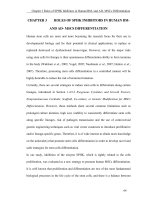
A total of 8 dogs infected with canine
babesiosis were diagnosed by clinical
examination and observation of intra
erythrocytic piroplasm within blood smears.
Giemsa-stained blood smears shows the
presence of small pear-shaped (Plate 1)
parasites.
DNA extraction
DNA isolation kits (QIAamp DNA Mini
Kit®, Qiagen) was used for the extraction of
parasite DNA from 200μl of blood sample
collected in EDTA vacutainers according to
the manufacturer’s instructions. Genomic
DNA isolated from the whole blood of
healthy dogs was used as negative control.
Animal infested with tick was observed (Plate
2). The most prevalent clinical abnormalities
were anorexia, Pale or icteric mucous
membranes (Plate 3a, 3b), lethargy (Plate 4),
fever, dark urine and ecchymosis on ventral
aspect of abdomen (Plate 5).
Table.1 PCR primers
Pathogen
Primer
Sequence (5’-3’)
Babesia
spp.
Ba103F
Ba721R
Ehr1401F
Ehr1780R
CCAATCCTGACACAGGGAGGTAGTGACA
CCCCAGAACCCAAAGACTTTGATTTCTCTCAAG 619 bp
CCATAAGCATAGCTGATAACCCTGTTACAA
380 bp
TGGATAATAAAACCGTACTATGTATGCTAG
E. canis
Product
Size
2129
Int.J.Curr.Microbiol.App.Sci (2019) 8(1): 2127-2132
Table.2 Hematological parameters and biochemical parameters in 8 dogs
infected with Canine babesiosis
Parameters
Hb (g/dl)
PCV (%)
RBC (× 106/mm3)
WBC (103/mm3)
Platelets (105/mm3)
Neutrophil %
Lymphocyte %
Monocyte %
Eosinophil %
BUN (mg/dl)
Creatinine (mg/dl)
Calcium (mg/dl)
Phosphorus (mg/dl)
ALT (U/L)
ALP (U/L)
Total bilirubin (mg/dl)
Direct bilirubin (mg/dl)
Total protein (g/dl)
Albumin (g/dl)
Glucose (mg/dl)
Mean±SE
4.29±1.63
13.03±0.58
2.12±0.08
8514.23±580.13
139750 ± 20490.39
73.88±0.44
17.5±0.54
6.25±0.36
2±0.07
69.88±12.16
1.6±0.30
10.23±0.22
5.05±0.09
113.76±11.44
280.25±33.89
0.96±0.12
0.49±0.036
5.93±0.22
1.71±0.07
88.88±1.97
2130
Int.J.Curr.Microbiol.App.Sci (2019) 8(1): 2127-2132
The finding revels that 8 dogs had the RBC, Hb,
PCV and platelets values below the reference
values and increase level of WBC value. In
biochemical examination, ALT, ALP, Total
bilirubin, BUN, Creatinine values are increase
than the reference values (Table 2). As shown
in (Plate.6) the primer Ba103F and Ba721R
successfully amplified an approximately 619 bp
DNA fragment from all 8 dogs infected with
canine babesiosis.
The goal of this study was to provide an
overview of molecular examination of
babesiosis and haematobiochemical changes.
In the present study, 8 cases infected with
Babesia were confirmed by mean of clinical
history, physical examination, hematological,
biochemical and multiplex PCR. Anemia and
thrombocytopenia were the most common
hematological alterations observed in the
present study which concurred with the report
of (Birkenheuer et al., 1999, Ayoob et al., 2010,
Irwin et al., 2005). The destruction of
circulating RBC by auto antibodies which are
directed against infected and non-infected red
cell membranes resulting in intravascular and
extravascular haemolysis. According to
Taboada and Lobetti, (2006), direct parasitic
damage contributes to anaemia. A low RBC
count, PCV and Hb concentration define
anemia. Severe microcytic-hypochromic anemia
may have been initiated by antibody mediated
cytotoxic destruction of erythrocytes and/or by
auto-antibody directed against components of
the membranes of infected and uninfected
erythrocytes which has also been reported
previously in B. gibsoni infection (Aysul et al.,
2013).
In the present study elevation in mean alanine
aminotransferase (ALT) was noticed, which
was correlated with Aysul et al., (2013)
findings. Aysul et al., (2013) reported that in
dogs suffering from babesiosis, elevation of
bilirubin, ALT and alkaline phosphatase levels
could be seen with hepatic hypoxia.
Acute renal failure in canine babesiosis
(Schoeman, 2009) might have resulted into
increase BUN and creatinine both B. gibsoni
and B. canis infected dogs. According to Amie
(2009) increased non-insulin mediated glucose
consumption believed to be induced by
inflammatory mediators, more especially in
macrophage-rich tissues like the spleen, liver
and the lungs was the cause of hypoglycaemia
in the affected dogs and at the same time
regarded it as a poor prognostic indicator.
Elevation of bilirubin, ALT and AKP are
indicative of hepatic hypoxia and increase BUN
and creatinine are indicative of degenerative
changes in kidneys. Results of this study
suggest that haemato-biochemical changes
could be beneficial in determination of the
severity of babesiosis in dogs.
2131
Int.J.Curr.Microbiol.App.Sci (2019) 8(1): 2127-2132
The use of molecular characterization to
identify Babesia species highlights the value of
procedure of PCR as an adjuvant to current
diagnostic methodology.
References
Amie, K., 2009. Hypoglycemia. Small anim.
Crit. Care Med. 69:295–299.
Ayoob, A.L., Hackner, S.G. and Prittie, J. 2010.
Clinical management of canine babesiosis.
J. Vet. Emerg. Critical Care. 20(1): 77-89.
Aysul, N., Ural, K., Ulutas, B., Eren, H. and
Karagenc, T. 2013. First detection and
molecular identification of Babesia gibsoni
in two dogs from the Aydin province of
Turkey. Turkish J. Vet. Anim. Sc, 37(2):
226-229.
Birkenheuer, A.J., Levy, M.G., Savary, K.C.M.,
Gager, R.B. and Breitschwerdt, E.B. 1999.
Babesia gibsoni infections in dogs from
North Carolina. J. Am. Anim. Hosp. Assoc.
35(2): 125-128.
Conrad, P., Thomford, J., Yamane, I., Whiting,
J., Bosma, L., Uno, T., Holshuh, H.J. and
Shelly, S.1991. Hemolytic anemia caused
by Babesia gibsoni infection in dogs. J.
Am. Vet Med. Assoc.199 (5):601-5.
Filipe, D.T. and Luciana, A.F. 2006. Canine
babesiosis: A Brazilian persepective.
Vet.Parasit. 141(3-4): 197-203.
Irwin,
P.J.,
2005.
Babesiosis
and
Cytauxzoonsis.
Arthropode-Borne
Infectious Diseases of Dogs and Cats,1st
edition.
Manson
Publishing
Ltd.,
Barcelona, Spain,
Jacobson, L.S. and Clark, I.A. 1994. The
pathophysiology of canine babesiosis: new
approaches to an old puzzle. J. South Afr.
Vet. Assoc. 65(3): 134-145.
Kledmanee,
K.,
Suwanpakdee,
S.,
Krajangwong, S., Chatsiriwech, J., Suksai,
P., Suwannachat, P., Sariya, L.,
Buddhirongawatr, R., Charoonrut, P. and
Chaichoun, K. 2009. Development of
multiplex polymerase chain reaction for
detection of Ehrlichia canis, Babesia spp
and Hepatozoon canis in canine blood.
South Asi. J. Trop. Med. Public Health. 40:
35–39.
Lobetti, R.G., 1998. Canine babesiosis.
Compend Contin. Educ. Pract. Vet. 20:418430.
Meinkoth, J.H., A.A.Kocan, S.D. Loud. and
M.D.Lorenz.
2002.
Clinical
and
hematologic effects of experimental
infection of dogs with recently identified
Babesia
gibsoni-like
isolates
from
Oklahoma. J. Am. Vet. Assoc. 220:185-189.
Schoeman, J.P., 2009. Canine babesiosis.
Onderstepoort. J. Vet. Res. 76:59-66.
Taboada, J. and S.R.Merchant. 1991. Babesiosis
of companion animals and man. Vet. Clin.
North Am. Small Anim. Pract. 21:103-123.
Taboada, J., 1998. Babesiosis. In: Greene CE
(Ed): Infectious diseases of the dog and cat.
Philadelphia, WB Saunders. Pp. 473-481.
Taboada, J. and Lobetti, R. 2006. Babesiosis.
In: Infectious Diseases of the Dog and Cat.
Greene, C.G. (Ed.), Elsevier, 3rd edition.
Wozniak, E.J., Barr, B.C., Thomford, J.W.,
Yaamanel, I., McDonough, S.P. Moore,
P.F., Naydan, D., Robinson. T.W. and
Conrad, P.A. 1997. Clinical, anatomic, and
immunopathologic characterization of
Babesia gibsoni infection in the domestic
dog (canis familiaris). J. parasitol. 83(4):
692-699.
How to cite this article:
Juripriya Brahma, D. Chandrasekaran, M.G. Jayathangaraj, S. Vairamuthu and Soundararajan, C.
2019. Clinical, Haemato-Biochemical and Molecular Findings of Babesiosis in Dogs.
Int.J.Curr.Microbiol.App.Sci. 8(01): 2127-2132. doi: />
2132