Evaluation of quorum quenching and probiotic activity of bacillus Thuringiensis QQ17 isolated from fish culture pond
Bạn đang xem bản rút gọn của tài liệu. Xem và tải ngay bản đầy đủ của tài liệu tại đây (311.04 KB, 16 trang )
Int.J.Curr.Microbiol.App.Sci (2019) 8(5): 1634-1649
International Journal of Current Microbiology and Applied Sciences
ISSN: 2319-7706 Volume 8 Number 05 (2019)
Journal homepage:
Original Research Article
/>
Evaluation of Quorum Quenching and Probiotic Activity of
Bacillus thuringiensis QQ17 Isolated from Fish Culture Pond
Divya V. Haridas1,2* and Devika Pillai1
1
Kerala University of Fisheries and Ocean Studies, Department of Aquatic Animal Health
Management, Centre for Aquatic Animal Health, Panangad P.O., Kochi, Kerala,
India, Pin- 682 506
2
Mahatma Gandhi University, School of Biosciences, Kottayam, Kerala, India, Pin- 686 560
*Corresponding author
ABSTRACT
Keywords
Quorum sensing,
Quorum quenching,
N-acyl-homoserine
lactones, Probiotic,
Bacillus
thuringiensis
Article Info
Accepted:
15 April 2019
Available Online:
10 May 2019
This work was aimed at isolating AHL degrading bacteria from fish culture pond soil, with
abilities appropriate for use as probiotic in aquaculture. The presence of an autoinducer
inactivation (aiiA) homologue gene and AHL-inactivation assay showed that
BacillusthuringiensisQQ17, which was one among the 20 isolates, could rapidly degrade
synthetic C6-HSL in vitro and hampered violacein production by Chromobacterium
violaceum. It had excellent biodegrading ability of natural N-AHL produced by
Aeromonas hydrophila, suggesting that it can be used as a potential quencher bacterium
for inhibiting the virulence of A. hydrophila. The isolate grew well at pH 3.0-7.0, was
resistant to high level of bile salts (0-0.9%) and 0.5 % of phenol. QQ17 also exhibited high
degree of auto-aggregation and co-aggregation, confirming that it possessed good probiotic
attributes. It was susceptible to all the 11 antibiotics tested and exhibited antagonistic
activity against A. hydrophila. Gold fish fed diet incorporated with 10 8 and 1010 CFU/g of
the QQ17 for 30 days showed 73.33-83.33% survival when challenged with pathogenic A.
hydrophila. The study indicates that the isolate B. thuringiensis QQ17 could be used as a
non- antibiotic feed additive in aquaculture to control bacterial diseases.
Introduction
Aquaculture is the rapidly expanding foodmanufacturing sector in the world. However,
the industry is hindered by unforeseeable
mortalities, many of which are generated by
infectious microorganisms. The intensive fish
farming has led to sudden occurrence of
various bacterial diseases, necessitating the
use of antibiotics in health management
policies (Fyzuland Austin, 2014). In the
beginning, use of antibiotic had been an
effective strategy, but the indiscriminate use
resulted in the emergence of antibiotic
resistance in fish pathogens and in the transfer
of these resistance genes to bacteria of
terrestrial animals and to human pathogens
(Verschuere et al., 2000). In addition to this,
there is a high risk of antibiotic residues in
human
food.
These
unfavourable
1634
Int.J.Curr.Microbiol.App.Sci (2019) 8(5): 1634-1649
circumstances
prompted
aquaculture
researchers to develop sustainable and ecofriendly approaches that are as equally
functional as antibiotics (Standen et al., 2013)
in controlling diseases. One such strategy is to
impede with the bacterial signaling pathways
controlling the production of virulence
factors.
It is evident that bacterial pathogenicity relies
on the quorum sensing (QS) process, where
gene expression is mediated by extracellular
signaling molecules called autoinducers (AIs).
Autoinducers
like
N-acyl-homoserine
lactones (AHLs) are responsible for the
regulation of virulence genes expression in
many Gram-negative pathogenic bacteria
(Federle and Bassler, 2003). Quorum
quenching (QQ) is the mechanism of
intercepting QS by inactivating signaling
molecules. This is achieved by small
molecule antagonists or signal degrading
enzymes and has been considered as a unique
approach to attenuate pathogenic bacteria
(Dong et al., 2000; De foirdt et al., 2007).
Quorum quenching enzymes, consisting
lactonase, acylase, oxidoreductase and
paraoxonase, have been recognized in quorum
sensing and non-quorum sensing microbes
(Dong et al., 2001; Lin et al., 2003).
As a more sustainable substitute to antibiotic,
the use of probiotic is gaining acceptance for
the control of bacterial pathogens in
aquaculture
too.
Probiotics
eliminate
pathogens by competition process and have
several mechanisms that provide health
benefits to the host. These beneficial
microorganisms have been discovered,
characterized and used in aquaculture during
the last three decades. In this context,
application of signal degrading (quorum
quenching) bacteria that can at the same time
act as probiotic would be a unique dual
strategy to control antibiotic-resistant
pathogens and to support the host in a positive
manner. Recently, some research works have
been reported in quorum quenching bacteria
isolated from gastrointestinal tract of aquatic
animals (Nhan et al., 2010; Ramesh et al.,
2014). It has also been shown that probiotic
bacteria such as Enterococcus durans and
Bacillus spp. inactivate the signal molecules
of pathogenic bacteria by enzymatic action
(Chu et al., 2010; Boopathi et al., 2017).
Bacillus thuringiensis is a spore forming soil
bacterium
that
naturally
synthesizes
insecticidal proteins and has been used for
insect control. They also occur in surfaces of
leaf, aquatic environments, animal fecal
matters, insect-rich environments etc. It has
been proven that many of the strains of
B.thuringiensisproduce
AHL-inactivating
enzymes and possesses quorum quenching
activity (Dong et al., 2001). Recently, studies
on the antagonistic and anthelmintic effect
of B.
thuringiensis strains
against fish
pathogens have also been reported (Bagde et
al., 2009; Luis et al., 2016). The study of
Chang et al., (2012) demonstrating the
probiotic potential of B. thuringiensis isolated
from cow milk is one of the very few studies
that looked at the probiotic properties of the
bacteria. The aim of this work was to study
the quorum quenching attributes and probiotic
properties of B. thuringiensis strain isolated
from fish culture pond and to explore its
potential use as a suitable biocontrol agent in
aquaculture. This could be a dual strategy to
control bacterial disease in aquaculture and
thus, prevent the indiscriminate use of
antibiotics.
Materials and Methods
Bacterial strains and growth conditions
CV026, a mini-Tn5 mutant derived from
Chromobacterium violaceum was used as a
biosensor to find out the presence of
exogenous AHLs (C6-HSL). It was purchased
1635
Int.J.Curr.Microbiol.App.Sci (2019) 8(5): 1634-1649
from Microbial Culture Collection (MCC),
NCCS, Pune. CV026 cannot synthesize AHL,
but it can detect and respond to exogenous
AHLs with acyl chain of four to eight
carbons, by production of the purple coloured
violacein pigment. CV026 strain was grown
in Luria-Bertani (LB) medium at 28°C
supplemented with 50µgmL−1 of kanamycin.
The
target
fish
pathogen Aeromonas
hydrophila used in this study was provided by
the National Bureau of Fish Genetic
Resources (ICAR, Kochi, India). It was
grown in LB broth (pH 7.2 ± 0.2) at 150 rpm
overnight at 30°C. Escherichia coli DH5α,
(Promega) also grown in LB medium at 370C,
served as negative control in AHLinactivation assay. All media used for AHLs
assay were buffered with 50 mmolL-13-[Nmorpholino] propane sulfonic acid (MOPS) to
pH 6.8, to prevent spontaneous degradation of
AHLs.
Isolation and identification of quorum
quenching bacteria from fish culture ponds
Soil samples were collected from tilapia
culture ponds located on the campus of the
Kerala University of Fisheries & Ocean
Studies (KUFOS), Kerala, India. A soil
suspension was prepared in sterile
physiological saline [(pH 7.4) 0.85% NaCl].
Samples were then enriched in minimal
medium (KG medium) with AHL as the sole
source of carbon and nitrogen. 100µL of the
soil suspension was inoculated into 100-mL
flask containing 10 mL of KG medium (pH
6.8) with 500 µg L-1 of C6-HSL, as
previously described (Chan et al., 2009) and
incubated at 300C, 150 rpm. After 24 hr, 1mL
of culture was transferred to fresh C6-HSL
containing KG medium for enrichment
culturing. At the third-time enrichment cycle,
a diluted soil suspension was plated onto LB
agar. Pure colonies were obtained by repeated
streaking on LB agar. The Selected bacterium
was identified following Bergey’s Manual of
Systematic Bacteriology (Ludwig et al.,
2009)in
accordance
with
different
biochemical and physiological characteristics.
Species level identification was carried out by
16S rDNA sequencing (SciGenom Labs,
India) using universal primers 27F and 1492R
and analyzed using NCBI nucleotide
database.
Screening of quorum quenching activity
PCR amplification of aiiA homologue gene
Initially, the quorum quenching activity of all
isolates was checked by screening for the
presence of aiiA (Autoinducer inactivation
homologue) gene by PCR. Total DNA was
extracted using HiPurA bacterial genomic
DNA purification Kit (Himedia, India). The
forward and reverse primers used were aiiA F
(5’-ATGGGATCCATGACAGTAAAGAAG
CTTTAT-3’)
and
aiiAR(5’GTCGAATTCCTCAACAAGATACTCCTA
-ATG-3’) respectively. PCR amplification
was performed in a thermal cycler (MJ MINI,
Biorad, USA), in 0.2 mL reaction tube
consisting of 25 μL total reaction volume
containing 9μL nuclease free water,
12.5μLGoTaq® Colorless Master Mix2X
(Promega, USA), 1.25 μL (10µM) of each
primer and 1 μL of template DNA (100ng).
The reaction consisted of an initial
denaturation of 94°C for 10 min, followed by
30 cycles of 94°C for 30 s, 52°C for 30s,
72°C for 1min and a final extension of 72°C
for 5 min. Samples electrophoresed in 1.5%
agarose gel at 70V were visualized using gel
documentation system (Biorad, USA).
Whole-cell AHL inactivation assay
The whole-cell AHL inactivation assay was
carried out as previously reported (Chan et
al., 2007) with minor modifications. Briefly,
randomly selected quorum quenching isolate
(isolate showing the presence of aiiA
1636
Int.J.Curr.Microbiol.App.Sci (2019) 8(5): 1634-1649
homologue gene) grown overnight at 300C in
LB medium was centrifuged at 5000rpm for
10 min at 40C. Cell pellet was washed two
times in 100 mM PBS (pH 6.8) and
resuspended in the same buffer to get OD600
of 1.0 (BIOPHOTOMETER, Eppendorf,
Germany). 10µg µL-1C6-HSL (a synthetic
AHL, Sigma-Aldrich, India) in absolute
ethanol was transferred to sterile micro
centrifuge tube and dried by evaporation
under aseptic conditions. The cell suspension
in PBS was added to rehydrate AHL to the
final concentration of 0.1µg µL-1. The mixture
was incubated at 300C with gentle shaking for
12 hr. C6-HSL inactivation was assessed at
3hr, 6hr and 12hr using CV026 as biosensor.
Heat-denatured reaction mixture (10 µL) at
above mentioned time periods was loaded
into the well of LB agar bioassay plate
overlaid with the biosensor CV026 and
incubated at 280C for 24 hr. E. coli strain
DH5α served as negative control. Absence of
violacein (purple zone) shown by CV026
indicated AHL degradation.
AHL degradation with culture supernatant
To find out whether the quorum quenching
factor is released out of the cell or is bound to
cell, an in vitro assay was carried out as
previously described by Chu et al (2010) with
minor modification. The isolate QQ17 grown
overnight at 300C in LB medium was
centrifuged for 10 min at 7000 rpm and the
filter-sterilized supernatant of the overnight
culture was taken for testing the AHL
degrading activity. 100µL of the supernatant
was mixed with an equal volume of 100 mM
PBS (pH 6.8) containing 0.2µg µL-1 C6-HSL.
Following that, the reaction mixture was
incubated at 300C for 24 hr with gentle
shaking, followed by incubation at 950C for 5
min to stop the reaction. 10 µL of the reaction
mixture was loaded into the well of a LB agar
plate seeded with the biosensor CV026 and
incubated at 280C for 24 hr.
Degradation of N-AHL
Aeromonas hydrophila
produced
by
Fish pathogen A.hydrophila was inoculated in
10 mL LB medium and incubated at 300C for
24hr. Bacterial cells were removed by
centrifugation at 12000 rpm for 5 min at 40C.
Filter sterilized cell free culture supernatant
was added to equal volume of fresh LB
medium and QQ17 was inoculated in this
medium. Bacterial culture was incubated at 30
°C for 48 hr and AHL inactivation was
assessed at 0 hr and 48 hr using CV026 as
biosensor.
Screening of probiotic activity
Bile salt and acid tolerance
The isolate QQ17 was tested for bile salt
tolerance and survival in acidic condition.
Bacterial strain was grown overnight in LB
media and 0.1 mL of culture suspension was
inoculated into tubes containing 10 mL of
autoclaved LB media with 0%, 0.3%, 0.6%,
and 0.9% bile salt (Himedia, India). The
inoculated tubes were incubated at 300C for
18 hr and the absorbance at 600 nm was
measured to evaluate growth. To determine
acidic tolerance of QQ17, 0.1 mL of actively
grown overnight culture at 300C in LB
medium was transferred to autoclaved LB
broth adjusted to pH 1-7 with HCl (Sigma,
India), which were then incubated at 300C for
18 hr followed by measurement of absorbance
at 600 nm.
Phenol tolerance assay
To check the phenol tolerance, actively
growing overnight culture of QQ isolate was
inoculated into LB media with concentration
of 0.2% and 0.5% phenol or without phenol.
Cell growth of the isolate was evaluated after
18 hr of incubation at 300C, by measurement
of absorbance at 600 nm.
1637
Int.J.Curr.Microbiol.App.Sci (2019) 8(5): 1634-1649
Auto-aggregation
assays
and
co-aggregation
To evaluate the probiotic potential of QQ17,
auto-aggregation and co-aggregation rate
were measured according to DelRe et al.,
(2000) with some modifications. Isolate was
grown for 18 hr at 300C in LB media. The
cells were harvested by centrifugation at 5000
rpm for 15 min at 40C, washed twice with
PBS (pH 7.2) and resuspended in the same
buffer. Absorbance (A600 nm) was adjusted to
0.2 in order to give viable counts of
approximately 108 CFU ml-1.
Cell suspension (5ml) was mixed by
vortexing for 10 s and the same suspension
was left to rest for 5 hr at room temperature
without vortexing. Auto-aggregation of cell
suspension was determined by taking 0.1 ml
of the upper suspension at every 1hr interval
to another tube with 4.9 ml of PBS and the
absorbance of suspension at 600 nm was
recorded. Cell auto-aggregation was measured
by decrease in absorbance and autoaggregation percentage is demonstrated as: 1(At/A0) X 100, where At represents the
absorbance at time t= 1, 2, 3, 4 or 5 hr and
A0the absorbance at t=0.
The method for preparing the cell suspension
for co-aggregation was the same as that for
auto-aggregation assay. QQ isolate prepared
as described above was mixed with equal
volume (2 ml) of the culture of fish pathogen
A.hydrophila and incubated at room
temperature without agitation.
In control tubes, 4 ml of each bacterial
suspension alone was added. After 5 hr of
incubation, the absorbance (A) at 600 nm of
the suspensions was measured. Coaggregation percentage was calculated using
the equation of Handley et al (1987). Coaggregation %=[( Apathog + AQQ)/2 - (Amix)
/(Apathog + AQQ)/2] X 100, where Apathog and
AQQ constitute the absorbance in the tubes
containing solely the pathogen or the quorum
quenching
bacteria
(control
tubes)
respectively, and Amix represents the
absorbance of the mixture.
Antibiotic sensitivity test
Antibiotic susceptibility test was performed
by disc diffusion method as stated by the
guidelines of the Clinical and Laboratory
Standard Institute (CLSI, 2002). Antibiotic
discs (Himedia, India) were placed onto
freshly plated QQ17 on the Muller-Hinton
agar (Himedia, India) and antibiotic resistance
was determined by measuring the diameter of
the inhibition zone after incubation of the
plate at 300C for 18 hr. The antibiotic discs
used in this test included ampicillin (10µg),
amikacin (30µg), erythromycin (15µg),
gentamycin (10 µg), neomycin (30 µg),
penicillin G (10 U), kanamycin (30 µg),
streptomycin (10µg), oxacillin (1 µg),
vancomycin (30 µg) and tetracycline (30 µg).
Antagonism test
Agar well-diffusion method was carried out
according to Schillinger and Lucke (1987)
with some modification, to detect the in vitro
antagonistic effect of the QQ17 against fish
pathogen A. hydrophila. 100µL of fresh,
actively growing pathogen was spread on
Mueller-Hinton agar plate. Well with a
diameter of 6 mm was prepared aseptically
and cell free supernatant of actively growing
QQ bacterial culture (75 µL/well) was loaded
into the well.
Plate was incubated at 300C for 24 hr and the
zone diameter of inhibition (ZDI) was
recorded. Inhibition zone of more than
20 mm, 10 to 20 mm, and less than 10 mm
was considered as strong, intermediate, and
low antimicrobial activity, respectively.
1638
Int.J.Curr.Microbiol.App.Sci (2019) 8(5): 1634-1649
In vivo study
Maintenance of experimental fish
To confirm the probiotic activity of QQ17, in
vivo study was carried out. Fingerlings of
goldfish Carassius auratus (Linnaeus, 1758)
of uniform size were initially acclimatized in
fibre reinforced plastic tanks of 300 L
capacity for three weeks before starting the
experiment. The fish were healthy, exhibited
no symptoms of disease (tested through the
examination of gills, fins and skin). The
pathogen-free status of the fish was also
confirmed by standard bacteriological
examination procedures in the laboratory.
During this period, a commercial fish feed
was given to fish twice daily. All tanks were
provided with proper aeration and water
temperature was maintained at 26 ± 1°C.
Safety of the QQ17
The pathogenicity of the QQ17 was also
ascertained before preparing probiotic feed.
Two groups of six gold fish (3.34-4.32 g
weight and 85.35-94.40 mm length), were
challenged with 0.1 mL of PBS with 1.0 x
107cells and 1.0 x 10¹⁰cells of QQ17
respectively by intraperitoneal injection. Gold
fish in control group were injected with
0.1mL of PBS. Fish were observed for
mortality for seven days. During this period
behaviour of fish was recorded daily. Before
conducting the challenge study, the infectious
dose of A. hydrophila was also determined by
50% lethal dose (LD50) determination.
Preparation of probiotic feed
The probiotic feed was prepared by
inoculating the QQ isolate in LB broth and
incubated at 300C for 24 h. The cells were
harvested by centrifugation at 3000 rpm for
15 min at 40C, washed twice with PBS (pH
7.2) and resuspended in the same buffer.
Afterwards, the concentration of bacterial
culture was adjusted to different cell densities
(104 CFU, 106 CFU, 108 CFU & 1010 CFU per
mL) using a spectrophotometer (Hach- DR
6000, Germany) and the suspension was
added at the rate of 1 mL of culture /g of feed
to incorporate 104 cells/g feed, 106 cells/g
feed, 108 cells/g feed & 1010 cells/g feed
respectively. A binder (Brand: Aqua one,
Salem Microbes Private limited, India) was
used @1mL/10g feed. Binder alone was
added in control feed. After proper mixing of
the ingredients, the feeds were air dried and
stored in screw capped glass bottles at room
temperature until used. To ensure a required
probiotic level in the supplemented feed, new
probiotic diets were made on a weekly basis.
Five groups of 10 gold fish each, C.auratus
were introduced into five glass tanks of 50 L
capacity. Four groups were fed with 104 CFU,
106 CFU, 108 CFU and 1010 CFU/g of
probiotic diet respectively, while the fifth
group was maintained as control group.
Feeding was done two times daily at the rate
of 3% of the body weight of C. auratus for 30
days. Continuous aeration and water flow
were maintained in all glass tanks. During the
study period, activity and behaviour of the
fish were monitored and recorded daily.
Bacterial challenge study
All fish were clinically healthy before
challenge. Control and probiotic fed fish were
challenged (10nos/group) via intraperitoneal
injection with 0.1mL of 1x 10⁶ cells (LD50
based on preliminary work) of A.hydrophila.
The fish were observed to determine
mortality, external signs of infection and
behavioural abnormalities for two weeks.
Dead fish were removed immediately for
bacteriological
examination.
Bacterial
isolation was carried out from hemorrhagic
and ulcerative lesions, and from dead fish’s
visceral organs.
1639
Int.J.Curr.Microbiol.App.Sci (2019) 8(5): 1634-1649
Statistical analysis
All the experiments were performed in
triplicate and the results were expressed as
mean ± standard deviation (SD) of triplicates.
Data were statistically processed by one way
ANOVA using SPSS (Version 21.0) to find
out whether there was significant difference
between the treatments in each of the
experiment.
Statistically
significant
differences were defined at p< 0.01.
Results and Discussion
Isolation and identification of quorum
quenching bacteria
20 bacterial isolates in the KG medium
containing C6-HSL were screened. Finally,
one representative isolate showing strong
AHL degrading activity was selected. It was
characterized
at
the
physiological,
biochemical and morphology levels. Based on
biochemical properties, the strain showed
close resemblance to Bacillus spp. To further
identify the strain, 16S rDNA sequencing was
carried out. Results showed QQ isolate shared
99% homology with B. thuringiensis species
(GenBank accession number AE017355).
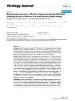
Detection of aiiA homologue gene
Autoinducer inactivation (aiiA) gene was
found in Gram-positive bacterium B.
thuringiensis QQ17. All the 20 bacterial
isolates were screened for presence of aii
Ahomologue gene by PCR and six bacteria
with aii Ahomologue gene were observed.
The expected amplicon size of approximately
800 base pairs was detected (Figure 1).
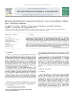
degraded after incubating with QQ isolate for
6 hr (Figure 2c), showing rapid AHL
degradation. Only leftover C6-HSL was
detected by CV026 when the reaction was
ceased after incubation for 3 hr (Figure 2b).
No visible AHL degradation was noticed in
DH5α that served as negative control (Figure
2a). The supernatant of QQ17 had no AHLinactivating activity, and the diameter of the
purple pigmented zone had no remarkable
difference with that of negative control DH5α
well (Data not shown). In order to confirm
AHL degrading activity of QQ isolate, crude
cell free culture supernatant of A.hydrophila
as natural N-AHL was used instead of
synthetic C6-HSL. Complete degradation of
natural N-AHL after 48 hr incubation with
QQ17 was observed (Data not shown). No
AHL degradation was observed and presence
of violacein (purple zone) was shown by
CV026 at 0 hr incubation. This result also
revealed the presence of natural N-AHL in
crude cell free culture supernatant of
A.hydrophila.
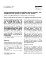
Bile salt, pH and phenol tolerance of B.
thuringiensis QQ17
B. thuringiensis QQ17 grew successfully in
all tested concentrations of bile (0-0.9%) after
18 hr of incubation. This data suggests that B.
thuringiensis QQ17 is resistant to high bile
salt concentration (Figure 3a). pH tolerance
studies showed that B. thuringiensis QQ17
grew at pH 3 or above but did not grow in
conditions less than pH 3 (Figure 3b). The
isolate grew well at 0 - 0.5 % of phenol in LB
media (Figure 3c).
Auto-aggregation
assays
and
Co-aggregation
Whole-cell AHL inactivation assay
B. thuringiensis QQ17 that possessed aiiA
homologue gene was selected for AHLinactivation assay. Almost all C6-HSL was
The result showed that B. thuringiensis QQ17
had excellent auto-aggregation property
[(81.94 ±0.13 %) (Figure 4)] and aggregation
values increased with time. B. thuringiensis
1640
Int.J.Curr.Microbiol.App.Sci (2019) 8(5): 1634-1649
QQ17 also exhibited very good coaggregation ability after 5 hr of incubation
with A. hydrophila, during which 41.6±0.04%
of QQ isolate was co-aggregated with
A.hydrophlila (Data not shown).
Antibiotic
resistance
thuringiensisQQ17
of
B.
Since antibiotic sensitive probiotics are most
preferred,
the
in
vitro
antibiotic
sensitivity/resistance of B. thuringiensis
QQ17 to 11 antibiotics was checked. Results
indicated that B. thuringiensis was susceptible
to antibiotics such as ampicillin, amikacin,
erythromycin,
gentamycin,
kanamycin,
neomycin,
oxacillin,
penicillin
G,
streptomycin, tetracycline, and vancomycin
(Table 1).
Antagonism test
B.thuringiensis QQ17 exhibited excellent
antimicrobial activity against fish pathogen
A.hydrophila (Figure 5) by producing growth
inhibition zone of 26±0.22 mm diameter in
agar well diffusion assay.
Safety of the B.thuringiensis QQ17
The administration of B.thuringiensisQQ17
even at the concentration of 1x10¹⁰ cells/fish
did not result in any unfavorable effect on fish
activity. All fish were clinically healthy and
behaved like control group. This result
suggested that the isolate B. thuringiensis
QQ17 is not virulent to fish.
Experimental challenge with A. hydrophila
The
administration
of
diet
(B.thuringiensis) afforded effective protection
against experimental A. hydrophila infection.
In control group, following challenge with A.
hydrophila, all fish showed severe skin
lesions and 50% mortality was observed in
two days. One fish each died in 104 CFU/g
feed and 106CFU/g feed in two days and
majority of the fish in both these treatments
showed mild skin lesions and haemorrhages.
In contrast, during the same time, there was
no mortality in the two groups fed with QQ
diet of 108 CFU/g feed and 1010 CFU/g feed
(Merely one fish in 108CFU/g out of the
entire lot of fish developed mild
haemorrhages). At the end of two weeks, the
highest survival rate was noticed in groups of
fish fed with 108 CFU/g (73.33%) and 1010
CFU/g (83.33%) probiotic diet. ANOVA
showed that there was significant difference
(p≤0.01) in the survival rates among different
concentrations. Post Hoc analysis using
Duncan’s Multiple Range Test grouped the
concentrations into three homogenous groups
viz; (1) Control (had only 13.33% survival)
(2) 104 CFU/g and 106 CFU/g probiotic feed
(had 43.33% survival) and (3) groups fed with
108 CFU/g (73.33% survival) and 1010 CFU/g
(83.33% survival) (Table 2). A. hydrophila
was isolated from haemorrhagic lesions of
both dead and survived fish.
The present study focused on soil bacteria B.
thuringiensis QQ17 that exhibited both
probiotic and quorum quenching ability. To
the best of our knowledge, there are hardly
any reports demonstrating the probiotic
activity of B. thuringiensis isolated from fish
culture pond soil that possess AHL degrading
activity. In this study, synthetic N-hexanoylL-homoserine lactone (C6-HSL) was used as
a test compound. The AHL-degrading ability
of isolated bacteria was initially screened by
PCR amplification of aiiA gene. Previous
studies by Dong et al (2000) revealed that the
aiiAgene is responsible for AHL degradation
in Bacillus sp. and is common among most
Bacillus strains. As the presence of
aiiAhomologue gene can only predict but
does not confirm the AHL degrading
function, the whole cell inactivation assay
was also carried out and finally we selected
1641
Int.J.Curr.Microbiol.App.Sci (2019) 8(5): 1634-1649
B.thuringiensis QQ17, which synthesize
AHL-degrading enzyme, based on its ability
to stop AHL-dependent violace in production
by the bio indicator CV026. In whole-cell in
vitro AHL-inactivation assay, nearly all
synthetic C6-HSL was degenerated after
incubating with B. thuringiensis QQ17 for 6
hr, indicating rapid and strong QQ activity.
Similar result was observed in a study by Chu
et al., (2010) in which the isolate QSI-1
(Bacillus spp.) degraded C6-HSL completely
within 6hr in whole-cell AHL-inactivation
assay. The supernatant of B. thuringiensis
QQ17 could not inactivate C6-HSL,
indicating that the degrading enzyme is not
discharged out of the cell, that agrees with the
reports by Molina et al., (2003) and Chu et al
(2010), suggesting that the signaling
molecules diffuse into the quorum quenching
bacterial cells where molecule inactivation
takes place. The efficacy evaluation of the
B.thuringiensis QQ17 for degradation of
natural N-AHL produced by A.hydrophila
resulted in the complete inactivation of NAHL within 48 hr of incubation. C4-HSL and
C6-HSL are the major autoinducers produced
by A.hydrophila(Swift et al., 1997) and can
be detected by CV026. This result suggests
that B.thuringiensis QQ17 can be used as
potential quencher bacterium in aquatic
environment very effectively for inhibiting
the virulence of A.hydrophila.
The results of the present study showed that
B.thuringiensis, in addition to possessing
excellent quorum quenching properties, has
very good probiotic properties such as bile
salts, acid and phenol resistance, auto
aggregation, co- aggregation, antibiotic
sensitivity and growth inhibitory effect
against fish pathogen A.hydrophila. Acid and
bile tolerance are two inevitable properties
that give a probiotic the potential to remains
alive in the upper gastrointestinal tract,
especially the acidic condition in the stomach
and the presence of bile in the small intestine
(Erkkila and Petaja, 2000). In the present
study, B. thuringiensis QQ17 tested for bile
salt tolerance exhibited growth even in 0.9%
bile salt at 18 hr of incubation, suggesting that
it has the capacity to withstand in fish as well
as in human gut. Many reports are found to
describe the bile salt tolerance of Bacillus sp.
(Verschuere et al., 2000; Chang et al., 2012).
Fish gastrointestinal pH shows great variation
among species with a range of 1.47 to 5.12
and the lowest value observed was 1.18
(Welliton et al., 2017). However, such
extreme low pH is transient. The pH value
raises to 3 and above in the presence of food
(Erkkila and Petaja, 2000). In the present
study, we found that B. thuringiensisQQ17
grew at pH 3 or above. These results suggest
that the QQ isolate B. thuringiensis given as a
probiotic diet will be able to survive the harsh
conditions of the gut environment and
colonize the intestinal tract, thereby will be
capable of imparting their benefits. In this
study the isolate could also grow and
persisted well at 0.5 % of phenol in LB
media. Phenol may be synthesized in the
intestine by bacterial deamination of various
aromatic amino acids derived from dietary or
endogenously derived protein (Suskovic et
al., 1997). Studies on different animal models
reveal that phenol has a bacteriostatic effect
against gut bacteria (Hoier, 1992). Since
probiotics should withstand the harsh gut
environment, tolerance to phenol is
considered as a mandatory probiotic property.
Auto-aggregation
and
co-aggregation
properties are considered as major
characteristics
of
probiotic
bacteria.
Assessment of auto-aggregation and potential
to co-aggregate with harmful intestinal
pathogens can be used for initial evaluation
and selection of the best probiotic strain. In
this study, the B.thuringiensis QQ17 exhibited
high degree of auto-aggregation (81.94
±0.13%)
and
co-aggregation
activity
(41.6±0.04%). Auto-aggregation property is
1642
Int.J.Curr.Microbiol.App.Sci (2019) 8(5): 1634-1649
responsible for the bacterial adhesion on to
the intestinal cell wall; an essential feature for
a good probiotic strain (Bao et al., 2010). Coaggregation abilities of probiotics might
become an obstacle that prevents colonization
of pathogenic bacteria in the gastrointestinal
tract (Garcia et al., 2014).
The antibiotic sensitivity of bacteria is
another important property to be considered
to formulate safe probiotic products for
aquaculture applications. Now, overuse of
antibiotics has become a serious health
problem and has led to the emergence of a
large number of antibiotic-resistant strains.
Antibiotic resistance in probiotic bacteria may
results in active transfer of antibiotic resistant
genes from probiotics to other intestinal
microflora and finally to opportunistic
pathogens that reside in the same harsh
environment. This may ultimately have
serious clinical ramifications (Imperial and
Ibana, 2016). In the present study, B.
thuringiensis QQ17 showed susceptibility to
all 11 antibiotics tested. This result supports
the possibility of the isolate to be developed
as probiotic. Recently, Chang et al (2012)
isolated B. thuringiensis strain from cow milk
that showed antibiotic susceptibility towards
all tested antibiotics.
The concept of antagonism in probiotics
against pathogenic bacteria has been well
studied. The antibacterial property has been
regarded as one of the important attributes in
selecting potential probiotics for inhibiting the
growth of pathogenic bacteria in the gut. The
antagonistic activity of beneficial bacteria
against pathogenic bacteria can be induced by
the production of carbon dioxide, organic
acids (mainly, lactic acids), hydrogen
peroxide, acetoin, ethanol, reutericyclin,
diacetyl,
acetaldehyde,
reuterin,
antimicrobials such as bacteriocins (Jin,
1996). This activity, along with the process of
competitive exclusion, in which probiotic
bacteria fight against intestinal pathogens for
food and attachment sites, would stop
colonization of pathogenic bacteria in the
gastrointestinal tract (Saulnier et al., 2009). In
the present study, the agar well diffusion
assay was used to find out the antagonistic
effect of cell-free supernatant. B.thuringiensis
QQ17 showed strong inhibitory effect
towards the tested pathogen A.hydrophila.
Earlier studies by Aly et al., (2008) showed
the growth inhibition of A. hydrophila using a
cell-free supernatant of three bacillus species
that were used as probiotic. Probiotic isolates
from the intestine of fresh water fishes
showed inhibitory activity against pathogenic
bacteria (Chemlal et al., 2012). Bagde et al.,
(2009) demonstrated antagonistic effect of
Bacillus thuringiensis sub. Sp. H12 on
pathogens from tilapia by agar well diffusion
method.
Since the bacterial pathogen A. hydrophila is
responsible for frequent disease occurrences
observed in aquariums and ornamental fish
culture, in the present work, we selected this
bacterium for bacterial challenge study. The
QQ isolate B. thuringiensis isolated in the
present study had no harmful effect on
goldfish and the probiotic diet supplemented
with 108CFU and 1010CFU for 30 days
protected the fish when challenged with A.
hydrophila. Lowest (13.33%) survival was
observed in the control (fed with basal diet)
compared with probiotic fed groups. Highest
survival of fish was recorded in the group fed
with probiotic diet of 1010 CFU/g feed and
108CFU/g feed (83.33% and 73.33%
respectively). Statistical analysis showed that
there was no significant difference in the
survival rate between these two groups (post
hoc analysis) suggesting that 108CFU/g may
be sufficient to afford protection to the fish
against A. hydrophila infection. These results
were comparable to findings by Brunt and
Austin (2005) where they used Aeromonas
sorbia GC2 at a dose of 5x 10⁷ cells / g feed
1643
Int.J.Curr.Microbiol.App.Sci (2019) 8(5): 1634-1649
as feed additive to control infection caused by
Lactococcus garvieae and Streptococcus iniae
in rainbow trout and the untreated control
experienced mortality of 75-100% when
challenged with L. garvieae and S.iniae. The
spore-forming bacterium, B. thuringiensis, is
widely known as a bio insecticide which
controls plant diseases. Previous studies have
demonstrated that Bt is basically non-toxic
and non-infectious to other living organisms
like birds, fish, and shrimp (Perez et al.,
2015). A great number of studies exist
suggesting that feeding of Bacillus spp.
significantly increases the resistance towards
bacterial infection in tilapia (Ghosh et al.,
2003) and brown trout (Balcazar et al., 2007).
Table.1 Antibiotic resistances of Bacillus thuringiensis QQ17
Zone of growth inhibition diameter (mm)
AM
AN
E
GM
K
N
OX
P
S
TE
VA
19±0.48 21±0.18 30±0.22 22±0.07 24±0.88 21±0.39 17±0.03 18±0.08 24±0.25 28±0.08 22±0.38
Foot note: AM: ampicillin (10 μg), AN: amikacin (30 μg), E: erythromycin (15 μg), GM: gentamycin (10 μg), K:
kanamycin (30 μg), N: neomycin (30 μg), OX: oxacillin (1 μg), P: penicillin G (10 U), S: streptomycin (10 μg), TE:
tetracycline (30 μg), VA: vancomycin (30 μg).The mean of three values of zone of growth inhibition of each
antibiotic are presented along with ±SD
Sensitive ≥20mm, Intermediate 15-19mm, Resistant ≤14
Table.2 Survival percentages of Carassius auratus (fed with different concentration of probiotic
diet) two weeks after the experimental infection with Aeromonas hydrophila
by intraperitoneal injection
Probiotic
concentration
Control
1 x 10⁴CFU
1 x 10⁶CFU
1 x 10⁸ CFU
1 x 10¹⁰ CFU
Survival
%
13.33±5.8a
43.33±5.8b
43.33±5.8b
73.33±5.8c
83.33±5.8c
Foot note:Values are mean±SD of triplicate observations. Values with different superscripts are significantly
different (p<0.01).
Fig.1 PCR detection of aiiA homologue gene. Lane A: 100 bp DNA ladder (Promega); lane B-G:
different isolates; lane H: negative control. Arrow shows the expected amplicon size of
approximately 800 bp
1644
Int.J.Curr.Microbiol.App.Sci (2019) 8(5): 1634-1649
Fig.2 AHL-degrading activity of Bacillus thuringiensis QQ17. QQ isolate was incubated with
C6-HSL for 3 hr (2b), 6 hr (2c) and 12 hr (2d). (2a) Escherichia coli DH5a (negative control).
Pigment formation indicates the presence of C6-HSL; degradation of C6-HSL is evident by loss
of pigment formation on the biosensor lawn. QQ isolate, quorum-quenching isolate;
AHL, acyl-homoserine lactone
Fig.3a,b&c Effect of bile salt (3a), pH (3b) and phenol (3c) on the growth of Bacillus
thuringiensis QQ17 at 30oC. To check bile-salt, pH and phenol resistance, the growth of
B.thuringiensis QQ17 in LB medium for 18 hr was determined by measuring OD at 600 nm after
adjusting the culture media to the specific pH, salt and phenol concentration. Values are
mean±SD of three different observations
1645
Int.J.Curr.Microbiol.App.Sci (2019) 8(5): 1634-1649
Fig.4 Auto-aggregation rate of quorum quenching isolate Bacillus thuringiensis QQ17.
Aggregation percentage increased every 1hr and highest rate was observed at 5 hr
Fig.5 The agar well diffusion assay to determine the antagonistic activity of Bacillus
thuringiensis QQ17. Zone of growth inhibition (26±0.22 diameter) indicates the antagonistic
activity of cell free supernatant of B. thuringiensis QQ17 towards A. hydrophila. This value is
the mean±SD of three different observations
Recently, there are reports of quorum
quenching probiotics that have the ability to
change the intestinal microflora structure by
degrading AHLs (Chu et al., 2010; Boopathi
et al., 2017). The results of the present study
clearly suggest that the significant increase in
survival of goldfish after challenging with
A.hydrophila is due to the combined effect of
quorum quenching ability of B. thuringiensis
QQ17 together with its probiotic activity.
Production of AHL degrading enzyme might
have inhibited the pathogenicity of
A.hydrophila, while, the probiotic potential of
the QQ isolate might have simultaneously
helped to out-compete A.hydrophila for
nutrients and space and exclude the
pathogenic bacteria through antagonistic
activity.
Acknowledgement
The authors thank the authorities of the
Kerala University of Fisheries and Ocean
Studies (KUFOS), Kochi, Kerala for the
facilities extended for carrying out this work
at the Centre for Aquatic Animal Health
Management, KUFOS. We thank Dr. Mathew
Sebastian, KUFOS for the statistical analysis
of results.
References
Aly, S. M., Ahmed, Y. A., Ghareeb, A. A.,
and Mohamed, M. F. 2008. Studies on
Bacillus subtilis and Lactobacillus
acidophilus, as potential probiotics, on
the immune response and resistance of
1646
Int.J.Curr.Microbiol.App.Sci (2019) 8(5): 1634-1649
Tilapia
Nilotica
(Oreochromis
niloticus) to challenge infections. Fish
and Shellfish Immunology, 25, 128 –
136.
Bagde, U.S., Bilolikar, B.V., and Pandit, R.S.
2009. Antagonistic effect of Bacillus
thuringiensis sub-species (h12) on
pathogens of tilapia species. Asian
Journal
of
Microbiology,
Biotechnology
&
Environmental
Sciences, 11,917-922.
Bandyopadhyay, P., and Mohapatra, P.K.D.
2009. Effect of a probiotic bacterium
Bacillus circulans PB7 in the
formulated
diets:
on
growth,
nutritional quality and immunity of
Catlacatla (Ham.). Fish Physiology
and Biochemistry, 35, 467-478.
Bao, Y., Zhang, Y., Zhang, Y., Liu, Y.,
Wang, S., Dong, X., Wang, Y., and
Zhang, H. 2010. Screening of
potential probiotic properties of
Lactobacillus fermentum isolated from
traditional dairy products. Food
Control, 21,695-701.
Boopathi, S., Selvakumar, N., and
Sivakumar,G.
2017.
Quorum
quenching potentials of probiotic
Enterococcus durans Lab38 against
methicillin resistant Staphylococcus
aureus.
Asian
Journal
of
Pharmaceutical
and
Clinical
Research, 10, 445-450.
Brunt, J.1., and Austin, B. 2005. Use of a
probiotic to control lactococcosis and
streptococcosis in rainbow trout,
Oncorhynchus mykiss (Walbaum).
Journal of Fish Disease, 28, 693-701.
Chan, K.G., Tiew, S.Z., and Ng, C.C. 2007.
Rapid isolation method of soil bacilli
and screening of their quorum
quenching
activity.
Asia-Pacific
Journal of Molecular Biology and
Biotechnology, 15, 153–156.
Chan, K.G., Yin, W.F., Sam, C.K., and Koh,
C.L. 2009. A novel medium for the
isolation
of
N-acylhomoserine
lactone-degrading bacteria. Journal of
Industrial
Microbiology
and
Biotechnology, 36,247–251.
Chang, H. K., Sang, Y. C., Hyog, Y. K., Eun,
H. K., Hyun, M. K., Jin, S. M., Geum,
C. J., Hee, S. L., Seung, W. K., Jong,
M. K., Suhkneung, P., and Dong, K.
R. 2012. Isolation, characterization,
and
evaluation
of
Bacillus
thuringiensis isolated from cow milk.
Korean Journal of Veterinary
Research, 52, 169-176.
Chemlal, K.F., Sahnouni, A., Matallah, B.,
and Boutiba, Z. 2012. The probiotic
potential of lactobacilli isolated from
Nile tilapia (Oreochromis niloticus)’s
intestine.
African
Journal
of
Biotechnology, 11,13220-13227.
Chu, W., Lu, F., Zhu, W., and Kang, C. 2010.
Isolation and characterization of new
potential probiotic bacteria based on
quorum-sensing system. Journal of
Applied Microbiology, 110, 202–208.
Clinical and Laboratory Standards Institute
(CLSI). Performance standards for
antimicrobial disk and dilution
susceptibility tests for bacteria isolated
from animals; approved standards. 2nd
ed. NCCLS document M32-A2, CLSI,
Wayne, 2002.
Defoirdt, T., Boon, N., Sorgeloos, P.,
Verstraete, W., and Bossier, P. 2007.
Alternatives to antibiotics to control
bacterial
infections:
luminescent
vibriosis in aquaculture as an example.
Trends in Biotechnology, 25, 472-479.
Del Re, B., Sgorbati, B., Miglioli, M., and
Palenzona, D. 2000. Adhesion, auto
aggregation and hydrophobicity of 13
strains of Bifidobacterium longum.
Letters in Applied Microbiology, 31,
438–442.
Dong, Y.H., Wang, L.H., Xu, J.L., Zhang,
H.B., Zhang, X.F., and Zhang, L.H.
2001. Quenching quorum sensing
1647
Int.J.Curr.Microbiol.App.Sci (2019) 8(5): 1634-1649
dependent bacterial infection by Nacyl homoserinelactonase. Nature,
411,813-817.
Dong, Y.H., Xu, J.L., Li, X.Z., and Zhang,
L.H. 2000. AiiA, an enzyme that
inactivates the acylhomoserine lactone
quorum sensing signal and attenuates
the virulence of Erwinia carotovora.
Procedings of the National Academy
of Sciences of the United States of
America, 97, 3526-3531.
Erkkila, S., and Petaja, E. 2000. Screening of
commercial meat starter cultures at
low pH and in the presence of bile
salts for potential probiotic use. Meat
Science, 55, 297-300.
Federle, M.J., and Bassler, B.L. 2003.
Interspecies
communication
in
bacteria.
Journal
of
Clinical
Investigation, 112, 1291-9.
Fyzul, N.A.I., and Austin, B. 2014.
Probiotics, immunostimulants, plant
products and oral vaccines, and their
role as feed supplements in the control
of bacterial fish diseases. Journal of
Fish Diseases, 38, 937–955.
Garcia,C.T., Korany, A.M., Bustos, I., Gomez
de,C.L.P., Requena, T., and Pelaez,
C.2014. Adhesion abilities of dairy
Lactobacillus
plantarum
strains
showing an aggregation phenotype.
Food Research International, 57, 44–
50.
Ghosh, K., Sen, S. K., and Ray, A. K. 2003.
Supplementation of an isolated fish
gut bacterium Bacillus circulans, in
formulated
diets
for
Rohu,
Labeorohita, fingerlings. The Israeli
Journal of Aquaculture Bamidgeh, 55,
13-21.
Handley, P.S., Harty, D.W.S., Wyatt, J.E.,
Brown, C.R., Doran, J.P., and Gibbs,
A.C.C. 1987. A comparison of the
adhesion, co aggregation and cellsurface hydrophobicity properties of
fibrillar and fimbriate strains of Streptococcus
salivarius. Journal of General
Microbiology, 133, 3207-3217.
Hoier, E. 1992. Use of probiotic starter
cultures in dairy products. In: The
25th Annual Convention, Australian
Institute of Food Science Technology,
Sydney, Australia.
Imperial, I. C., and Ibana, J. A.
2016. Addressing
the
antibiotic
resistance problem with probiotics:
reducing the risk of its double-edged
sword
effect. Frontiers
in
Microbiology, 7, 1-10.
Jin, L.Z. 1996. Studies on the mechanisms
and utilization of probiotics (direct-fed
microbial) in broilers [Ph.D. thesis]
Kuala Lumpur, University Putra
Malaysia, Malaysia.
Lin, Y.H., Xu, J.L., Hu, J., Wang, S. L., and
Ong, J.R. 2003. Acyl-homoserine
lactone acylase from ralstonia strain
XJ12B represent a novel and potent
class of quorum quenching enzymes.
Molecular Microbiology, 47,849-860.
Luis, J.M., Victor, M.H., Ivan, A.S.,
Fernando, I.F., Jorge, M.M., and
Guadalupe, P.C. 2016. Anthelmintic
Effect of Bacillus thuringiensis Strains
against the Gill Fish Trematode
Centrocestus formosanus. BioMedical
Research International, 20, 1-9.
Molina, L., Constantinescu, F., Michel, L.,
Reimmann, C., Duly, B., and De fago,
G. 2003. Degradation of pathogen
quorum-sensing molecules by soil
bacteria: a preventive and curative
biological control mechanism. FEMS
Microbiology Ecology, 45, 71–81.
Nhan, D.T., Cam, D.T.V., Wille, M.,
Defoirdt, T., Bossier, P., and
Sorgeloos,
P.
2010.
Quorum
quenching
bacteria
protect
Macrobrachium rosenbergii larvae
from Vibrio harveyi infection. Journal
1648
Int.J.Curr.Microbiol.App.Sci (2019) 8(5): 1634-1649
of Applied Microbiology, 109, 10071016.
Perez, J., Bond, C., Buhl, K., and Stone, D.
2015. Bacillus thuringiensis (Bt)
General Fact Sheet. National Pesticide
Information Center, Oregon State
University
Extension
Services.
Retrieved
from
/>ml.
Ramesh, K., Natarajan, M., Sridhar, H., Uma
Vanitha, M., and UmaMaheswar, S.
2014. Feasibility of shrimp gut
probionts with anti-vibrio and anti-QS
in penaeid culture. International
Journal of Fisheries and Aquatic
Studies, 51, 26-34.
Saulnier, D.M., Spinler, J.K., Gibson, G.R.,
and Versalovic, J. 2009. Mechanisms
of
probiosis
and
prebiosis:
considerations for enhanced functional
foods.
Current
Opinion
in
Biotechnology, 20, 135-41.
Schillinger, U., and Lucke, F.K. 1987.
Identification of lactobacilli from
meat and meat products. Food
Microbiology, 4,199-208.
Standen, B.T., Rawling, M.D., Davies, S.J.,
Castex, M., Foey, A., and Gioacchini,
G. 2013. Probiotic Pediococcus
acidilactici modulates both localised
intestinal and peripheral immunity in
tilapia (Oreochromis niloticus). Fish
and Shellfish Immunology, 35, 1097104.
Suskovic, J., Brkic, B., Matosic, S., and
Maric, V. 1997. Lactobacillus
acidophilus M92 as potential probiotic
strain. Milk science international, 52,
430-435.
Swift, S.1., Karlyshev, A.V., Fish, L., Durant,
E.L., Winson, M.K., Chhabra, S.R.,
Williams, P., Macintyre, S., and
Stewart, G.S. 1997. Quorum sensing
in Aeromonas hydrophila and
Aeromonas salmonicida: identification
of the LuxRI homologs AhyRI and
AsaRI and their cognate Nacylhomoserine
lactone
signal
molecules. Journal of Bacteriology,
179, 5271-81.
Verschuere, L., Rombaut, G., Sorgeloos, P.,
and Verstraete, W. 2000. Probiotic
bacteria as biological control agents in
aquaculture.
Microbiology
and
Molecular Biology Reviews, 64, 655671.
Welliton, G.F., Tania, C.P., Fabricio, M.D.,
Frank, L., Eduardo, L., Cupertino, B.,
and Leandro, P. 2017. Gastrointestinal
tract pH measurement in juveniles
Pacu
Piaractus
mesopotamicus
(Characiformes: Characidae). PanAmerican journal of Aquatic Sciences,
12,254-258.
How to cite this article:
Divya V. Haridas and Devika Pillai. 2019. Evaluation of Quorum Quenching and Probiotic
Activity of Bacillus thuringiensis QQ17 Isolated from Fish Culture Pond.
Int.J.Curr.Microbiol.App.Sci. 8(05): 1634-1649. doi: />
1649