Rapid in vitro clonal propagation of Herbal Spice, Mentha piperita L. using shoot tip and nodal explants
Bạn đang xem bản rút gọn của tài liệu. Xem và tải ngay bản đầy đủ của tài liệu tại đây (757.91 KB, 8 trang )
Research in Plant Sciences, 2017, Vol. 5, No. 1, 43-50
Available online at />©Science and Education Publishing
DOI:10.12691/plant-5-1-5
Rapid in vitro Clonal Propagation of Herbal Spice,
Mentha piperita L. Using Shoot Tip and Nodal Explants
A. T. M. Rafiqul Islam1,*, Md. Monirul Islam2, M. Firoz Alam3
1
Department of Botany, Faculty of Bio-Sciences, University of Barisal, Barisal, Bangladesh
Institute of Food and Radiation Biology, Bangladesh Atomic Energy Commission, Dhaka, Bangladesh
3
Department of Botany, University of Rajshahi, Rajshahi, Bangladesh
*Corresponding author:
2
Abstract A high frequency efficient protocol for rapid propagation of the herbal spice Mentha piperita L. from
shoot tip and nodal explants was established by using full and half strength of Murashige and Skoog (MS) medium
supplemented with various concentrations of 6-benzyl amino purine (BAP; 1.0-5.0 mg/L) and kinetin (Kn; 1.0-5.0 mg/L).
The highest number of shoots (42.0) with 100% frequency was obtained from nodal explants in the full strength of
medium containing 3.0 mg/L BAP. For further elongation, microshoots were transferred to MS medium containing
different concentrations of gibberellic acid (GA3; 0.5-2.0 mg/L). The highest shoot length (13.1 cm) with 100%
frequency was achieved on medium containing 1.0 mg/L GA3. In vitro proliferated shoots were then excised from
the shoot clumps and transferred to the rooting medium containing different concentrations of indole butyric acid
(IBA; 0.5-2.0 mg/L) and indole acetic acid (IAA; 0.5-2.0 mg/L) alone. Among these, the highest root proliferation
was obtained in the medium containing 1.5 mg/L IBA. The rooted plantlets were hardened on MS basal liquid
medium and subsequently in polycups containing sterile soil and vermiculite (1:1) and finally transferred to the field.
The survival rate was 100% after 25 days.
Keywords: in vitro, clonal propagation, Mentha piperita L., shoot tip, node, medicinal plant
Cite This Article: A. T. M. Rafiqul Islam, Md. Monirul Islam, and M. Firoz Alam, “Rapid in vitro Clonal
Propagation of Herbal Spice, Mentha piperita L. Using Shoot Tip and Nodal Explants.” Research in Plant
Sciences, vol. 5, no. 1 (2017): 43-50. doi: 10.12691/plant-5-1-5.
1. Introduction
Medicinal plants have been using for health care
reasons in all over the world through Ayurvedic, Unani,
and the folk medicinal systems since ancient times, and
still are widely used as remedies in modern therapeutic
practices.
The genus Mentha belonging to family Lamiaceae
includes large number of species that differ widely in their
characteristics and ploidy level. Mentha piperita L. is a
perennial plant that is found in various countries of the
world as both cultivated and wild, and it could be
multiplied in nature by reproductive and vegetative means
as well [1]. Members of this family possess great
pharmacological
and
commercial
significance.
Pepperment oil is usually obtained from the leaves of M.
piperita and M. arvensis. Menthol is used in a variety of
food and medicinal products [2]. Essential oils e.g.
Limonene, cineol, polygon, piperitone in the genus
Mentha, have anti-feeding, insecticidal [3] antiviral,
antibacterial, immuno modulating [4] and anti-aging
properties [5]. According to the German Commission E
monographs [6], peppermint oil (as well as peppermint
leaf) has been used internally as an antispasmodic (upper
gastrointestinal tract and bile ducts) and to treat irritable
bowel syndrome, catarrh of the respiratory tract, and
inflammation of the oral mucosa and applied externally as
for myalgia and neuralgia. According to Commission E,
peppermint oil may also act as a carminative, cholagogue,
antibacterial, and secretolytic, and it has a cooling action.
Mentha piperita is a sterile natural hybrid of
M. aquatica (2n = 96) × M. spicata (2n = 48) which is
allohexaploid (2n = 72) and produces the typical
peppermint cyclic monoterpenes, menthol and menthone.
Due to sterility it is not amenable to improvement by
sexual crosses [7]. Moreover, due to pollen-sterility and
high ploidy number, conventional breeding methods are
often difficult even unsuccessful in peppermint species. In
one previous report it has been demonstrated that 18,000
peppermint floral spikes containing more than 2.75
million ovules have only 6 viable seeds [8].
In vitro clonal propagation therefore could be a
beneficial technique for large-scale production of fresh
and disease free M. piparita plantlets for production of
medicine and other industrial products. In addition, this
technique has the potential to introduce genetic variability
in peppermint genotypes through somaclonal variants,
somatic hybrids and transgenic plants as well [9].
However a prerequisite to applied plant biotechnology is
the development of a suitable and reproducible plant
regeneration system under least cost [1]. In this case, plant
tissue culture technology seems to be a very useful and
promising tool to overcome this problem and can play a
vital role in the rapid mass clonal multiplication,
44
Research in Plant Sciences
germplasm production and conservation, secondary
metabolite production and sustainable use of this plant.
A number of researcher have earlier been successfully
cultured Mentha piperita [1,10-18], and other species of
mint including M. viridis [19,20], M. spicata [22,23], M.
arvensis, M. pulegium, and M. suaveolens in vitro using
leaf disc, node, inter node, shoot tip, and other propagule
as explants either by direct organogenesis or through
callogenesis on different strength of MS medium [24]
with or without use of plant growth regulators. However,
some of the serious limitations in the above mentioned
protocols were low regeneration frequency, low survival
rate after acclimatization, unstable and little number of
shoots and roots as well as appearance of callus phase
during organogenesis.
Here, the present investigations reports direct in vitro
rapid clonal propagation of Mentha piperita L. using shoot
meristems and nodal explants on full and half strength MS
salts and vitamins supplemented medium with various
concentrations of BAP or Kn alone either in combination of
both. The aims and objectives of the study was to develop
a rapid, convenient regeneration and micropropagation
procedures of M. piperita L. that could ensure high
frequency of regeneration within a short time and high
survival rate of plants after acclimatization.
2. Materials and Methods
detergent for 5 min. Disinfection was done by a quick dip
in 70% alcohol and surface sterilization was done with
0.1% HgCl2 solution for 3–5 min. Three washings were
done with sterilized double distilled water.
2.2. Culture Medium and Culture Conditions
After surface sterilization, the explants were excised
into small pieces (1cm long) and cultured individually on
full and half strength MS medium [24] containing 0.8%
(w/v) agar supplemented with different concentrations
(0.5-3.0 mg/L) of benzyl amino purine (BAP), and kinetin
(Kn) singly or in combination to induce multiple shoots.
The pH of the entire medium used was adjusted to 5.8
before autoclaving at 1.06 kg/cm2. All the cultures were
maintained in a growth room with a 16 h photoperiod
(cool, white fluorescent light – 3000 lux light intensity)
and the temperature was maintained at 25 ± 2°C, with
50 - 80% relative humidity.
2.3. Sub Culturing
In vitro initiated mass of proliferated shoots from both
the explants were sub cultured after 14 days and cultured
on fresh MS basal medium , supplemented with 0.5, 1.0,
1.5, and 2.0 mg/L of gibberellic acid (GA3) , only for
shoot proliferation and elongation.
2.4. Rooting and Acclimation
2.1. Plant Material and Surface Sterilization
Healthy juvenile Mentha piperita L. plants were
obtained from Oushodhigram (Medicinal Village), Natore
district, situated in northern region of Bangladesh and
raised in pots containing soil and farm yard manure (1:1)
under greenhouse condition at Plant Biotechnology and
Microbiology Lab., Department of Botany, University of
Rajshahi, Rajshahi-6205, Bangladesh. Shoot tips and
nodal segments were used as explants in the present study
and collected from potted plants and processed for aseptic
culture (Figure 1). Explants were washed in running tap
water for 30 minutes and then in a solution of mild liquid
In vitro elongated shoots (5-6 cm long) bearing at least
4-5 internodes were excised from the mass of proliferated
shoots and transferred to rooting medium containing 0.52.0 mg/L of either indole butyric acid (IBA) or indole
acetic acid (IAA). Rooted plantlets were carefully washed
with tap water and transferred to polycups containing
sterile soil and vermiculite (1:1) and covered with plastic
bag to maintain humidity. Subsequently, the plantlets were
transferred to greenhouse after one month and planted in
the soil. Plantlets, thus, developed were successfully
established and finally transferred to the field. The
survival rate was 100 per cent after 25 days.
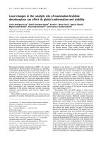
Figure 1. Materials used for In vitro propagation. A: Mentha piperita L. twig. B&C: Shoot tip and nodal explants
Research in Plant Sciences
45
Rauwolfia serpentina, Emblica officinalis, Holarrhena
antidysenterica and Enicostemma hyssopifolium [10].
2.5. Experimental Design
A completely randomized experimental design was
performed in this study. In all experiments, each treatment
had at least three replicates, and there were 20 explants
per replicate (per dish). The explants in all experiments
were sub cultured at 2-week intervals. The data pertaining
to number of multiple shoots, shoot elongation and rooting
were subjected to analysis of variance (ANOVA) test.
Mean separation was done using Duncan’s Multiple
Range Test (DMRT) (P< 0.05) and were presented as the
mean ± standard error (SE).
3. Results and Discussion
3.1. Explants Superiority
Shoot regeneration was highly influenced by the explants
type of Mentha piperita. It was observed that nodal
explants were more superior for multiple shoot regeneration
as compared to shoot tip explants (Figure 2). In comparison
between shoot tips and nodal explants for multiple shoot
regeneration, nodal explants regenerated a significantly
larger average number of shoots than that of shoot tip
explants (Table 1 and Table 2). The maximum number of
shoot regeneration 100% with 42.00 ± 4.42 shoots per
explants with an average mean length of 9.45 ± 4.13 cm
from nodal segments was recorded. Whereas, 80% frequency
of shoot regeneration was recorded from shoot tip explants
with mean number of 37.35 ± 5.82 shoot per explants with
an average mean length of 7.11 ± 4.35 cm on same
environment, where all exogenous growth regulator and
nutrient medium were equalized. Shoot buds emerged on
7th and 13th day of culture (Figure 1) from nodal and shoot
tip explants, respectively. However, shoot started proliferating
after 21 and 25 days respectively. Shoot meristems and
nodes are more potent for shoot regeneration as compared
to internodes and petiole explants was reported by Sarwar
et. al., [1]. Single node explants elicited more numbers of
multiple shoots as compared to shoot tip explants was also
reported by Ghanti et. al., [10]. The proliferation efficiency
of nodal explants from healthy plants was significantly
higher than that of shoot tip explants was also reported by
Raja and Arockiasamy [19]. Nodal explants as the best
source of multiple shoot induction have also been
suggested in case of other medicinal plants, such as
3.2. Medium Strength on Shoot and Root
Formation
The shoot tip and nodal explants were cultured on both
full and half strength of MS medium containing with BAP
and Kn at different concentrations (1.0, 2.0, 3.0, 4.0 and
5.0 mg/L) for production of multiple shoots (Table 1 and
Table 2). In both explants, shoot proliferation was highly
achieved in full strength MS medium supplemented with
BAP either Kn as compared to half strength of MS
medium. The highest frequency of shoot regeneration
100% with mean number of 42.00 ± 4.42 shoots per
explants with an average mean length of 9.45 ± 4.13 cm
was recorded on full strength MS medium in case of nodal
explants. Whereas, only 50% shoot regeneration was
recorded in case of half strength MS medium. Moreover,
in half strength MS medium, explants remained vitrified
with no proliferation of off-shoots and callus formation
was also started at the base of the shoots. The full or half
strength of MS medium without any PGR was failed to
induce rooting of regenerated shoots. There is extremely
few information that define direct organogenesis from
various explants on half strength MS medium [25,26]. But
addition of various plant growth regulators in the medium
at appropriate level may influence organogenesis from any
type of cells [1]. Sarwar et. al., [1] reported that, varying
shoot regeneration was achieved from different explants of
Mentha piperita on half strength MS media but shooting
response was not as high which may be due to use of half
strength MS medium. Paques and Boxus [27] have shown
in some species that media rich in mineral nutrients such
as MS [24] were shown to promote vitrification, while
half strength MS salts improved plant development and
provide regeneration of highest number of shoots and
induction of roots per explants was reported in Mentha
spicata [23] and Mentha piperita [1]. Using media with
lower levels of minerals or only half of the MS salts
improved carnation and cucumber plant development
[28,29]. In this investigation, high frequency of shoot
regeneration was achieved on full strength MS medium in
combination with different plant growth regulator while
half strength MS medium showed less number of shoot
proliferation.
Table 1. Effect of basal medium and BAP on shoot proliferation from shoot tip and nodal explants of Mentha piperita L.
Basal
medium
Full
strength
MS
Half
strength
MS
Conc. Of
BAP
(mg/L)
1.0
2.0
3.0
4.0
5.0
1.0
2.0
3.0
4.0
5.0
Response
(%)
30
55
80
50
45
15
20
35
25
30
Shoot tip explant
No. of
shoots/explants
(mean ±SD)
15.50 ± 9.37
26.80 ± 6.81
37.35 ± 5.82
27.35 ± 6.58
16.75 ±9.61
12.50 ± 1.35
13.25 ± 2.37
15.65 ± 5.39
11.35 ± 3.39
10.65 ± 5.19
Shoot length/explants
(cm)
(mean ±SD)
2.90 ± 0.99
6.70 ± 4.28
7.11 ± 4.35
2.85 ± 3.82
6.85 ± 6.71
3.19 ± 1.25
2.55 ± 1.82
5.15 ± 3.35
3.65 ± 1.95
3.15 ± 1.45
Response
(%)
25
35
100
70
55
20
35
50
30
25
Nodal explant
No. of
Shoot length/explants
shoots/explants
(cm)
(mean ±SD)
(mean ±SD)
13.85 ± 8.10
3.20 ± 0.63
26.40 ± 9.65
8.00 ± 4.27
42.00 ± 4.42
9.45 ± 4.13
19.35 ±12.41
3.95 ± 3.89
17.45 ±11.51
5.85 ± 3.79
15.19± 2.13
2.85 ± 3.45
19.25 ± 3.13
3.65 ± 2.45
21.65 ± 4.23
6.85 ± 5.79
16.29± 3.13
2.99 ± 2.45
13.25± 2.29
1.99 ± 1.05
**Twenty explants were used for each treatment and data (Mean ± SD) recorded three – four weeks after culture.
46
Research in Plant Sciences
Table 2. Effect of basal medium and Kn on shoot proliferation from shoot tip and nodal explants of Mentha piperita L.
Shoot tip explant
Basal
medium
Full
strength
MS
Half
strength
MS
Conc. Of
Kn
(mg/L)
Response
(%)
No. of
shoots/explants
(mean ±SD)
1.0
25
28.65 ± 6.08
Nodal explant
Shoot
length/explants
(cm)
(mean ±SD)
3.60 ± 1.89
Response
(%)
No. of shoots/explants
(mean ±SD)
Shoot length/explants
(cm)
(mean ±SD)
20
15.15 ± 8.44
2.80 ± 0.91
2.0
45
28.75 ± 4.96
3.70 ± 3.28
30
27.65 ± 7.34
5.60 ± 4.05
3.0
60
32.01 ± 4.70
3.10 ± 3.26
75
34.01 ± 4.02
6.50 ± 4.20
4.0
40
20.10 ± 8.38
3.85 ± 3.02
50
19.35 ±9.41
2.50 ± 2.68
3.55 ± 2.59
5.0
35
15.75 ±9.51
2.75 ± 2.41
55
15.35 ±6.51
1.0
20
11.51 ± 2.35
3.01 ± 2.25
15
11.15 ± 6.45
2.05 ± 3.15
2.0
35
12.55 ± 2.07
2.05 ± 1.02
25
12.15 ± 3.03
1.60 ± 1.45
3.0
40
13.65 ± 3.39
3.25 ± 2.25
50
15.69 ± 3.25
5.15 ± 4.38
4.0
30
11.05 ± 3.09
3.05 ± 2.98
25
13.25± 5.15
2.09 ± 2.15
5.0
35
11.55 ± 2.09
2.25 ± 1.25
30
11.05± 3.20
2.98 ± 1.65
**Twenty explants were used for each treatment and data (Mean ± SD) recorded three – four weeks after culture.
On the other hand, roots were developed two weeks
after the transfer of individual shoots on both full and half
strength of MS medium containing with various
concentrations of plant growth regulators. But when
individual shoots were trans-cultured in half or full
strength MS medium free from PGR, poor and few
numbers of roots were developed with low frequency.
Fadel et. al., [23] was observed that there has a significant
effect of the half strength of MS culture medium in
combination with plant growth regulators (PGR) on
root and shoot formation over the full strength of MS
medium in case of in vitro organogenesis of spearmint
(Mentha spicata L.). He reported that the maximum
number of shoots and roots induced per explants as well
as the maximum average shoot length was observed on
half-strength MS medium.
3.3. Growth Regulators Promotion on
Multiple Shoot Induction
Cytokinins, especially BAP, were reported to overcome
apical dominance, release lateral buds from dormancy,
and promote shoot formation [30]. In this investigation,
different concentrations of BAP and Kn were evaluated on
shoot initiation and further proliferation. For multiple
shoot initiation, the nodal and shoot tip explants were
inoculated on full strength and half strength MS medium
containing different concentrations of BAP and Kn in the
range of 1.0-5.0 mg/L and showed enhanced shoot
proliferation. Comparative analysis of the results on the
various cytokinins used indicated that proliferation of shoots
was more effective in most of the BAP concentrations.
BAP at its 3.0 mg/L concentration showed high frequency
and highest number of shoot proliferation in both nodal
(100%) and shoot tip (80%) explants (Table 1). Further
increase in the concentration of BAP reduced the
frequency and number of shoots in both explants. On the
other hand, when the explants were cultured on Kn based
medium only 20-75% of them responded to proliferation.
In this treatment the highest number of shoots per explants
and average shoot length were 34.01 ± 4.02 and 6.50 ±
4.20 cm for nodal explants, 32.01 ± 4.70 and 3.10 ± 3.26
cm for shoot tip explants, respectively. The percentage of
explants showing proliferation and the number of shoots
per culture increased gradually with an increase of
cytokinins concentration from 1.0 to 3.0 mg/L. When the
concentration of cytokinins increased to above
3 mg/L, shoot regeneration frequency decreased and
vitrification occurred. Similar results were also reported
in Mentha viridis [19] Prosalia corylifolia [31] and
Terminalia arjuna Roxb. [32].The results of this
experiment also indicate that 3.0 mg/L BAP was more
suitable than 3.0 mg/L Kn for shoot proliferation (Table 1).
Superior effect of BAP over Kn has been documented in
Mentha piperita itself [10]. Similar results of efficacy of
BAP over Kn were reported for the axillary proliferation
in many medicinal plants of Lamiaceae like M. spicata, M.
arvensis, and Lavandula viridis [33,34,35]. In contrast to,
superior effect of Kn over BAP has been documented in
Mentha piperita itself [11,36].
Besides this, incorporation of NAA or IAA in
combination with BAP improved bud proliferation but the
shoots remained stunted (Data not shown). After initial
proliferation of shoots on medium containing 3.0 mg/L
BAP were sub-cultured on same fresh medium in every 21
days later. On the other hand, Kn showed little response
for multiple shoot initiation as compared to BAP in both
explants. Inoculation of BAP or Kn into MS medium for
multiple shoot initiation in culture, BAP showed better
performance than Kn and the maximum number of shoot
was obtained on its 3.0 mg/L concentration. When BAP
was used in combination with Kn, a fluctuate number of
responses were observed (Data not shown). But highly
effective response was observed on medium containing
0.5 mg/L BAP + 2.0 mg/L Kn (Average number of shoots
3.41 +0.37, shoot length 7.56 + 0.32 cm).
3.4. Shoot Elongation
Separated single shoots from proliferated multiple
shoots were transferred to MS medium containing with
different concentration of GA3 in the range of 0.5-2.0mg/L
for shoot elongation. The highest shoot length (13.1cm)
with 100% frequency was recorded on medium containing
1.0 mg/L GA3. Similar results were also reported by
other workers [10,37,38,39,40]. However, shoot length
and frequency gradually decreased in other higher
concentration of GA3.
Research in Plant Sciences
Table 3. Effect of GA3 on in vitro shoot elongation of Mentha piperita L.
Plant growth regulator
GA3 (mg/L)
0.5
1.0
1.5
2.0
Response (%)
Shoot length/explant (cm)
95
100
90
85
9.49 ± 1.15
13.1 ± 0.55
9.01 ± 1.25
8.99 ± 1.15
**Twenty explants were used for each treatment and data (Mean ± SD)
recorded three – four weeks after culture.
3.5. Root Initiation and Elongation
Generally, roots were not initiated during the culture
inoculation for shoot formation and shoot proliferation in
cytokinin regime. But when individual shoots were
trans-cultured in half or full strength MS medium free
from PGR, poor and few numbers of roots were developed
47
with low frequency. Root induction was enhanced in the
in vitro regenerated well elongated shoots by culturing
them on MS medium with supplementation of different
concentrations of IBA and IAA separately in the range of
0.5-2.0 mg/L. However in the present study, the best
rooting response was observed on medium containing 1.5
mg/L IBA (Figure 2). Incorporation of 1.5 mg/L IBA in
MS medium enhanced the rate of rhizogenesis in both
frequency and number of roots.
Maximum number of roots (35.01±1.99) were produced
in 1.5 mg/L IBA and mean root length was found found to
be 5.45±1.05 cm (Table 4). Similar results were also
reported in Mentha viridis [19] Ocimum amaricannum
[41] Hybanthus enneaspermus [42] Tylophora asthmatica
[43]. Roots formed in IBA were thick, long and dark
coloured, whereas those in IAA were thin short and white
coloured.
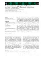
Figure 2. (A-F) – In vitro propagation of Mentha piperita. A: Initiation of multiple shoots from shoot tip explants on MS medium containing 3.0 mg/L
BAP after 12 days. B: High frequency of multiple shoot formation from shoot tip explants on MS medium containing 3.0 mg/L BAP after 25 days of
culture. C: Initiation of multiple shoots from nodal explants on MS medium containing 3.0 mg/L BAP after 6 days. D: High frequency of multiple shoot
production from nodal explants on MS medium containing 3.0 mg/L BAP after 21 days of culture. E &F: Rooting of regenerated shoots on MS medium
containing 1.5 mg/L IBA after 25 days
48
Research in Plant Sciences
Table 4. Effect of different concentrations of IBA and IAA on root induction from in vitro grown microshoots of Mentha piperita L.
Plant growth regulator (mg/L)
% of response
No. of roots/explants
Root length/explants (cm)
Days to emergence of roots
IBA
0.5
82
27.75±1.05
3.15±0.09
15-18
1.0
85
30.50±1.11
5.15±0.15
13-16
1.5
100
35.01±1.99
5.45±1.05
11-15
2.0
75
25.25±0.75
3.50±0.75
13-17
IAA
0.5
55
11.70±1.15
2.05±0.15
14-20
1.0
70
25.75±1.50
5.01±0.35
15-18
1.5
85
27.75±0.75
5.05±.031
13-15
2.0
60
13.50±.086
2.50±0.15
14-17
**Twenty explants were used for each treatment and data (Mean ± SD) recorded three – four weeks after culture.
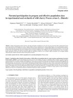
Figure 3. (A-G). A: In vitro- raised Peppermint plant after 2 weeks transplantation. B&C: In vitro grown plantlets, 4 weeks after acclimatization. D&E:
Hardened plantlets in growth chamber’s artificial environment. F&G: Hardened plantlets in ex vitro condition, showing branching-6 weeks old
Research in Plant Sciences
3.6. Hardening and Field Transfer
After 3 week, the rooted plantlets were transferred to
polycups (Figure 3) containing sterile soil and vermiculate
(1:1). These plantlets were acclimatized well and
transferred to green house and planted in the soil with
100% survivability. There was an increase in length of
shoots and new leaves emerged which expanded quickly
(Figure 3).
[11] Sunandakumari C, Martin KP, Chithra M, Sini S, Madhusoodanan
[12]
[13]
[14]
4. Conclusion
The above protocol describes high frequency shoot
propagation along with ex vitro rooting enables to provide
disease free planting propagules at low cost and within a
short time, which will attract small scale farmers to
mediculture and it can ensure a stable supply of this
medicinally important oil yielding plant and may serve as
a better source for biological active compounds.
Furthermore, in vitro propagules can be used for
interspecific hybridization and genetic transformation.
[15]
[16]
[17]
[18]
[19]
Acknowledgements
[20]
The authors are highly grateful to Plant Biotechnology
and Microbiology Laboratory, Department of Botany,
University of Rajshahi, Rajshahi-6205, Bangladesh for
providing the laboratory facilities required for conducting
this research work during the study as well as for
providing congenial environment.
References
[21]
[22]
[23]
[1]
Sarwar S, Zia M, Rehman RU, Fatima Z, Sial RA, Chaudhary MF
(2009). In vitro direct regeneration in mint from different explants
on half strength MS medium. African Journal of Biotechnology
8(18): 4667 - 4671.
[2] Foster S (1996). Peppermint: Mentha piperita. American
Botanical Council- Botantical Series 306: 3-8.
[3] Hori M (1999). Antifeeding, settling inhibitory and toxic activities
of labiate essential oils against the green peach aphid, Myzus
persicae (Sulzer) (Homoptera: Aphididae). Appl. Entomol. Zoo.
34: 113-118.
[4] Juergens UR, Stober M, Vetter H (1998). The anti-inflammatory
activity of L-menthol compared to mint oil in human monocytes in
vitro: a novel perspective for its therapeutic use in inflammatory
diseases. Eur. J. Med. Res. 3: 539-545.
[5] Ali MA, Saleem M, Ahmad W, Parvez M, Yamdagni R (2002). A
chlorinated monoterpene, ketone, acylated β-sitosterol glycosides
and a flavanone glycoside from Mentha longifolia (Lamiaceae).
Phytochem. 59: 889-895.
[6] Blumenthal M, Brusse WR, Goldberg A, Gruenwald J, Hall T,
Riggins CW, Rister RS (1998). The Complete German
Commission E Monographs. Therapeutic Guide to Herbal
Medicines. Austin, Tex: American Botanical Council: 136-138.
[7] Tucker AO (1992). The truth about mints. Herb Companion 4:
51-52.
[8] Veronese P, Li X, Niu X, Weller SC, Bressan RA, Hasegawa PM
(2001). Genetic bioengineering for mint crop improvement. Plant
Cell Tiss. Org. Cult. 64: 133-144.
[9] Jullien F, Diemer F, Colson M, Faure O (1998). An optimising
protocol for protoplast regeneration of three peppermint cultivars
(Mentha piperita). Plant Cell Tiss. And Org. Cult. 54: 153-159.
[10] Ghanti K, Kaviraj CP, Venugopal RB, Jabeen FTZ, Rao S (2004).
Rapid Regeneration of Mentha piperita L. from shoot tip and
nodal explants. Indian J Biotechnology 3:594-598.
49
[24]
[25]
[26]
[27]
[28]
[29]
[30]
[31]
[32]
[33]
PV (2004). Rapid axillary bud proliferation and ex vitro rooting of
herbal spice, Mentha piperita L. Indian J Biotechnology. 3: 108112.
Wang X, Gao Z, Wang Y, Bressan RA, Weller SC, Li X (2009).
Highly efficient in vitro adventitious shoot regeneration of
peppermint (Mentha x piperita L.) using internodal explants. In
Vitro Cellular & Developmental Biology- Plant 45:435-440.
Vasile L, Maria Z, Simona V, Eliza A (2011). Use of nodal
explants in "in vitro" micropropagation of Mentha piperita L.
Fascicula Protecţia Mediului. 16: 247-251.
Mehta J, Naruka R, Sain M, Dwivedi A, Sharma D, Mirza J
(2012). An efficient protocol for clonal micropropagation of
Mentha piperita L. (Peppermint). Asian Journal of Plant Science
and Research 2(4): 518-523.
Bolouk SG , Kazemitabar SKA, Sinaki JM (2013). In vitro Culture
of the Peppermint Plant (Mentha piperita) without the use of
Hormones. International Journal of Agriculture and Crop Sciences
6(18):1279-1283.
Van Eck JM, Kitto SL (1992). Regeneration of peppermint and
orange mint from leaf disks. Plant Cell Tiss. Org. Cult. 30: 41-46.
Sato H, Enomoto S, Oka S, Hosomi K, Ito Y (1993). Plant
regeneration from protoplasts of peppermint (Mentha piperita L.).
Plant Cell Rep. 12: 546-550.
Caissard JC, Faure O, Jullien F, Colson M, Perrin A (1996). A
direct regeneration in vitro and transient GUS expression in
Mentha piperita. Plant Cell Rep.16: 67-70.
David RH, Arockiasamy DI (2008). In vitro Propagation of
Mentha viridis L. from Nodal and Shoot tip Explants. Plant Tissue
Cult. & Biotech. 18(1): 1-6.
Senthil K, Kamraj M (2012). Direct shoot regeneration from inter
nodal explants of Mentha viridis L. International Journal of
Pharmaceutical Sciences and Research 3(4): 1101-1103.
Rahman MM, Ankhi UR, Biswas A (2013). Micropropagation of
Mentha viridis L.: An aromatic medicinal plant. International
Journal of Pharmacy & Life Sciences 4 (9): 2926-2930.
Ozdemir FA (2017). Effects of 6-benzylaminopurine and αnaphthalene acetic acid on micropropagation from ten days old
cotyledon nodes of Mentha spicata subsp. Spicat. Romanian
Biotechnological Letters 22(3):12554-12559.
Fadel D, Kintzios S, Economou SA, Moschopoulou G,
Constantinidou HA (2010). Effect of Different Strength of
Medium on Organogenesis, Phenolic Accumulation and
Antioxidant Activity of Spearmint (Mentha spicata L.) The Open
Horticulture Journal 3: 31-35.
Murashige T, Skoog F (1962). A revised medium for rapid growth
and bioassays with tobacco tissue cultures. Physiol. Plant 15:
473-497.
Nhut DN, Le BV, Tanaka M, Scientia KTT (2001). Shoot
induction and plant regeneration from receptacle tissues of Lilium
longiflorum. Horticulturae. 87: 131-138.
Martin KP (2004). Plant regeneration through somatic
embryogenesis in medicinally important Centella asiatica L. In
Vitro Cell. Dev. Biol. Plant. 40(6): 586-591.
Paques M, Boxus P (1987). A model to learn “vitrification”, the
rootstock apple M-26 present results. Acta Hortic. 212: 193-210.
Ziv M, Schwarts A, Fleminger D (1987a). Malfunctioning stomata
in vitreous leaves of carnation (Dianthus caryophyllus) plants
propagated in vitro; implications for hardening. Plant Sci. 52:
127-134.
Ziv M, Gadasi G (1986). Enhanced embryogenesis and plant
regeneration from cucumber (Cucumis sativus L.) callus by
activated charcoal in solid/liquid double-layer cultures. Plant Sci.
47:115-122.
George FF (1993). Plant propagation by tissue culture. Part 1: the
technology. The Edington Technology Exegetics Ltd.
Jebakumar M, Jayabalan M (2000). An efficient method for
regeneration of plantlets from nodal explants of Prosalea
corydifolia Linn. Plant Cell Biotec. Mol. Biol. 1(1&2): 37-40.
Varghese T, Rema Shree AB, Naheesa E, Neelakandan N,
Nandakumar S (2003). In vitro propagation of Terminalia arjuna
Roxb. multipurpose tree. Plant cell Biotech. Mol. Biol. 4 (1&2):
95-98.
Hirata T, Murakami S, Ogihara K, Suga T (1990). Volatile
monoterpenoid constituents of the plantlets of Mentha spicata
produced by shoot tip culture, Phytochemistry, 29: 493-495.
50
Research in Plant Sciences
[34] Kukreja AK, Dhawan OP, Mathur AK, Ahuja PS, Mandal S
[39] Vangadesan G, Ganapathi A, Prem Anand R, Ramesh AV (2000).
(1991). Screening and evaluation of agronomically useful
somaclonal variations in Japanese mint (Mentha arvensis),
Euphytica 53: 183-191.
Dias MC, Almeida R, Romano A (2002). Rapid clonal
multiplication of Lavandula viridis L’ Her through in vitro axillary
shoot proliferation, Plant Cell Tissue Organ Cult, 68(1):99-102.
Nadaska M, Erdelský K, Čupka P (1990). Improving the
Czechoslovakian Mentha piperita L.cv. Perpeta by in vitro
micropropagation and stabilizing the component, Biologia
(Bratislava) 45(11):955-959.
Kulkarni VM, Ganapathi TR, Suprasanna P, Bapat VA, Rao PS
(1997). In vitro propagation in Ensete superbum. A species closely
related to Musca, Indian J Exp Biol, 3:596-98.
Nirmal BK, Anu A, Ramashree AB, Praveen K (2000). Micro
propagation of curry leaf tree, Plant Cell Tissue Organ Cult.
61:199-203.
In vitro organogenesis and plant formation in Acacia sinuate,
Plant Cell Tissue Organ Cult. 61:23-28.
Xie D, Hong Y (2001). In vitro regeneration of Acacia mangium
through organogenesis, Plant Cell Tissue Organ Cult. 66:167-173.
Pathnaik SK, Chand PK (1996). In vitro propagation of the
medicinal herbs Ocimum americanum. Syn. Ocimum sims Hoary
Basil and Ocimum sanctum (Holly Basil). Plant Cell Report.15:
846-860.
Natarajan E, Arockiasamy D, John BS (1999). Regeneration of
plantlets from the callus of stem explants of Hybabanthes
enneaspermus (L.) F. Muell, Plant Tissue Cult. 9: 167-172.
Mukundan U, Sivaram U, Kumar A (2002). Micropropagation of
Tylophora ashumatica and Uararia picta. Plant Cell. Biotech. and
Mol. Biol. 3(1&2): 73-76.
[35]
[36]
[37]
[38]
[40]
[41]
[42]
[43]