A simple, efficient and universal method for the extraction of genomic DNA from bacteria, yeasts, molds and microalgae suitable for PCR-based applications
Bạn đang xem bản rút gọn của tài liệu. Xem và tải ngay bản đầy đủ của tài liệu tại đây (1.59 MB, 9 trang )
Life Sciences | Biotechnology
A simple, efficient and universal method for the extraction
of genomic DNA from bacteria, yeasts, molds
and microalgae suitable for PCR-based applications
Van Tuan Tran1,2*, Thi Binh Xuan Loc Do2, Thi Khuyen Nguyen2, Xuan Tao Vu2, Bich Ngoc Dao2, Hoai Ha Nguyen3
1
Faculty of Biology, University of Science, Vietnam National University, Hanoi
National Key Laboratory of Enzyme and Protein Technology, University of Science, Vietnam National University, Hanoi
3
Institute of Microbiology and Biotechnology, Vietnam National University, Hanoi
2
Received 2 August 2017; accepted 30 November 2017
Abstract
The extraction of genomic DNA from microbial cells plays a significant role
in PCR-based applications such as molecular diagnosis, microbial taxonomy,
screening of genetically engineered microorganisms, and other such PCRbased applications. Currently, many methods for extraction of genomic
DNA from microorganisms have been developed. However, these methods
either require hazardous chemicals or consist of time-consuming steps for
effective execution. In this study, we have established a simple and universal
genomic DNA extraction method for different microorganisms including
bacteria, yeasts, molds, and microalgae. Our method does not require harmful
reagents such as phenol and chloroform for the extraction process to minimize
the generation of hazardous wastes. The obtained genomic DNA products
displayed high concentrations and represented a good purity level with the
average 260 nm/280 nm absorbance ratios (A260/280) that range from 1.6 to
2.0. The DNA molecules further remained considerably intact when analyzed
on agarose gels. More importantly, these DNA products were qualified
through successful PCR amplifications of 16S rRNA gene, rDNA internal
transcribed spacer (ITS), or 18S rRNA gene from genomes of bacteria, fungi,
and microalgae respectively. Furthermore, with the extracted genomic DNA
products, the processes of the identification of the haploid and diploid states of
the Saccharomyces yeast strains or detection of putative strains of Aspergillus
oryzae and Aspergillus flavus that have been isolated from infected food
materials through PCR analyses are facilitated. The genomic DNA extraction
method established in this study is easy to manage, time saving and costeffective, and environmentally friendly.
Keywords: bacteria, microalgae, molds, PCR, simple genomic DNA extraction,
yeasts.
Classification number: 3.5
Introduction
Across
natural
processes,
microorganisms play important roles
in nutritional cycles that are involved
in the maintenance of the balance in
ecological systems. In the context
of applied microbiology, numerous
microbial species are utilized for the
production of foods, beverages, drugs,
biofertilizers, or for the applications
of environmental pollution treatment
[1, 2]. The accurate identification of these
microorganisms for specific purposes
is usually performed on the basis of
barcode ribosomal DNA sequences that
include bacterial 16S rRNA, fungal ITS
(internal transcribed spacer) region, and
microalgal 18S rRNA
*Corresponding author: Email:
66
Vietnam Journal of Science,
Technology and Engineering
December 2017 • Vol.59 Number 4
[3-6]. Furthermore, the selected useful
microorganisms can be subjected to
further genetic improvement to enhance
beneficial traits [1, 2, 7]. Consequently,
the development of efficient genomic
DNA extraction methods with respect
to different microbial species is always
considered a central step in PCR-based
molecular biology applications that
include molecular taxonomy, molecular
diagnosis, recombinant DNA cloning
studies, etc. These DNA extraction
methods were developed according to
either chemical reagents or commercial
kits [8-12]. However, commercial kits
employed for microbial genomic DNA
extraction are expensive for large-scale
screening experiments in laboratories,
while conventional genomic DNA
extraction methods are usually developed
for a specific microbial group or require
hazardous reagents such as phenol and
chloroform for the cleanup step [8]. In this
study, we have successfully established
a simple and universal method for the
rapid extraction of genomic DNA from
different microbial species including
bacteria, yeasts, molds, and microalgae.
The extracted genomic DNA samples
displayed superior quality and were
determined as suitable for specific PCRbased applications.
Materials and methods
Microbial strains and cultivation
conditions
All of the microbial strains and PCR
primers are listed in Table 1 and Table 2
Life Sciences | Biotechnology
Table 1. Microbial strains used in this study.
Name
Description
Source
Escherichia coli DH5α
The laboratory Gram-negative bacterial strain
Our collection
Agrobacterium tumefaciens AGL1
The laboratory Gram-negative bacterial strain employed for genetic
transformation of plants and fungi
Our collection
Burkholderia vietnamiensis LU4.4
A Gram-negative bacterial strain isolated from rice rhizosphere displaying
antifungal activity
Our collection
Lactobacillus fermentum H7
A Gram-positive lactic acid bacterial strain isolated from a fermented
pickle
Our collection
Bacillus subtilis PY79
The laboratory Gram-positive bacterial strain
Our collection
Saccharomyces cerevisiae BY4741 The laboratory haploid yeast strain (MATa)
Euroscarf
Saccharomyces cerevisiae BY4742 The laboratory haploid yeast strain (MATα)
Euroscarf
Saccharomyces cerevisiae BY4743 The laboratory diploid yeast strain (MATa/MATα)
Euroscarf
Saccharomyces boulardii NOM
A probiotic yeast strain isolated from the commercial product Normagut
(Germany)
Our collection
Saccharomyces boulardii PE
A probiotic yeast strain isolated from the commercial product Perenterol
(Germany)
Our collection
Saccharomyces boulardii BIO
A probiotic yeast strain isolated from the commercial product Bioflora
(France)
Our collection
Candida albicans JCM2070
An opportunistic yeast-causing candidasis in human
JCM, Japan
Candida glabrata RN4
A yeast strain of Candida glabrata isolated from a fermented sticky rice
product
Our collection
Pichia anomala BMH9
A yeast strain isolated from a traditional yeast cake
Our collection
Hanseniaspora thailandica Y39
A yeast strain isolated from the peel of a red apple fruit
Our collection
Aspergillus oryzae RIB40
The laboratory strain used for the research of food production
Our collection
Aspergillus flavus NRRL3357
The laboratory strain used for the research of mycotoxin biosynthesis
Our collection
Aspergillus niger N402
The laboratory strain used for the research of production of enzymes and
organic acids
Our collection
A fungal strain used for the research of penicillin production
VTCC, Vietnam
A fungal pathogen that causes the rice blast disease isolated in Southern
Vietnam
Our collection
The fungal strains isolated from mold-infected rice seeds in Hanoi
Our collection
The fungal strains isolated from mold-infected peanut seeds in Hanoi
Our collection
Chlorella sp. PT01
A freshwater microalgal strain
VTCC, Vietnam
Chlorella sp. PT02
A marine microalgal strain
VTCC, Vietnam
Penicillium chrysogenum
VTCC-F1172
Magnarporthe oryzae MN1
Aspergillus sp. A1
Aspergillus sp. A2
Aspergillus sp. A3
Aspergillus sp. A4
Aspergillus sp. A5
December 2017 • Vol.59 Number 4
Vietnam Journal of Science,
Technology and Engineering
67
Life Sciences | Biotechnology
respectively.
Four bacterial species that include
Escherichia coli, Bacillus subtilis,
Agrobacterium
tumefaciens,
and
Burkholderia vietnamiensis were grown
in the LB medium (1% peptone, 0.5%
yeast extract, 0.5% NaCl). One lactic
acid bacterium Lactobacillus fermentum
was cultivated in the MRS medium (1%
sucrose, 1% peptone, 1% yeast extract,
0.02% MgSO4.7H2O, 0.005% MnSO4,
0.5% CH3COONa, 0.2% K2HPO4, 0.2%
NaH2PO4, 0.5% CaCO3, 0.1% Tween 80,
pH 6.5).
Six yeast species that include
Saccharomyces
cerevisiae,
Saccharomyces
boulardii, Candida
albicans, Candida glabrata, Pichia
anomala, and Hanseniaspora thailandica
were cultivated in the YPG medium (1%
yeast extract, 1% peptone, 2% glucose,
1.8% agar). A single colony of each
microbial strain (Table 1) was grown
in a conical flask that contained 10 ml
of a suitable medium at 30°C, 200 rpm
until the OD600 value reached 1.5-2.0
and the respective cell biomass was then
harvested.
Five different mold species including
Aspergillus
oryzae,
Aspergillus
flavus, Aspergillus niger, Penicillium
chrysogenum, Magnaporthe oryzae,
and five putative strains of A. oryzae
and A. flavus that were isolated from
mold-infected rice seeds and moldinfected peanut seeds (Table 1) were
cultivated in the potato dextrose medium
(Himedia, India) or Czapek-Dox medium
(comprising 3% sucrose, 0.3% NaNO3,
0.1% KH2PO4, 0.05% MgSO4, 0.05%
KCl, 0.001% FeSO4) at 30oC for 3-7 days.
Two microalgal strains Chlorella sp.
PT01 and PT02 were cultivated in 100 ml
conical flasks that contained 50 ml BBM
(Bold’s Base medium) [13]. The flasks
were incubated at room temperature
under white light of 2,000 lux intensity,
subjected to a lighting cycle of 12 h/12 h
(light/dark).
Preparation of the extraction buffer
This genomic DNA extraction
protocol requires only a unique
extraction buffer referred to as the
68
Vietnam Journal of Science,
Technology and Engineering
GX buffer (2.5% SDS, 200 mM TrisHCl, 250 mM NaCl, 25 mM EDTA,
0.2% β-mercaptoethanol). The buffer
composition was adapted from certain
published reports [8, 10, 14-17]. It
is observed that the stock solutions,
including 1 M Tris-HCl (pH 8.0),
0.25 M EDTA (pH 8.0), 2.5 M NaCl,
can be autoclaved and stored at room
temperature for subsequent use. Further,
SDS (sodium dodecyl sulfate) should
be added to the buffer after the other
components. This buffer provided better
results for genomic DNA extraction
when freshly prepared. Alternatively, the
ready extraction buffer can also be stored
in the dark at room temperature for 2-3
weeks, and it requires to be heated at
60°C for 10 min before its application.
Genomic DNA extraction
The genomic DNA extraction method
was adapted from some previously
published protocols for fungi [7, 10, 14]
with suitable modifications for each
microorganism employed in this study.
For bacterial cells, the following
procedure was performed: 2 ml of each
bacterial culture with the OD600 values
of 1.5-2.0 was centrifuged at 12,000
rpm for 1 min to harvest the cells. The
cell pellet was resuspended in 70 µl TE
buffer [10 mM Tris-HCl (pH 8), 1 mM
EDTA (pH 8)] and the tube was strongly
vortexed for 15 s. Subsequently, 30 µl
of lysozyme (10 mg/ml) was added
to the tube. The resultant mixture was
incubated at room temperature for 10
min. In the subsequent step, 600 μl
of GX buffer and 3 µl proteinase K
(20 mg/ml) were added to the tube.
The tube was gently vortexed for
15 s and incubated at 60ºC for 30 min.
To achieve neutralization, 300 μl of a
3 M sodium acetate solution (pH 5.2)
was added to the tube. The supernatant
phase (600-700 μl) obtained from a
Table 2. Primers used in this study.
Name
Sequence (5’-3’)
Target
sequence
Reference
16SfD1
AGAGTTTGATCCTGGCTCAG
Bacterial 16S
Weisburg, et
16SrP1
ACGGTTACCTTGTTACGA
rRNA gene
al. (1991) [4]
ITS1
TCCGTAGGTGAACCTGCGG
Fungal rDNA
White, et al.
ITS4
TCCTCCGCTTATTGATATGC
ITS
(1990) [6]
18S1
TACCTGGTTGATCCTGCCAG
Microalgal 18S
Honda, et al.
18S12
CCTTCCGCAGGTTCACCTAC
rRNA gene
(1999) [6]
ScMAT
AGTCACATCAAGATCGTTTATGG
ScMATa
ACTCCACTTCAAGTAAGAGTTTG
ScMATα
GCACGGAATATGGGACTACTTCG
Saccharomyces
mating-type
Illuxley, et al.
genes MATa,
(1990) [18]
MATα
Specific to the
AO-ITSuni-F
ATGGCCGCCGGGGGCTCT
rDNA ITS1 of
Chiba, et al.
A. oryzae and A.
(2013) [19]
flavus
Specific to
AFB-F
AAGCAAACCAAGACCAACAAG
AFB-R
AACAAGTCTTTTCTGGGTTCTA
December 2017 • Vol.59 Number 4
aflatoxin
biosynthesis
gene cluster in A.
flavus
Chiba, et al.
(2013) [19]
Life Sciences | Biotechnology
centrifugation at 12,000 rpm, 4ºC for
20 min was transferred to a new 1.5
ml microcentrifuge tube. The genomic
DNA was precipitated with 700 μl of
cold isopropanol before it was subjected
to centrifugation at 12,000 rpm, 4ºC
for 20 min. The obtained pellet was
washed with 500 μl of 70% ethanol
and recollected by centrifugation. The
DNA pellet was subsequently dried in a
SpeedVac machine (Thermo Scientific,
USA) and dissolved in 50 μl of TE
buffer. This genomic DNA product
was treated with 3 μl of RNase A
(10 mg/ml) at 60ºC for 30 min for the
removal of RNA and stored at -20oC for
ensuing applications.
For yeasts and microalgae, the
following processes were performed:
Yeast cells were collected from 2 ml
of each culture obtained through a
centrifugation at 4,000 rpm for 5 min,
while microalgal cells were harvested
at 8,000 rpm for 15 min. To break the
cells, 600 µl of GX buffer and 150 mg
of 0.1 mm diameter glass beads (Carl
Roth, Germany) were added to the tube.
The tube was strongly vortexed for 30
s and subsequently added with 3 µl of
proteinase K (20 mg/ml). Subsequently,
the tube was incubated at a temperature
of 60°C for 30 min. The remaining
steps of the extraction procedure were
performed as those described above for
the extraction of genomic DNA from
bacteria.
For molds: 1 ml of each fungal
spore suspension (106 spores/ml)
was added to a 250 ml conical flask
containing 100 ml of potato dextrose
broth or Czapek-Dox liquid. The flask
was subjected to a shaking incubator
at 200 rpm, at a temperature of 30°C
for 3 days. Fungal mycelium was
collected by filtration through Miracloth
(Calbiochem, Germany), and 200 mg of
the obtained biomass was distributed to
a 2 ml microcentrifuge tube. The fungal
biomass was crushed directly in the
tube for 1 min using a clean glass rod.
Subsequently, 600 μl of GX buffer and
3 µl of proteinase K (20 mg/ml) were
added to the tube. The tube was vortexed
for 15 s and incubated at 60ºC for 30
min. The next steps of the extraction
procedure were performed as described
above for the extraction of genomic
DNA from bacteria.
Analysis of the extracted genomic
DNA products
The genomic DNA products
were analyzed on 0.7% agarose gels
through electrophoresis and the DNA
concentrations were measured with a
NanoDrop spectrophotometer (Thermo
Scientific, USA) for the 260/280 nm
absorbance ratios (A260/280).
Verification of genomic DNA quality
by PCR
All genomic DNA products were
diluted to the concentration of 100 ng/
µl as DNA template for PCR. Taq DNA
polymerase as GoTaq® Green MasterMix
(Promega, USA) was utilized for all
PCR amplifications in accordance
to the manufacturer’s instruction.
The universal primer pairs including
16SfD1/16SrP1 [4], ITS1/ITS4 [6], and
18S1/18S12 [5] (Table 2) were employed
for specific amplifications of bacterial
16S rRNA gene, fungal rDNA ITS, and
microalgal 18S rRNA gene, respectively.
The thermal cycling parameters were
determined as follows: 94°C (6 min);
30 cycles of 94°C (30 s), 58°C (30 s),
72°C (40 s to 1.5 min); 72°C (10 min);
4°C (∞). The obtained PCR products
were analyzed on 0.7% agarose gels and
visualized under UV light of the Gel Doc
XR System (Bio-Rad, USA).
Determination of haploid and
diploid states in Saccharomyces yeast
strains: Three strains of the baker’s
yeast S. cerevisiae, including BY4741
(haploid, MATa), BY4742 (haploid,
MATα), BY4743 (diploid, MATa/MATα),
and three strains of the commercial
probiotic S. boulardii, including NOM,
PE, BIO (Table 1) were cultivated in
the YPG liquid medium for genomic
DNA extraction. The yeast ploidy states
were determined through the PCR by
employing the primer pairs ScMAT/
ScMATa and ScMAT/ScMATα (Table
2) that are known to specifically amplify
the mating-type genes MATa and MATα
respectively [18]. The thermal cycling
parameters are as follows: 94°C (6 min);
30 cycles of 94°C (30 s), 58°C (30 s),
72°C (30 s); 72°C (10 min); 4°C (∞). Each
yeast strain was examined separately for
the genes MATa and MATα. Thereafter,
the obtained PCR products were mixed
together for a comparative analysis on a
0.7% agarose gel.
Detection of A. oryzae and A. flavus
strains by singleplex and multiplex PCR:
The genomic DNA samples extracted
from the fungal isolates including A.
oryzae RIB40, A. flavus NRRL3357,
and Aspergillus sp. (A1, A2, A3, A4,
A5) were employed for singleplex PCR
using five different primers in pairs
that include ITS1/ITS4, AO-ITS-uni-F/
ITS4, and AFB-F/AFB-R (Table 2).
The universal primer pair ITS1/ITS4 is
widely employed the amplification of the
ITS region of rDNA in fungi [6], whereas
the primer pair AO-ITS-uni-F/ITS4 was
designed for specific amplification of
the rDNA ITS in A. flavus and A. oryzae
[19]. The primer pair AFB-F/AFB-R
was designed to amplify the specific
sequence located between aflR and aflJ
of the aflatoxin biosynthesis gene cluster
in A. flavus [19]. For multiplex PCR, five
primers were applied simultaneously in a
single reaction with the thermal cycling
parameters as follows: 94°C (6 min); 30
cycles of 94°C (30 s), 60°C (30 s), 72°C
(1.5 min); 72°C (10 min); 4°C (∞). Two
standard strains A. oryzae RIB40 and A.
flavus NRRL3357 were employed as the
reference controls. The obtained PCR
products were analyzed on 1.2% agarose
gels.
December 2017 • Vol.59 Number 4
Vietnam Journal of Science,
Technology and Engineering
69
Life Sciences | Biotechnology
Results and discussions
Establishment of a universal
genomic DNA extraction method for
different microorganisms
In this study, a unique procedure has
been established for the extraction of
genomic DNA from several microbial
species including bacteria, yeasts,
molds, and microalgae. Only the first
step of the microbial biomass treatment
is specific for each cell type. For lysis of
bacterial cells, lysozyme was utilized to
break down the peptidoglycan layer of
the bacterial cell wall. Since this enzyme
works more effectively in the presence of
EDTA [20, 21], the bacterial cells in our
procedure were treated with lysozyme
in the TE (Tris-EDTA) buffer. The cells
of yeasts and microalgae were broken
mechanically in the extraction buffer
(GX buffer) with glass beads, while the
mycelia of the molds were crushed by
hand with a glass rod. The overview of
the genomic DNA extraction procedure
is illustrated in Fig. 1.
The results revealed that the
established method worked effectively
for both Gram-positive and Gramnegative bacteria, including Escherichia
coli,
Agrobacterium
tumefaciens,
Burkholderia
vietnamiensis,
Lactobacillus fermentum, and Bacillus
subtilis (Fig. 2A). To test the efficacy
of this method for yeasts, five different
yeast species including Saccharomyces
cerevisiae,
Candida
albicans,
Candida glabrata, Pichia anomala
and Hanseniaspora thailandica were
employed. Since the yeast cell wall is
easily disrupted with glass beads through
the process of vortexing [22], we added
glass beads with a diameter of 0.1 mm
and GX buffer to a 2 ml microcentrifuge
tube containing the yeast biomass, and
subsequently, the tube was vortexed
strongly to break the cells. Following the
subsequent steps for the genomic DNA
extraction (Fig. 1), the results indicated
that the genomic DNA products
extracted from the yeasts as well as from
70
Vietnam Journal of Science,
Technology and Engineering
Fig. 1. The universal procedure of genomic DNA extraction for different
microorganisms.
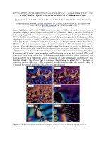
Fig. 2. Extraction of genomic DNA from bacteria, yeasts and microalgae.
(A) The genomic DNA (gDNA) products extracted from five bacteria and the
PCR products of the 16S rRNA genes on agarose gels. (B) The genomic DNA
samples extracted from five yeasts and the PCR products of the rDNA ITS on
agarose gels. (C) The analysis of the genomic DNA products extracted from
two microalgae and the respective PCR products of the 18S rRNA genes on
agarose gels.
December 2017 • Vol.59 Number 4
Life Sciences | Biotechnology
the bacteria displayed sharp bands with
lesser amounts of smearing of DNA on
agarose gels (Figs. 2A, 2B). Particularly,
these DNA products exhibited high
concentrations that ranged from 753 to
6,059 ng/μl and superior purity with the
A260/280 values ranging from 1.81 to 2.02
(Table 3). When the same procedure
as that for yeasts was applied to the
microalgal strains including Chlorella
sp. PT01 and PT02, the results revealed
that this method also worked suitably
for these green microalgae (Fig. 2C). In
comparison to the bacteria and yeasts,
the genomic DNA products extracted
from the microalgae exhibited lower
concentrations (99-177 ng/μl) with
the A260/280 values ranging from 1.57 to
1.87 (Table 3). More importantly, all
the extracted genomic DNA products
could be employed productively as the
DNA template for PCR amplifications of
the bacterial 16S rRNA gene, the yeast
rDNA ITS sequence or microalgal 18S
rRNA gene using the respective primer
pair (Fig. 2, Table 2).
For genomic DNA extraction from
molds, we crushed fungal biomass
directly in a 2 ml microcentrifuge
tube with a glass rod (Fig. 1). Five
mold species including Aspergillus
oryzae, Aspergillus flavus, Aspergillus
niger, Penicillium chrysogenum and
Magnaporthe oryzae (Fig. 3A, Table 1)
were utilized to test this procedure. The
obtained genomic DNA products were
superior in quality with the A260/280 values
ranging from 1.86 to 1.96 and high DNA
concentrations of 1,466-6,528 ng/µl
(Fig. 3B, Table 3). It is worth mentioning
that the crushing of fungal cells in the
tubes with a clean glass rod facilitates
the prevention of cross-contamination
among fungal samples and reduces the
cost when compared to the grinding of the
fungal biomass in liquid nitrogen using a
mortar and a pestle. The obtained fungal
genomic DNA products were evaluated
for quality by PCR. The universal primer
pair ITS1/ITS4 (Table 2) was utilized for
amplification of the ITS region of fungal
rDNA. The results indicated that the ITS
Table 3. The concentration and purity of the extracted genomic DNA products.
DNA concentration (ng/
µl)
A260/280
Escherichia coli DH5α
1,183 ± 203
1.81
Bacillus subtilis PY79
2,288 ± 139
1.92
Agrobacterium tumefaciens AGL1
859 ± 170
1.85
Lactobacillus fermentum H7
753 ± 208
1.90
Burkholderia vietnamiensis LU4.4
1,135 ± 52
1.92
Yeasts
Saccharomyces cerevisiae BY4743
858 ± 61
1.94
Candida albicans JCM2070
6,056 ± 55
1.98
Candida glabrata RN4
2,701 ± 239
1.84
Pichia anomala BMH9
1,762 ± 276
2.02
Hanseniaspora thailandica Y39
2,341 ± 38
1.95
Aspergillus oryzae RIB40
4,766 ± 91
1.87
Aspergillus flavus NRRL3357
2,669 ± 291
1.96
Aspergillus niger N402
6,177 ± 543
1.89
Penicillium chrysogenum VTCC-F1172
6,528 ± 711
1.86
Magnaporthe oryzae MN1
1,466 ± 104
1.90
Microalgae
Chlorella sp. PT01
177 ± 12
Chlorella sp. PT02
99 ± 23
Microbial species
Bacteria
Molds
region was successfully amplified from
the genomes of all five fungal species
(Fig. 3C).
Although the genomic DNA
extraction method established in
this study works well for numerous
microbial species, it does not always
work suitably for all microorganisms.
In fact, we tested this method for the
Gram-positive pathogenic bacterium
Staphylococcus aureus, but no DNA
bands appeared on the agarose gel
(data not shown). The reason behind
this is that the cell wall of S. aureus
1.87
1.57
is highly resistant to the digestion of
lysozyme [23]. Additionally, we tested
this method for some other fungal
species. It also worked rather well
for the citrus postharvest pathogen
Penicillium digitatum, the antagonistic
fungus Trichoderma asperellum, and
the opportunistic human pathogenic
fungus Aspergillus fumigatus. However,
this method did not prove to work
efficiently for the extraction of genomic
DNA from the model filamentous
fungus Aspergillus nidulans, the plant
pathogen Curvularia lunata, and
December 2017 • Vol.59 Number 4
Vietnam Journal of Science,
Technology and Engineering
71
Life Sciences | Biotechnology
are extremely closely related to each
other and share similar morphology
and genome homology amounting
to 99.5%, their recognition is easily
confused [2, 27].
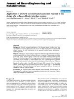
Fig. 3. Extraction of genomic DNA from different molds. (A) The morphology
of the tested molds on the PDA medium at 30°C for 3-7 days. (B) The extracted
genomic DNA (gDNA) products on a 0.7% agarose gel. (C) Analysis of the PCR
products of the ITS on a 0.7% agarose gel.
the medicinal mushroom Cordyceps
militaris, although the obtained DNA
products were still functional for
successful PCR amplifications (data not
shown). Therefore, this method requires
to be improved for certain specific
microorganisms.
Simple identification of haploid and
diploid states in Saccharomyces yeast
strains by PCR
The baker’s yeast S. cerevisiae can
exist as diploid strains that possess
MATa and MATα mating-type genes or
haploid strains that carry only MATa or
MATα gene [24]. The probiotic yeast S.
boulardii is employed commonly for
the treatment of antibiotic-associated
diarrhea caused by Clostridium difficile
infection in human. This probiotic
yeast and S. cerevisiae share almost
identical genomes [25]. In this study,
we demonstrated that the ploidy states
of three S. boulardii strains that were
isolated from the commercial probiotic
yeast products (Table 1) could be rapidly
identified through PCR amplifications.
Three standard S. cerevisiae strains,
including BY4741 (haploid, MATa),
BY4742 (haploid, MATα) and BY4743
(diploid, MATa/MATα), were adopted as
controls and three S. boulardii isolates
named NOM, PE, BIO were cultivated
in the YPG liquid medium for genomic
72
Vietnam Journal of Science,
Technology and Engineering
DNA extraction adhering to the above
established method. The genomic
DNA products extracted from all six
yeast strains displayed high quality as
indicated on an agarose gel (Fig. 4A).
The extracted DNA products were
utilized as the template for PCR with
the specific primer pairs (Table 2); and
further, the obtained data indicated that
the haploid strains BY4741 and BY4742
possess either MATa (544 bp) or MATα
(404 bp) gene respectively. Conversely,
the diploid strain BY4743 carries both
MATa and MATα genes (Fig. 4B). These
results are consistent with the results
previously reported [26]. Interestingly,
all three probiotic strains (NOM, PE,
BIO) of S. boulardii exist as diploids
that carry both the mating-type genes
MATa (544 bp) or MATα (404 bp)
like the diploid strain BY4743 of S.
cerevisiae (Fig. 4B).
Quick detection of Aspergillus
oryzae and Aspergillus flavus strains
by PCR
A. oryzae and A. flavus play
significant roles in the food industry
and food safety. A. oryzae has been
commonly employed for the industrial
production of soy sauce, miso, sake,
soybean sauce paste in Asian countries,
while A. flavus produces the carcinogenic
aflatoxins. Since these fungal species
December 2017 • Vol.59 Number 4
In this study, we cultured five
isolates Aspergillus sp. (A1, A2, A3, A4,
A5) that share similar phenotypes of A.
oryzae and A. flavus for genomic DNA
extraction. The extracted genomic DNA
products were good in quality displaying
sharp bands on the agarose gel (Fig. 4C).
With the utilization of singleplex PCR
with the universal primer pair ITS1/
ITS4, we amplified successfully the
ITS region of rDNA with the same size
of 595 bp from the genomes of all five
Aspergillus sp. isolates, as well as from
the genomes of the standard strains A.
oryzae RIB40 and A. flavus NRRL3357.
For the specific amplifications of the
ITS region from A. oryzae and A. flavus,
the primer pair AO-ITS-uni-F/ITS4
was utilized. The primer AO-ITS-uni-F
was designed to bind only to the ITS1
sequence of A. oryzae and A. flavus
[19]. The PCR with this primer pair
resulted in a DNA band of 486 bp for all
tested strains that include the reference
strains A. oryzae RIB40 and A. flavus
NRRL3357. To discriminate between
A. oryzae and A. flavus, the primer
pair AFB-F/AFB-R that specifically
binds to the aflatoxin biosynthesis gene
cluster of A. flavus was utilized [19].
With this PCR, only a DNA band of
116 bp appeared for the A. flavus strains
(Fig. 4D). From the obtained results,
we suggested that the strains A1, A2,
A4, A5 belong to A. flavus and A3 is
A. oryzae. Furthermore, these results
were additionally confirmed through the
performance of multiplex PCR in which
all five primers (ITS1, AO-ITS-uni-F,
ITS4, AFB-F, AFB-R) were combined
in a single reaction. The multiplex PCR
resulted in three bands (116 bp, 486
bp, 595 bp) for the A. flavus strains
(NRRL3357, A1, A2, A4, A5) and only
two bands (486 bp, 595 bp) for the
A. oryzae strains (RIB40, A3) on an
agarose gel (Fig. 4D).
Life Sciences | Biotechnology
Lam University, Ho Chi Minh city) for
kindly providing the required microbial
strains. We are indebted to Thi Viet Anh
Nguyen and Thi Hanh Vo (the former
members of the Genomics Unit, National
Key Laboratory of Enzyme and Protein
Technology, University of Science,
Vietnam National University, Hanoi) for
their technical assistance. This work was
funded by the National Foundation for
Science and Technology Development
of Vietnam (NAFOSTED) under grant
number 106-NN.04-2014.75.
REFERENCES
[1] B.K. Singh (2010), “Exploring microbial
diversity for biotechnology: The way forward”,
Trends Biotechnol., 28(3), pp.111-116.
[2] M. Machida, O. Yamada, K. Gomi
(2008), “Genomics of Aspergillus oryzae:
Learning from the history of Koji mold and
exploration of its future”, DNA Res., 15(4),
pp.173-183.
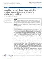
Fig. 4. Genetic identification of the closely related fungal species by PCR. (A)
Genomic DNA products extracted from three standard strains of the baker’s
yeast S. cerevisiae and three newly isolated strains of the probiotic yeast S.
boulardii. (B) Determination of haploid and diploid states in the Saccharomyces
yeasts with the utilization of singleplex PCR amplifications with the primer
pairs ScMAT/ScMATa and ScMAT/ScMATα specific to the yeast mating-type
genes MATa and MATα respectively. (C) The genomic DNA products extracted
from the molds A. oryzae RIB40, A. flavus NRRL3357, and Aspergillus sp.
(A1-A5). (D) Quick detection of putative strains of A. oryzae and A. flavus
isolated from infected food materials through singleplex and multiplex PCR
amplifications.
In summary, the genomic DNA
products obtained with our genomic
DNA extraction method are completely
suitable for PCR-based applications.
The method described in this study is
significantly uncomplicated in terms of
execution and considerably more secure,
since it does not employ toxic chemicals
such as phenol and chloroform like
other DNA extraction methods [17, 2830]. Our method further provides higher
DNA concentrations as compared to the
simple method reported by Cenis (1992)
[10], and even the DNA concentrations
are 10 times higher in comparison to
genomic DNA concentrations obtained
from the phenol-chloroform method
employed by Umesha (2016) [30].
Conclusion
The genomic DNA extraction method
established in this study is universal,
simple to handle, safe and cost-effective
for the extraction of high-quality
genomic DNA from various microbial
cell types. The obtained genomic DNA
products can be utilized for different
research purposes, especially for PCRbased applications.
ACKNOWLEDGMENTS
We are grateful to Prof. Dr. Thi Van
Anh Nguyen, Prof. Dr. Thi Viet Ha Bui,
Dr. Thi Dam Linh Mai (University of
Science, Vietnam National University,
Hanoi) and Dr. Bao Quoc Nguyen (Nong
[3] C.L. Schoch, K.A. Seifert, S. Huhndorf,
V. Robert, J.L. Spouge, C.A. Levesque, W. Chen,
Fungal Barcoding Consortium (2012), “Nuclear
ribosomal internal transcribed spacer (ITS)
region as a universal DNA barcode marker
for Fungi”, Proc. Natl. Acad. Sci., 109(16),
pp.6241-6246.
[4] W.G. Weisburg, S.M. Barns, D.A.
Pelletier, D.J. Lane (1991), “16S ribosomal
DNA amplification for phylogenetic study”, J.
Bacteriol., 173(2), pp.697-703.
[5] D. Honda, T. Yokochi, T. Nakahara, S.
Raghukumar, A. Nakagiri, K. Schaumann, T.
Higashihara (1999), “Molecular phylogeny of
Labyrinthulids and Thraustochytrids based on
the sequencing of 18S ribosomal RNA gene”, J.
Eukaryot. Microbiol., 46(6), pp.637-647.
[6] T.J. White, T. Bruns, S. Lee, J.
Taylor (1990), “Amplification and direct
sequencing of fungal ribosomal RNA genes
for phylogenetics”, PCR protocols: A Guide to
Methods and Applications, 18(1), pp.315-322.
[7] K.T. Nguyen, Q.N. Ho, T.H. Pham,
T.N. Phan, V.T. Tran (2016), “The construction
and use of versatile binary vectors carrying
pyrG auxotrophic marker and fluorescent
reporter genes for Agrobacterium-mediated
transformation of Aspergillus oryzae”, World J.
Microbiol. Biotechnol., 32(12), p.204.
[8] G.S. Mahuku (2004), “A simple
extraction method suitable for PCR-based
analysis of plant, fungal, and bacterial DNA”,
Plant Mol. Biol. Rep., 22(1), pp.71-81.
[9] M. Greco, C.A. Saez, M.T. Brown, M.B.
December 2017 • Vol.59 Number 4
Vietnam Journal of Science,
Technology and Engineering
73
Life Sciences | Biotechnology
Bitonti (2014), “A simple and effective method
for high quality co-extraction of genomic DNA
and total RNA from low biomass Ectocarpus
siliculosus, the model brown alga”, PLoS One,
9(5), pp.e96470.
[17] A. Kalia, A. Rattan, P. Chopra (1999),
“A method for extraction of high-quality
and high-quantity genomic DNA generally
applicable to pathogenic bacteria”, Anal.
Biochem., 275(1), pp.1-5.
[10] J.L. Cenis (1992), “Rapid extraction
of fungal DNA for PCR amplification”, Nucleic
Acids Res., 20(9), p.2380.
[18] A. Velegraki, M. Kambouris, A.
Kostourou, G. Chalevelakis, N. Legakis (1999),
“Rapid extraction of fungal DNA from clinical
samples for PCR amplification”, Med. Mycol.,
37(1), pp.69-73.
[11] N. Mahmoudi, G.F. Slater, R.R.
Fulthorpe (2011), “Comparison of commercial
DNA extraction kits for isolation and
purification of bacterial and eukaryotic
DNA from PAH-contaminated soils”, Can. J.
Microbiol., 57(8), pp.623-628.
[19] K. Wilson (1997), “Preparation
of genomic DNA from bacteria”, Current
Protocols in Molecular Biology, chapter 2.4.12.4.5
[12] M.K. Lee, H.S. Park, K.H. Han, S.B.
Hong, J.H. Yu (2017), “High molecular weight
genomic DNA mini-prep for filamentous
fungi”, Fungal Genet. Biol., 104, pp.1-5.
[20] S.T. Harrison (1991), “Bacterial cell
disruption: a key unit operation in the recovery
of intracellular products”, Biotechnol. Adv.,
9(2), pp.217-240.
[13] J.R. Stein (1973), Handbook of
Phycological Methods: Culture Methods and
Growth Measurements, Cambridge University
press, p.448.
[21] L. Chitarra, P. Breeuwer, R. Van Den
Bulk, T. Abee (2000), “Rapid fluorescence
assessment of intracellular pH as a viability
indicator of Clavibacter michiganensis subsp.
michiganensis”, J. Appl. Microbiol., 88(5),
pp.809-816.
[14] C. Illuxley, E.D. Green, I. Dunham
(1990), “Rapid assessment of S. cerevisiae
mating type by PCR”, Trends Genet., 6(8),
p.236.
[15] T. Chiba, Y. Takahashi, K. Sadamasu,
A. Nakama, A. Kai (2013), “Discrimination
of Aspergillus flavus group fungi using
phylogenetic tree analysis and multiplex PCR”,
Shokuhin eiseigaku zasshi. Journal of the Food
Hygienic Society of Japan, 55(3), pp.135-141.
[16] S. Aljanabi, I. Martinez (1997),
“Universal and rapid salt-extraction of
high quality genomic DNA for PCR-based
techniques”, Nucleic Acids Res., 25(22),
pp.4692-4693.
74
Vietnam Journal of Science,
Technology and Engineering
[22] C.C. Stowers, E.M. Boczko (2007),
“Reliable cell disruption in yeast”, Yeast,
24(6), pp.533-541.
[23] A. Bera, S. Herbert, A. Jakob, W.
Vollmer, F. Götz (2004), “Why are pathogenic
staphylococci so lysozyme resistant? The
peptidoglycan O-acetyltransferase OatA is the
major determinant for lysozyme resistance
of Staphylococcus aureus”, Mol. Microbiol.,
55(3), pp.778-787.
[24] J.E. Haber (2012), “Mating-type
genes and MAT switching in Saccharomyces
cerevisiae”, Genetics, 191(1), pp.33-64.
December 2017 • Vol.59 Number 4
[25] L. Edwards-Ingram, P. Gitsham,
N. Burton, G. Warhurst, I. Clarke, D. Hoyle,
S.G. Oliver, L. Stateva (2007), “Genotypic
and
physiological
characterization
of
Saccharomyces boulardii, the probiotic strain
of Saccharomyces cerevisiae”, Appl. Environ.
Microbiol, 73(8), pp.2458-2467.
[26] T. Schmidlin, M. Kaeberlein, B.A.
Kudlow, V. MacKay, D. Lockshon, B.K.
Kennedy (2008), “Single-gene deletions that
restore mating competence to diploid yeast”,
FEMS Yeast Res., 8(2), pp.276-286.
[27] C. Rank, M.L. Klejnstrup, L.M.
Petersen, S. Kildgaard, J.C. Frisvad, C. Held
Gotfredsen, T. Ostenfeld Larsen (2012),
“Comparative Chemistry of Aspergillus
oryzae (RIB40) and A. flavus (NRRL 3357)”,
Metabolites, 2(1), pp.39-56.
[28] S. De, G. Kaur, A. Roy, G. Dogra,
R. Kaushik, P. Yadav, R. Singh, T.K. Datta,
S.L. Goswami (2010), “A simple method
for the efficient isolation of genomic DNA
from Lactobacilli isolated from traditional
Indian fermented milk (dahi)”, Indian J. Med.
Microbiol., 50(4), pp.412-418.
[29] J.A. Van Burik, R. Schreckhise,
T.C. White, R. Bowden, D. Myerson (1998),
“Comparison of six extraction techniques for
isolation of DNA from filamentous fungi”, Med.
Mycol., 36(5), pp.299-303.
[30] S. Umesha, H. Manukumar, S.
Raghava (2016), “A rapid method for isolation
of genomic DNA from food-borne fungal
pathogens”, 3 Biotech., 6(2), pp.1-9.