The combination of weak expression of PRDX4 and very high MIB-1 labelling index independently predicts shorter disease free survival in stage I lung adenocarcinoma
Bạn đang xem bản rút gọn của tài liệu. Xem và tải ngay bản đầy đủ của tài liệu tại đây (2.24 MB, 10 trang )
Int. J. Med. Sci. 2018, Vol. 15
Ivyspring
International Publisher
1025
International Journal of Medical Sciences
2018; 15(10): 1025-1034. doi: 10.7150/ijms.25734
Research Paper
The Combination Of Weak Expression Of PRDX4 And
Very High MIB-1 Labelling Index Independently Predicts
Shorter Disease-free Survival In Stage I Lung
Adenocarcinoma
Akihiro Shioya1, Xin Guo1, Nozomu Motono2, Seiya Mizuguchi3, Nozomu Kurose1,3, Satoko Nakada1,3,
Akane Aikawa1,3, Yoshitaka Ikeda4, Hidetaka Uramoto2, Sohsuke Yamada1,3
1.
2.
3.
4.
Department of Pathology and Laboratory Medicine, Kanazawa Medical University, Ishikawa.
Department of Thoracic Surgery, Kanazawa Medical University, Ishikawa.
Department of Pathology, Kanazawa Medical University Hospital, Ishikawa.
Division of Molecular Cell Biology, Department of Biomolecular Sciences, Saga University Faculty of Medicine, Saga, Japan.
Corresponding author: Sohsuke Yamada, M.D., Ph.D., Department of Pathology and Laboratory Medicine, Kanazawa Medical University, 1-1 Daigaku,
Uchinada, Ishikawa, 920-0293, Japan. Tel: 81-76-218-8264; Fax: 81-76-286-1207; and E-mail:
© Ivyspring International Publisher. This is an open access article distributed under the terms of the Creative Commons Attribution (CC BY-NC) license
( See for full terms and conditions.
Received: 2018.02.26; Accepted: 2018.05.25; Published: 2018.06.14
Abstract
Background: Oxidative stress plays pivotal roles in the progression of lung adenocarcinoma (LUAD)
through cell signaling related closely to cancer growth. We previously reported that peroxiredoxin 4
(PRDX4), a secretory-type antioxidant enzyme, can protect against the development of various diseases,
including potential malignancies. Since many patients with early-stage LUAD develop recurrence, even
after curative complete resection, we investigated the association of the PRDX4 expression with the
clinicopathological features and recurrence/prognosis using post-surgical samples of stage I-LUAD.
Methods: The expression of PRDX4 and MIB-1, a widely accepted Ki67 protein, was
immunohistochemically analysed in 206 paraffin-embedded tumour specimens of patients with stage
I-LUAD. The PRDX4 expression was considered to be weak when less than 25% of the adenocarcinoma
cells showed positive staining.
Results: A weak PRDX4+ expression demonstrated a significantly close relationship with pathologically
poor differentiation, highly invasive characteristics and recurrence. The decrease in PRDX4-positivity
potentially induced cell growth in LUAD, which was correlated significantly with a very high MIB-1
labelling index (≥17.3%). Univariate/multivariate analyses revealed that the subjects with both weak
PRDX4+ expression and a very high MIB-1 index had significantly worse disease-free survival rates than
other subjects.
Conclusions: The combination of weak PRDX4 expression and a very high MIB-1 index can predict high
proliferating activity and recurrence with a potential poor prognosis, especially in post-operative stage
I-LUAD patients.
Key words: lung adenocarcinoma (LUAD); stage I; PRDX4; MIB-1; recurrence.
Introduction
Lung cancer is one of the most common fatal
malignancies in developed countries [1,2] and it has
been the number-one cause of cancer-related deaths
among Japanese for two decades. Up to 105,000 new
cases of lung cancer were diagnosed in 2013, and in
2016, more than 50,000 patients died of it in Japan
alone
( />summary.html). More than 85% of lung cancer cases
are classified as non-small cell lung cancer (NSCLC),
and lung adenocarcinoma (LUAD) is the most
well-known histopathological subtype of NSCLC in
Japan [3]. The 5-year overall survival rate is
Int. J. Med. Sci. 2018, Vol. 15
reportedly less than 20% for NSCLC, including LUAD
[4], and surprisingly, up to 30% of patients develop
recurrence within 5 years, even in cases of stage
I-LUAD after curative complete surgical resection
[5,6]. The potential cell growth of LUAD, regardless
occult metastases at the time of operation, is
suggested to be primarily responsible for its
recurrence with a subsequent poor prognosis [7].
Therefore, predicting which patients are prone to
develop recurrence after surgery is critical, even with
early-stage LUAD. Indeed, clinicopathological
elements,
such
as
the
differentiation
or
tumour-node-metastasis (TNM) stage of LUAD, can
strongly suggest the risk of recurrence and/or the
prognosis [8,9], but no molecular or genetic factors
have yet been identified, and the clinical significance
of such biological markers is still under evaluation.
Oxidative stress, induced by reactive oxygen
species (ROS), can function as a crucial and diverse
pathophysiological regulator of cellular signalling
pathways, such as the response to inflammatory and
growth factor stimulation [10]. Accumulating
evidence also suggests that the dysregulation of
oxidant and antioxidant redox signalling might cause
or accelerate a host of various human diseases,
including malignancies [11]. In this vein, the aberrant
expressions of oxidative stressors and antioxidant
properties play pivotal roles in the initiation of the
progression of LUAD through cell signalling
pathways related closely to cancer growth [12].
Peroxiredoxin 4 (PRDX4) is a member of the
PRDX antioxidant enzyme family, which consists of at
least six distinct PRDX genes, expressed in mammals
(PRDX1–6) [13]. In contrast to the merely intracellular
localization of other family members, PRDX4 is the
only secretory form, and significant levels of this
enzyme have been noted, particularly in cultured
medium [14]. According to our serial in vivo studies,
the elevated expression of PRDX4 has been
recognized in not only endoplasmic reticulum but
serum and various tissues of mice and human with
chronic inflammatory diseases, manifesting as
metabolic syndrome and potential malignancies
[15,16]. The overexpression of PRDX4 in mice can
markedly suppress the local and systemic levels of
ROS and protect various tissues against oxidative
damage by reducing the inflammatory response and
apoptosis and/or growth factor stimulation in the
intra-/extra-cellular space [17]. Furthermore, a
growing body of evidence suggests that apoptotic
and/or proliferative activities might be significantly
correlated with the PRDX4 expression [18,19].
Given the above, we hypothesize that PRDX4 not
only regulates basic cellular functions of LUAD but is
a parameter of cell growth, similar to the
1026
widely-accepted Ki67 (MIB-1) protein [20,21].
Furthermore, PRDX4 might be a promising clinical
biomarker for the recurrence/prognosis of LUAD and
be a target for early diagnoses and therapies for
LUAD. However, no studies have explored possible
associations between the PRDX4 expression,
especially in
early-stage
LUAD,
and
the
clinicopathological characteristics of a lesion,
including its differentiation and invasiveness or
patients’ recurrence/prognosis.
In the current study, using an original, specific
rabbit polyclonal PRDX4 antibody generated against
the recombinant PRDX4 protein [22], we evaluated
the expression of PRDX4 in post-surgical specimens
using stage I-LUAD patients’ clinicopathological data,
demonstrating that PRDX4 was weakly expressed in
most invasive human LUAD specimens, especially
those with poor differentiation, pleural involvement,
recurrence, and an MIB-1 labelling index exceeding
17.3% (i.e. very high proliferating activity). These
findings suggest that the combination of weak
PRDX4+ expression and a very high MIB-1 index is
significantly correlated with a poor disease-free
survival (DFS; i.e. recurrence) of stage I-LUAD.
Materials and methods
Patients and tissue specimens
Surgically resected stage I-LUAD tissues were
evaluated in the present study. Pathological reports
were reviewed to identify patients who underwent
lobectomy (170 patients), partial resection (4 patients),
or segmentectomy (32 patients) for LUAD between
January 2005 and December 2015 at the hospital of
Kanazawa Medical University. All materials in this
article were approved by the Ethical Committee of
Kanazawa Medical University (I159). Patients who
suffered perioperative deaths, defined as death
during the patient’s initial hospitalization or within 30
days of surgery, were excluded. A total of 206 patients
with available follow-up data comprised the cohort of
this retrospective study after further excluding those
with the following characteristics: (a) other prior or
concomitant malignant tumours, (b) coexisting
medical problems of sufficient severity to shorten the
life expectancy, and (c) adjuvant chemotherapies or
radiotherapies prior to the surgery.
Three pathologists examined all resected
specimens to confirm their histopathological features,
including the differentiation. Revisions in the
International System for Staging Lung Cancer was used
for the final staging [23], and all lung
adenocarcinomas were further classified based on the
histological
classification
system
from
the
International Association for the Study of Lung
Int. J. Med. Sci. 2018, Vol. 15
Cancer (IASLC)/American Thoracic Society (ATS)/
European Respiratory Society (ERS)/International
Multidisciplinary
Classification
of
Lung
Adenocarcinoma [24].
In accordance with this IASLC/ATS/ERS
classification system [24], adenocarcinoma in situ
(AIS) cases were selected using haematoxylin and
eosin (H&E)-stained sections according to the
following criteria: localized lesion (≤3 cm) with
growth of neoplastic cells along pre-existing alveolar
structures, lack of stromal invasion, absence of
papillary or micropapillary patterns, and absence of
intra-alveolar
tumour
cells.
Tumours
were
subclassified as minimally invasive adenocarcinoma
(MIA) in cases with a solitary adenocarcinoma (≤3 cm)
with a predominantly lepidic growth pattern and ≤5
mm invasion in the greatest dimension of any one
focus. The invasive component to be measured in
MIA was defined as follows: histological subtypes
other than a lepidic pattern (i.e. acinar, papillary,
micropapillary, or solid) or tumour cells infiltrating
myofibroblastic stroma. The invasive component was
measured morphometrically, and a 5-mm cut-off
value was used to distinguish MIA from
lepidic-predominant invasive adenocarcinoma (LPA).
For cases that contained multiple tumour foci, only
the largest focus was examined. Elastica van Gieson
(EVG) stains were also performed if necessary. MIA
was excluded if the tumour invaded the lymphatics,
blood vessels, pleura, or contained tumour necrosis.
LPA and non-lepidic adenocarcinomas with invasion
that were >5 mm in diameter were classified as
invasive adenocarcinoma and divided further into
acinar (APA), papillary (PPA), solid (SPA), mucinous
adenocarcinoma (MA), and micropapillary (MPA)
based on their predominant invasive pattern in H&E
sections.
Clinical information was gathered from patients’
records. The disease-free survival (DFS) and
disease-specific survival (DSS) were defined as the
interval from the date of surgery to recurrence and
from the date of surgery to death, except for patients
who died from causes other than LUAD, or the most
recent clinic visit, respectively. Patients were followed
up and prospectively evaluated every month within
the first postoperative year and at approximately twoto four-month intervals thereafter using chest X-ray,
thoracic and abdominal computed tomography (CT),
brain magnetic resonance imaging (MRI), serum
biochemistry, or measurements of tumour markers.
CT, MRI, and bone scintigraphy were performed
every six months for three years after surgery.
Additional examinations were performed if any
symptoms or signs of recurrence were recognized.
Formalin-fixed, paraffin-embedded tissue blocks
1027
came from our Department of Pathology & laboratory
medicine. EVG and immnohistochemical D2-40
(Nichirei Bioscience Co., Tokyo, Japan, diluted 1:1)
staining very clearly revealed pleural involvement
(pl) and vascular invasion (v) in the former, and
lymphatic invasion (ly) in the latter, respectively.
Preparation of antibodies against PRDX4 and
secondary antibodies, and
immunohistochemistry of tissue samples
A rabbit anti-PRDX4 IgG was produced as
previously described [22]. Immunohistochemical
staining was performed by the antibody-linked
dextran polymer method for antibody-bridge
labelling,
with
haematoxylin
counterstaining
(EnVision; Dako Cytomation, Co., Glostrup,
Denmark). Deparaffinized and rehydrated 4-µm
sections were incubated in 10% H2O2 for 5 min to
block the endogenous peroxidase activity. The
sections were then rinsed and incubated with rabbit
polyclonal anti-PRDX4 (diluted 1:1000) and mouse
monoclonal MIB-1 (Ki67; Dako Cytomation, Co.,
diluted 1:50) antibodies for 2 h and 30 min,
respectively [19,21]. The second antibody-peroxidaselinked polymers were then applied, and the sections
were incubated with a solution consisting of 20 mg of
3.3’-diaminobenzidine tetrahydrochloride, 65 mg of
sodium azide, and 20 ml of 30% H2O2 in 100 ml of
Tris-HCL (50 mM, pH7.6). After counterstaining with
Meyer’s haematoxylin, the sections were observed
under a light microscope. The sections were first
scanned at a low power for all fields (original
magnification: × 40) with tumour and non-tumour
tissues to account for the heterogeneity of
distribution. The number of cells showing positive
staining and the pattern of staining were recorded.
Necrotic tissues, stromal cells, and lymphoid cells
were not included in the recording.
The evaluation of the immunohistochemical
results by scoring
The immunoreactivity for PRDX4 in each case
was assessed semi-quantitatively by evaluating the
proportion of positive cells compared to the total
neoplastic LUAD cells. We selected and validated the
immunohistochemical cut-off scores for PRDX4
positivity (25%) and the MIB-1 labelling index (17.3%)
based on the performance of a receiver operating
characteristic (ROC) curve analysis [25]. All patients
were divided into two groups based on the PRDX4
expression as follows: strong when the PRDX4
staining was ≥25% and weak when the staining was
<25%.
All histological and immunohistochemical slides
were evaluated by two independent observers
Int. J. Med. Sci. 2018, Vol. 15
1028
(certified surgical pathologists in our department;
A.S. and N.K.) using a blind protocol design
(observers blinded to the clinicopathological data).
The agreement between the observers was excellent
(more than 90%) for all antibodies investigated, as
measured by the interclass correlation coefficient. For
the few (less than 1%) instances of disagreement, a
consensus score was determined by a third
board-certified pathologist (S.Y.) in our department
[21,26,27].
Table 1. Detailed patients'clinicopathological characteristics
Characteristic
Age (years)
Average
Median
Range
>60
≤60
Sex
Male
Female
Brinkman index (BI)
≥400
<400
Months after surgery
Average
Median
Range
Tumour differentiation
Well
Moderately
Poorly
Histopathological subtype
AIS
MIA
LPA
APA
PPA
MA
MPA
SPA
Tumour size (mm)
Average
Median
Range
CEA(μg/L)
≥5
<5
Patients (n=206)
67
68
33-83
166
40
104
102
78
128
51
49
2-145
112
78
16
19
38
52
29
49
3
3
13
23.5
22
6-50
65
141
AIS = adenocarcinoma in situ; MIA = minimally invasive adenocarcinoma; LPA =
invasive adenocarcinoma, lepidic predominant; APA = invasive adenocarcinoma,
acinar predominant; PPA = invasive adenocarcinoma, papillary predominant; SPA
= invasive adenocarcinoma, solid predominant; MA = invasive mucinous
adenocarcinoma; MPA = invasive adenocarcinoma, micropapillary predominant
Statistical analyses
The significance of correlations was determined
using Fisher’s exact test or χ2 test, where appropriate,
in order to assess the relationships between the
immunohistochemical
expression
and
the
clinicopathological features [27]. Survival curves were
plotted with the Kaplan-Meier method and compared
with the log-rank test. Hazard ratios and 95%
confidence intervals (95% CIs) were estimated using
univariate or multivariate Cox proportional hazard
models [21,26-29]. All statistical tests were two-tailed,
with values of P < 0.05 considered to be significant.
All of the above statistical analyses were
performed with the EZR (Saitama Medical Center,
Jichi Medical University, Japan) graphical user
interface for the R software program (The R
Foundation for Statistical Computing, version 2.13.0)
[27,28,30]. More precisely, it is a modified version of R
commander (version 1.6-3) that incorporates the
statistical functions frequently used in biostatistics.
Results
Patient characteristics
The clinicopathological features of the 206
patients with stage I-LUAD who were able to be
evaluated are summarized in Table 1. The range of
age at surgery was 33–83 years (average and median
were 67 and 68 years, respectively). More than half of
patients (128/206) had a Brinkman index (BI) under
400; the remaining patients (78/206) were ≥ 400 BI.
The median tumour size was 22 mm (range: 6–50
mm). The tumour grading included 112 welldifferentiated (54.4%), 78 moderately differentiated
(37.9%), and 16 poorly differentiated adenocarcinoma
(7.8%). According to further histopathological
analyses with the IASLC/ATS/ERS classification
system (Travis et al., 2011), 19 (9.2%) patients had AIS,
38 (18.4%) MIA, 52 (25.2%) LPA, 29 (14.1%) APA, 49
(23.8%) PPA, 3 (1.5%) MA, 3 (1.5%) MPA, and 13
(6.3%) SPA. Postoperative follow-up was available for
all 206 patients (average: 51 months; range: 2–145
months). The median postoperative DFS was 42
months with a 1-year recurrence rate of 2.9%, 2-year
recurrence rate of 11.2%, 5-year recurrence rate of 17%,
and total recurrence rate of 20%.
Association of the PRDX4 expression with the
clinicopathological variables and DFS
Based on the cut-off points for the PRDX4 and
MIB-1 expression, all subjects were divided into two
groups for each parameter: a weak and strong PRDX4
group and a low and high MIB-1 group (Figure 1). To
clarify the association of PRDX4 expression (weak vs.
strong PRDX4+) (Figure 2) with the clinicopathological characteristics of the cohort, the variables were
split as shown in Table 2. There were no significant
differences between the patients with weak and
strong PRDX4+ tumour expressions in terms of the
age, gender, and BI (P > 0.05). The moderately to
poorly differentiated tumour rate in the strong
PRDX4+ samples was 30/103 (29.1%), but the rate
was 65/103 (63.1%) in weak PRDX4+ samples.
Furthermore, the highly invasive (APA/PPA/MA/
MPA/SPA) adenocarcinoma rate was 30/103 (29.1%)
Int. J. Med. Sci. 2018, Vol. 15
1029
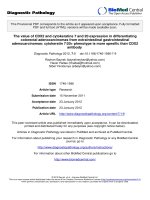
Figure 1. The results of the receiver operating characteristic (ROC) curve analyses for selecting and validating the immunohistochemical cut-off points for PRDX4 and MIB-1
expression. We selected the cut-off values of PRDX4 and MIB-1 using ROC and the area under the curve (AUC), as an effective measure of accuracy has been considered a
meaningful interpretation. We selected 25 and 17.3, respectively, as the cut-off points for PRDX4 and MIB-1, since the AUC for recurrence was the highest among all
clinicopathological variables.
in strong PRDX4+ samples but 67/103 (65%) in weak
PRDX4+ samples. Weak PRDX4 expression was
closely associated with moderate to poor
differentiation (P < 0.0001), highly invasive subtypes
(APA/PPA/MA/MPA/SPA) (P < 0.0001), and a high
(≥17.3%) MIB-1 labelling index (P = 0.0018, r = -0.172)
but not with the tumour size or presence of v and ly (P
> 0.05). PRDX4 expression was apparently detectable
in the adjacent non-neoplastic bronchioloalveolar
epithelium (Figure 2). On immunohistochemistry,
PRDX4 and MIB-1 displayed intracytoplasmic and
nuclear expression patterns, respectively (Figure 2).
Furthermore, the PRDX4 stain status was significantly
correlated with the presence of pl (P = 0.017). The rate
of PRDX4 expression in an intracytoplasmic pattern
was much lower in invasive LUAD areas, including pl
(+), than in non-invasive ones (Figure 3).
In a Kaplan–Meier analysis, lung adenocarcinoma patients with weak PRDX4+ expression had a
significantly shorter postoperative DFS than those
with strong PRDX4+ expression (P = 0.004, Figure
4A). Lung adenocarcinoma patients with weak
PRDX4+ and a high MIB-1 index had a markedly
shorter postoperative DFS than other patients (P <
0.0001, Figure 4B). However, the PRDX4 expression
was not associated with the postoperative DSS in the
present study.
The combination of weak PRDX4 expression
and a high MIB-1 labelling index represents a
significant independent prognostic indicator
for lung adenocarcinoma
Cox proportional-hazards model was created in a
forward fashion including only covariates that had
statistically significant correlations with the DFS,
using an inclusion threshold of P < 0.05 (Table 3). A
univariate analysis showed that the tumour size (> 2
cm), tumour grade, and presence of pl, ly, and v and
both weak PRDX4+ and a high MIB-1 labelling index
status, were significant predictors of a poor survival
(P = 0.021, < 0.0001, < 0.0001, = 0.0002, < 0.001, and <
0.0001, respectively). Furthermore, a multivariate
analysis showed that, after correction for confounding
variables, the combination of weak PRDX4+
expression and a high MIB-1 index remained an
independent prognostic indicator for the DFS (P =
0.013), as well as the tumour grade (P = 0.0009).
Discussion
In the present large cohort, we showed that weak
PRDX4 expression was closely correlated with
various critical clinicopathological features of 206
patients with post-surgical LUAD especially in stage
I, using a unique polyclonal antibody raised against
the distinctive, recombinant PRDX4 protein. The
current findings have indicated, for the first time, that
the combination of weak PRDX4+ expression and a
very high MIB-1 labelling index is novel and powerful
independent marker for post-operative recurrence
with a potential poor outcome in stage I-LUAD
patients.
To assess whether or not the PRDX4 expression
was an independent predictor of postoperative DFS, a
Int. J. Med. Sci. 2018, Vol. 15
1030
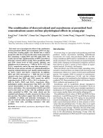
Figure 2. Representative images of immunohistochemical analyses of PRDX4 and MIB-1 in human stage I-LUAD (strong PRDX4 with low MIB-1; weak PRDX4 with high MIB-1).
Intracytoplasmic staining pattern of PRDX4 was confirmed in LUAD cells (inset). (Original magnification: ×100; inset, ×400). Bar = 200 µm (×100)
Table 2. Detailed correlations between the PRDX4 expression
and clinicopathological variables
Age
>60 years
≤60 years
Gender
Male
Female
Brinkman index (BI)
≥400
<400
Tumour differentiation
Well
Moderately
Poorly
Histopathological subtype
AIS
MIA
LPA
APA
PPA
MA
MPA
SPA
Tumour size
>2cm
Strong expression
(n=103)
Number (%)
Weak expression
(n=103)
Number (%)
P
88 (85.4)
15 (14.6)
81 (78.6)
22 (21.4)
0.276
46 (44.7)
57 (55.3)
58 (56.3)
45 (43.7)
0.125
36 (35.0)
67 (65.0)
45 (43.7)
58 (56.3)
0.254
73 (70.9)
25 (24.3)
5 (4.9)
38 (36.9)
54 (52.4)
11 (10.7)
<0.0001
13 (12.6)
28 (27.2)
32 (31.1)
6 (5.8)
18 (17.5)
1 (1.0)
2 (1.9)
3 (2.9)
6 (5.8)
10 (9.7)
20 (19.4)
23 (22.3)
31 (30.1)
2 (1.9)
1 (1.0)
10 (9.7)
<0.0001
52 (50.5)
63 (61.2)
0.161
≤2cm
pl
(+)
(-)
ly
(+)
(-)
v
(+)
(-)
MIB-1 index
≥17.3% (high)
<17.3% (low)
Recurrence
(+)
(-)
Strong expression
(n=103)
Number (%)
51 (49.5)
Weak expression
(n=103)
Number (%)
40 (38.8)
P
11 (10.7)
92 (89.3)
25 (24.3)
78 (75.7)
0.017
32 (31.1)
71 (68.9)
37 (35.9)
66 (64.1)
0.555
25 (24.3)
78 (75.7)
37 (35.9)
66 (64.1)
0.095
18 (17.5)
85 (82.5)
39 (37.9)
64 (62.1)
0.0018
9 (8.7)
94 (91.3)
31 (29.2)
72 (70.8)
0.0002
AIS = adenocarcinoma in situ; MIA = minimally invasive adenocarcinoma; LPA =
invasive adenocarcinoma, lepidic predominant; APA = invasive adenocarcinoma,
acinar predominant; PPA = invasive adenocarcinoma, papillary predominant; SPA
= invasive adenocarcinoma, solid predominant; MA = invasive mucinous
adenocarcinoma; MPA = invasive adenocarcinoma, micropapillary predominant;
pl = pleural involvement; ly = lymphatic invasion; v = vascular invasion.
Int. J. Med. Sci. 2018, Vol. 15
1031
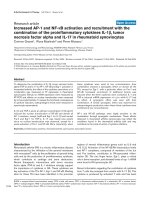
Figure 3. Representative pictures for H&E, elastica van Gieson (EVG) and immunohistochemical analyses of PRDX4 in stage I-LUAD tissue with pleural involvement (pl). EVG
staining very clearly reveals elastic fibres of the visceral pleura (pl(+)). An intracytoplasmic staining pattern of PRDX4 was confirmed in LUAD cells (inset). (Original magnification:
Bar = 2 mm (×12.5) or 200 µm (×100); inset, ×400).
Figure 4. Kaplan–Meier curves of the disease-free survival (DFS) in patients with lung adenocarcinoma after surgery according to the PRDX4 expression. Weak PRDX4
expression alone as well as weak PRDX4/high MIB-1 is associated with a significantly shorter postsurgical DFS in stage I-LUAD patients.
Int. J. Med. Sci. 2018, Vol. 15
1032
Table 3. Univariate and multivariate analyses of the survival in 206 patients with stage I-LUAD, according to the clinicopathological
variables and a low PRDX4 expression and high MIB-1 labelling index
Weak PRDX4+high MIB-1
Tumour size
Differentiation
pl (+)
ly (+)
v (+)
Univariate
Hazard ratio
6
2.33
14.22
4.6
3.77
3.99
95% CI
3.16-11.39
1.14-4.78
5.05-40.01
2.43-8.73
1.88-7.59
2.02-7.87
Recurrence in LUAD patients after curative
surgery remains a significant problem and can
significantly affect the clinical course and survival of
these patients [6,31]. Accumulated data suggest that
weak PRDX4+ expression in stage I-LUAD is closely
associated with pathological poorly differentiated
characteristics and further invasive/aggressive
behaviours, including pleural involvement or
recurrence; furthermore, lesions with a weak PRDX4+
expression often co-express a very high MIB-1
labelling index (≥ 17.3%), resulting in potential cell
growth (i.e. high proliferating activity) of even
early-stage LUAD. We were able to prove a critical,
key specific antioxidant molecule, PRDX4, which
should
be
poorly
differentiated,
invasive/
proliferative and recurrent tumour markers or
therapeutic targets especially for stage I-LUAD.
However, some limitations associated with the
present study warrant mention. First, this is a
cohort-based, retrospective study at a single
institution, even though we conducted thorough
control
through
the random selection of
post-operative stage I-LUAD patients and adherence
to strict exclusion criteria. Second, we only conducted
immunohistochemical and not detailed molecular
analyses. Further in-depth follow-up in much larger
cohorts of stage I-LUAD patients, along with detailed
molecular investigations using LUAD cell culture
lines, will be required to confirm the intriguing
correlation of weak PRDX4+ expression and very high
Ki67 expression with recurrence and a subsequent
poor survival in post-surgical stage I-LUAD patients.
The mechanism underlying how PRDX4 is involved
in cellular signalling pathways, including the
response to growth factor stimulation, should also be
examined in a future research article. However,
despite these limitations, the PRDX4 and/or Ki67
expression patterns in both post-/pre-operative tissue
and serum samples of LUAD may allow for improved
patient selection of candidates for adjuvant/
neoadjuvant systemic therapy as well as the early
prediction of the clinical post-operative course. In
addition, since secretory-type PRDX4 can appear in
body fluids, it might be a quantitative soluble,
tumour-specific marker for LUAD.
P-value
<0.0001
0.021
<0.0001
<0.0001
0.0002
<0.0001
Multivariate
Hazard ratio
2.56
2.14
13.1
1.45
1.32
0.89
95% CI
1.21-5.42
0.94-4.89
2.88-59.67
0.70-2.99
0.61-2.89
0.39-1.99
P-value
0.013
0.068
0.0009
0.316
0.478
0.769
We suspected that PRDX4 might have a
significant function of inhibiting ROS-related
carcinogenesis of LUAD as a tumour suppressor
through oxidant and antioxidant redox signalling
pathways associated with cancer growth. Some of our
present findings are in line with those of previous
studies of several other human malignancies. For
example, acute promyelocytic leukemia showed
significantly reduced PRDX4 expression along with
the control of granulocyte colony-stimulating factor
(i.e. growth factor) responses [32]. Furthermore, our
unpublished data suggest that human hepatocellular
carcinoma specimens with a low expression of PRDX4
tend to have a highly malignant phenotype with a
poor overall survival. However, other findings of ours
disagree with those of other groups with regard to
PRDX4 immunohistochemistry in squamous cell
carcinomas (SCCs) [33,34]. These authors found that
patients with an increased PRDX4 expression, which
was closely associated with greater progressive
activity, had a significantly shorter post-operative
DSS in cases of oral cavity SCC [33] and DFS in cases
of early-stage lung SCC [34] than those with a low
expression. These discrepancies may be due in part to
not only the heterogeneity of malignancies but also
the methodology of assessment in each study, such as
the size of the cohort, differences in the antibodies
used against each PRDX4, and the arbitrary or strict
selection and validation of the immunohistochemical
cut-off scores for PRDX4, which were occasionally not
based on any ROC curve analyses. Further
experiments are necessary to address methodology
standardization for PRDX4 in clinical specimens after
collecting and investigating a much larger number of
surgical cases.
Conclusion
Our observations suggest that weak PRDX4+
expression in primary stage I-LUAD is very closely
related to pathological phenotypes with a poor
outcome, e.g. those with poor differentiation, highly
invasive characteristics and recurrence, or a very high
MIB-1 labelling index, reflecting a background of
marked cancer cell growth/proliferation. Furthermore, the DFS of LUAD patients with both weak
PRDX4+ and a very high MIB-1 index was
Int. J. Med. Sci. 2018, Vol. 15
significantly shorter than that of other patients. These
analyses suggest for the first time that the
combination of weak PRDX4 and high MIB-1 may be
a novel and useful independent predictor of
recurrence with a poor prognosis in patients with
primary stage I-LUAD.
Abbreviations
LUAD,
lung
adenocarcinoma;
PRDX4,
peroxiredoxin 4; NSCLC, non-small cell lung cancer;
TNM, tumour-node-metastasis; ROS, reactive oxygen
species; EVG, Elastica van Gieson; AIS, adenocarcinoma in situ; MIA, minimally invasive
adenocarcinoma; LPA, invasive adenocarcinoma,
lepidic predominant; APA, invasive adenocarcinoma,
acinar predominant; PPA, invasive adenocarcinoma,
papillary predominant; SPA, invasive adenocarcinoma, solid predominant; MA, invasive mucinous
adenocarcinoma; MPA, invasive adenocarcinoma,
micropapillary predominant; DFS, disease-free
survival; DSS, disease-specific survival; ROC, receiver
operating characteristic and SCC, squamous cell
carcinoma.
1033
Science Foundation of Hebei Province (No.
H2016206170) (to X.G.), and High level talent support
project of Hebei Province (No. CG2015003011) (to
X.G.).
Competing Interests
The authors have declared that no competing
interest exists.
References
1.
2.
3.
4.
5.
6.
7.
8.
Declarations
9.
Ethics approval
10.
All materials including consent to participate in
this article were approved by the Ethical Committee
of Kanazawa Medical University (I159).
11.
12.
Consent for publication
13.
Written informed consent was obtained from the
patient the patient and his family on admission for the
publication of this case report and any accompanying
images.
14.
Availability of data and materials
The dataset supporting the findings and
conclusions of this research is included within the
article.
Acknowledgments
We would like to thank Yuka Hiramatsu, Mariko
Nakano and Manabu Yamashita for their expert
technical assistance.
Funding
This work was supported in part by
Grants-in-Aid for Scientific Research 16K08750 to S.Y.
and 25462202 to H.U.) from the Ministry of Education,
Culture, Sports, Science and Technology, Tokyo,
Japan; a grant from the MSD Life Science Foundation,
Public Interest Incorporated Foundation, Japan (to
S.Y.); and grants from National Natural Science
Foundation of China (No. 81402490) (to X.G.), Natural
15.
16.
17.
18.
19.
20.
21.
22.
Mitsudomi T, Suda K, Yatabe Y. Surgery for NSCLC in the era of personalized
medicine. Nat Rev Clin Oncol. 2013; 10: 235–244.
Uramoto H. Current Topics on Salvage Thoracic Surgery in Patients with
Primary Lung Cancer. Ann Thorac Cardiovasc Surg. 2016; 22: 65–68.
Lemjabbar-Alaoui H, Hassan OU, Yang YW, Buchanan P. Lung cancer:
Biology and treatment options. Biochim Biophys Acta. 2015; 1856: 189-210.
Jemal A, Siegel R, Xu J and Ward E. Cancer statistics, 2010. CA Cancer J Clin.
2010; 60: 277-300.
Asamura H, Goya T, Koshiishi Y, Sohara Y, Eguchi K, Mori M, Nakanishi Y,
Tsuchiya R, Shimokata K, Inoue H, Nukiwa T, Miyaoka E; Japanese Joint
Committee of Lung Cancer Registry. A Japanese Lung Cancer Registry study:
prognosis of 13,010 resected lung cancers. J Thorac Oncol. 2008; 3: 46–52.
Uramoto H, Yamada S, Tanaka F. Angiogenesis of lung cancer utilizes existing
blood vessels rather than developing new vessels using signals from
carcinogenesis. Anticancer Res. 2013; 33: 1913–1916.
Yamashita T, Uramoto H, Onitsuka T, Ono K, Baba T, So T, So T, Takenoyama
M, Hanagiri T, Oyama T, Yasumoto K. Association between
lymphangiogenesis-/micrometastasis- and adhesion-related molecules in
resected stage I NSCLC. Lung Cancer. 2010; 70: 320–328.
Spiro SG, Silvestri GA. One hundred years of lung cancer. Am J Respir Crit
Care Med. 2005; 172: 523–529.
Ou SH, Zell JA. Validation study of the proposed IASLC staging revisions of
the T4 and M non-small cell lung cancer descriptors using data from 23,583
patients in the California Cancer Registry. J Thorac Oncol. 2008; 3: 216–227.
Finkel T. Signal transduction by reactive oxygen species. J Cell Biol. 2011; 194:
7–15.
Fruehauf JP, Meyskens FL Jr. Reactive oxygen species: a breath of life or
death? Clin Cancer Res. 2007; 13:789-94.
Okumura N, Yoshida H, Kitagishi Y, Nishimura Y, Iseki S, Matsuda S. Against
Lung Cancer Cells: To Be, or Not to Be, That Is the Problem. Lung Cancer Int.
2012: 659365.
Fujii J, Ikeda Y, Kurahashi T, Homma T. Physiological and pathological views
of peroxiredoxin 4. Free Radic Biol Med. 2015; 83: 373–379.
Okado-Matsumoto A, Matsumoto A, Fujii J, Taniguchi N. Peroxiredoxin IV is
a secretable protein with heparin-binding properties under reduced
conditions. J Biochem. 2000; 127:493-501.
Guo X, Yamada S, Tanimoto A, Ding Y, Wang KY, Shimajiri S, Murata Y,
Kimura S, Tasaki T, Nabeshima A, Watanabe T, Kohno K, Sasaguri Y.
Overexpression of peroxiredoxin 4 attenuates atherosclerosis in
apolipoprotein E knockout mice. Antioxid Redox Signal. 2012; 17: 1362–1375.
Nabeshima A, Yamada S, Guo X, Tanimoto A, Wang KY, Shimajiri S, Kimura
S, Tasaki T, Noguchi H, Kitada S, Watanabe T, Fujii J, Kohno K, Sasaguri Y.
Peroxiredoxin 4 protects against nonalcoholic steatohepatitis and type
2diabetes in a nongenetic mouse model. Antioxid Redox Signal. 2013; 19:
1983–1998.
Yamada S, Ding Y, Sasaguri Y. Peroxiredoxin 4: Critical roles in inflammatory
diseases. J UOEH. 2012; 34: 27–39.
Ding Y, Yamada S, Wang KY, Shimajiri S, Guo X, Tanimoto A, Murata Y,
Kitajima S, Watanabe T, Izumi H, Kohno K, Sasaguri Y. Overexpression of
peroxiredoxin 4 protects against high-dose streptozotocin-induced diabetes by
suppressing oxidative stress and cytokines in transgenic mice. Antioxid Redox
Signal. 2010; 13: 1477–1490.
Nawata A, Noguchi H, Mazaki Y, Kurahashi T, Izumi H, Wang KY, Guo X,
Uramoto H, Kohno K, Taniguchi H, Tanaka Y, Fujii J, Sasaguri Y, Tanimoto A,
Nakayama T, Yamada S. Overexpression of peroxiredoxin 4 affects intestinal
function in a dietary mouse model of nonalcoholic fatty liver disease. PLoS
One. 2016; 11: e0152549.
Burger PC, Shibata T, Kleihues P. The use of the monoclonal antibody Ki-67 in
the identification of proliferating cells: application to surgical neuropathology.
Am J Surg Pathol. 1986; 10: 611–617.
Kawatsu Y, Kitada S, Uramoto H, Li Z, Takeda T, Kimura T, Horie S, Tanaka F,
Sasaguri Y, Izumi H, Kohno K, Yamada S. The combination of strong
expression of ZNF143 and high MIB-1 labelling index independently predicts
shorter disease-specific survival in lung adenocarcinoma. Br J Cancer. 2014;
110: 2583–2592.
Ito R, Takahashi M, Ihara H, Tsukamoto H, Fujii J, Ikeda Y. Measurement of
peroxiredoxin-4 serum levels in rat tissue and its use as a potential marker for
hepatic disease. Mol Med Rep. 2012; 6: 379–384.
Int. J. Med. Sci. 2018, Vol. 15
1034
23. Vallières E, Shepherd FA, Crowley J, Van Houtte P, Postmus PE, Carney D,
Chansky K, Shaikh Z, Goldstraw P. International Association for the Study of
Lung Cancer International Staging Committee and Participating Institutions.
The IASLC Lung Cancer Staging Project: proposals regarding the relevance of
TNM in the pathologic staging of small cell lung cancer in the forthcoming
(seventh) edition of the TNM classification for lung cancer. J Thorac Oncol.
2009; 4: 1049–1059.
24. Travis WD, Brambilla E, Noguchi M, Nicholson AG, Geisinger KR, Yatabe Y,
Beer DG, Powell CA, Riely GJ, Van Schil PE, Garg K, Austin JH, Asamura H,
Rusch VW, Hirsch FR, Scagliotti G, Mitsudomi T, Huber RM, Ishikawa Y, Jett
J, Sanchez-Cespedes M, Sculier JP, Takahashi T, Tsuboi M, Vansteenkiste J,
Wistuba I, Yang PC, Aberle D, Brambilla C, Flieder D, Franklin W, Gazdar A,
Gould M, Hasleton P, Henderson D, Johnson B, Johnson D, Kerr K, Kuriyama
K, Lee JS, Miller VA, Petersen I, Roggli V, Rosell R, Saijo N, Thunnissen E,
Tsao M, Yankelewitz D. International association for the study of lung
cancer/american thoracic society/european respiratory society international
multidisciplinary classification of lung adenocarcinoma. J Thorac Oncol. 2011;
6: 244–285.
25. Hanley JA. Receiver operating characteristic (ROC) methodology: the state of
the art. Crit Rev Diagn Imaging. 1989; 29: 307–335.
26. Harada Y., Izumi H., Noguchi H., Kuma A., Kawatsu Y., Kimura T., Kitada S.,
Uramoto H., Wang K.Y., Sasaguri Y., Hijioka H., Miyawaki A., Oya R.,
Nakayama T., Kohno K. and Yamada S. Strong expression of polypeptide
N-acetylgalactosaminyltransferase 3 independently predicts shortened
disease-free survival in patients with early stage oral squamous cell
carcinoma. Tumour Biol. 2016; 37: 1357–1368.
27. Hiraki T., Yamada S., Higashi M., Hatanaka K., Yokoyama S., Kitazono I.,
Goto Y., Kirishima M., Batra S.K., Yonezawa S. and Tanimoto A.
Immunohistochemical expression of mucin antigens in gallbladder
adenocarcinoma: MUC1-positive and MUC2-negative expression is associated
with vessel invasion and shortened survival. Histol. Histopathol. 2017; 32:
585–596.
28. Kitada S, Yamada S, Kuma A, Ouchi S, Tasaki T, Nabeshima A, Noguchi H,
Wang KY, Shimajiri S, Nakano R, Izumi H, Kohno K, Matsumoto T, Sasaguri
Y. Polypeptide N-acetylgalactosaminyl transferase 3 independently predicts
high-grade tumours and poor prognosis in patients with renal cell carcinomas.
Br J Cancer. 2013; 109: 472–481.
29. Honjo K, Hiraki T, Higashi M, Noguchi H, Nomoto M, Yoshimura T, Batra SK,
Yonezawa S, Semba I, Nakamura N, Tanimoto A, Yamada S.
Immunohistochemical expression profiles of mucin antigens in salivary gland
mucoepidermoid carcinoma: MUC4- and MUC6-negative expression predicts
a shortened survival in the early postoperative phase. Histol Hitopathol. 2018;
33: 201–213.
30. Kanda Y. Investigation of the freely available easy-to-use software 'EZR' for
medical statistics. Bone Marrow Transplant. 2013; 48: 452–458.
31. Motono N, Matsui T, Machida Y, Usuda K, Uramoto H. Prognostic
significance of histologic subtype in pStage I lung adenocarcinoma. Med
Oncol. 2017; 34:100.
32. Palande KK, Beekman R, van der Meeren LE, Beverloo HB, Valk PJ, Touw IP.
The antioxidant protein peroxiredoxin 4 is epigenetically down regulated in
acute promyelocytic leukemia. PLoS One. 2011; 6: e16340.
33. Chang KP, Yu JS, Chien KY, Lee CW, Liang Y, Liao CT, Yen TC, Lee LY,
Huang LL, Liu SC, Chang YS, Chi LM. Identification of PRDX4 and P4HA2 as
metastasis-associated proteins in oral cavity squamous cell carcinoma by
comparative tissue proteomics of microdissected specimens using iTRAQ
technology. J Proteome Res. 2011; 10: 4935–4947.
34. Hwang JA, Song JS, Yu DY, Kim HR, Park HJ, Park YS, Kim WS, Choi CM.
Peroxiredoxin 4 as an independent prognostic marker for survival in patients
with early-stage lung squamous cell carcinoma. Int J Clin Exp Pathol. 2015; 8:
6627–6635.