Impulse control disorder and response-inhibition alterations in Parkinson’s disease. A rare case of totally absent functionality of the medial-prefrontal cortex and review of literature
Bạn đang xem bản rút gọn của tài liệu. Xem và tải ngay bản đầy đủ của tài liệu tại đây (710.21 KB, 4 trang )
Journal of Advanced Research 8 (2017) 713–716
Contents lists available at ScienceDirect
Journal of Advanced Research
journal homepage: www.elsevier.com/locate/jare
Case Report
Impulse control disorder and response-inhibition alterations in
Parkinson’s disease. A rare case of totally absent functionality of the
medial-prefrontal cortex and review of literature
Sara Palermo a, Rosalba Morese b,c,⇑, Maurizio Zibetti a, Francesca Dematteis a, Stefano Sirgiovanni d,
Mario Stanziano d, Maria Consuelo Valentini d, Leonardo Lopiano a
a
Department of Neuroscience, University of Turin, Via Cherasco 15, 10126 Turin, Italy
Department of Psychology, University of Turin, Via Verdi 10, 10123 Turin, Italy
c
Faculty of Communication Sciences, Università della Svizzera Italiana, Via Buffi 13, CH-6904, Lugano, Switzerland
d
Azienda Ospedaliera Universitaria ‘‘Città della Salute e della Scienza di Torino”, Department of Neuroradiology, Corso Bramante 88/90, 10126 Turin, Italy
b
a r t i c l e
i n f o
Article history:
Received 3 April 2017
Revised 20 September 2017
Accepted 21 September 2017
Available online 21 September 2017
Keywords:
Parkinson’s disease
ICD
Response-inhibition
Action monitoring
fMRI
ACC
a b s t r a c t
This report illustrates a Parkinson’s disease (PD) patient with impulse-control disorder (ICD) and selective
impairment in response-inhibition abilities as revealed by the performance in a functional magnetic resonance imaging (fMRI) anterior cingulate cortex - sensitive GO-NOGO task. In line with hypothesis on the
role of response-inhibition disabilities in the arising of impulsivity in PD, the patient completely failed
the GO-NOGO task. Moreover, fMRI acquisition revealed absent task-sensitive activity in the anterior cingulate cortex, medial prefrontal, and orbitofrontal cortices for the contrast NOGO versus GO, which signifying
that a hypo-function of this network could be associated with ICD. A fronto-striatal and cingulo-frontal
dysfunction may reflect impairment in metacognitive-executive abilities (such as response-inhibition,
action monitoring, and error awareness) and promote compulsive repetition of behavior. Responseinhibition tasks may be useful in PD post-diagnostic phase, to better identify individuals at risk of developing ICD with dopaminergic medication.
Ó 2017 Production and hosting by Elsevier B.V. on behalf of Cairo University. This is an open access article
under the CC BY-NC-ND license ( />
Introduction
Parkinson’s disease (PD) is the second most prevalent neurodegenerative disease in the world. PD is characterized by resting tremor, bradykinesia, rigidity, postural instability. Secondary motor
symptoms could be freezing of gait, micrographia, mask-like
expression and unwanted accelerations. PD can also be associated
with neurobehavioral disorders, cognitive impairment, and autonomic dysfunctions. Functional changes in basal ganglia circuitry
are responsible for the major clinical features of PD. Core structures
of the basal ganglia are the striatum and globus pallidus, which
have close functional associations with the subthalamic nucleus,
substantia nigra and ventral thalamic nuclei. Indeed, a frontostriatal network disruption affects motor and non-motor dysfunctions in PD [1]. The loss of dopaminergic neurons impacts on the
functioning of four fronto-striatal circuits involved in different
motor, cognitive, affective, and motivational aspects of behavior:
Peer review under responsibility of Cairo University.
⇑ Corresponding author.
E-mail address: (R. Morese).
the supplementary motor area, the dorsolateral prefrontal, the
orbitofrontal, and the anterior cingulate loops. Each of them arises
from a specific region of the frontal cortex and innervates different
levels of the striatum before being relayed back to its cortical origin, via the thalamus [2]. The subthalamic nucleus (STN) has been
regarded as a significant modulator of basal ganglia output and it
has been studied because of its dual role in movement and in
non-motor behaviors. In particular, the STN has been implicated
in impulse control and related construct of valence processing
[3]. STN transmits to two fronto-striatal circuits of particular interest with regard to non-motor symptomatology in PD: (i) the
orbito-frontal cortex (OFC), associated with decision-making,
impulse control, mood expression and perseveration; (ii) the anterior cingulate cortex (ACC), associated with conflict monitoring,
intention, response initiation/inhibition [1]. Dopamine replacement treatment and dopamine-agonists have been implicated in
impulse-control disorder (ICD) development, since they can induce
alterations in those fronto-striatal networks that manage reward
and mediate impulse monitoring and control [4]. Indeed, tonic
stimulation of dopamine receptors may damage inhibitory control
mechanisms and reward processing, while promoting compulsive
repetition of behavior [4]. Voon et al. pointed out an enriched
/>2090-1232/Ó 2017 Production and hosting by Elsevier B.V. on behalf of Cairo University.
This is an open access article under the CC BY-NC-ND license ( />
714
S. Palermo et al. / Journal of Advanced Research 8 (2017) 713–716
bottom-up ventral-striatal dopamine release to incentive cues,
gambling tasks and reward prediction, and possible inhibition of
top-down orbito-frontal influences [5]. Indeed, dopamine
agonist-related ventral-striatal hypo-functionality seems to be
consistent with impaired risk evaluation [5]. An inability to resist
an impropriate drive - usually of a hedonistic nature - and the consequent repetition of behaviors characterize ICD [6]. Pathological
gambling; punding; hedonistic homeostatic behavioral disorder;
hypersexuality; compulsive shopping; and binge eating are typical
manifestations of ICD in PD [6,7]. The incidence of ICD in PD is as
high as 40% of patients on dopamine agonist therapy and approximately 15% of patients overall [6]. An Italian non-interventional,
prospective study on more than 1000 patients (ICARUS) has previously demonstrated that prevalence of ICD behaviors was relatively stable across the 2-year observational period (point
prevalence: 28.6% at baseline, 29.3% at year 1, 26.5% at year 2)
[7]. In this study, the most prevalent ICD subtypes were in line with
literature. Moreover, authors have found that ICD-positive patients
had more severe depression, poorer sleep quality and reduced
quality of life [7]. Several risk factors are considered: younger
age at onset, male sex, single status, a family/personal history of
addictive behaviors, dopamine agonist medication in combination
with levodopa treatment, high doses of dopaminergic medication,
longer disease duration, long duration of pharmacological treatments, and a personality profile characterized by impulsiveness
[6,7]. ICD appears to have some clinical overlap with compulsive
behaviors (such as the compulsion for repetitive actions and the
inability to inhibit intrusive thoughts) [8]. Moreover, ICD seems
to share several features with drug addiction: (i) repetitive engagement in a behavior despite adverse consequences; (ii) diminished
control over it; (iii) an appetitive urge/craving state prior to
engagement; and (iv) a hedonic feeling experienced during the
performance of the problematic behavior. All these features have
led to a description of ICD as behavioral addiction [9]. The authors
therefore hypothesized that this kind of ‘‘impulsivity” could be
related with ‘‘disinhibition of prepotent responses”.
The neuropsychological approach considers two measurable
functions from which ICD can be detected: (i) integration of
reward/punishment contingencies in individual choices, whose
neural substrate is located in the orbito-prefrontal cortex; and
(ii) response-inhibition, whose neural substrate is located in the
inferior portion of the prefrontal cortex. impulsivity often develops
from disturbed inhibitory control, a function mainly regulated by
c-Aminobutyric acid (GABA) levels in the anterior cingulate cortex
(ACC) and the fronto-striatal system [10,11]. Interestingly, Li and
colleagues identified the rostral cingulate as the area underlying
poor performance in a response-inhibition task in cocaineaddicted subjects, with greater impulsivity correlating with ACC
hypo-functionality [12]. Considering the above, the primary aims
of this case report were: (i) to quantify psychometric (trait) and
behavioral impulsivity in a PD patient with ICD; (ii) to evaluate
the association between impulsivity and both response-inhibition
and neural correlates of impulsivity measures. At the time of
patient’s examination, the authors hypothesized to obtain findings
similar to the one proposed by Li et al. [10].
Neurological assessment was performed using the Movement
Disorder Society - Unified Parkinson’s Disease Rating Scale (MDSUPDRS). Motor features and disease severity were evaluated in
On-/Off- conditions and scored using MDS-UPDRS part III and
UPDRS total scores, respectively. Hoen and Yahr’s (H-Y) was used
to stage the disease. Neurological examination was negative except
for bilateral bradykinesia and tremor of the upper limbs (MDSUPDRS-III ON = 33; H-Y = 2).
The neuropsychological assessment was performed in the beston phase, immediately after the neurological examination and the
approval by the treating neurologist. The evaluation was based on
the guidelines of the Task Force commissioned by the Movement
Disorder Society to identify Mild Cognitive Impairment. These criteria provide an operational scheme based on two levels of assessment of the cognitive profile differing in their methods of
evaluation and diagnostic certainty. Specifically, for the case
described here, the first level of evaluation was applied. The assessment included the Mini-Mental State Examination (MMSE) and the
Addenbrooke’s Cognitive Examination – Revised version (ACE-R) to
detect the presence of a general cognitive deterioration; attention,
perceptual tracking of a sequence and speeded performance were
analysed using the Attentional Matrices (AM) and Trail Making
Test (TMT) part A; abstract reasoning and fluid intelligence using
Table 1
Neuropsychiatric and neuropsychological assessment in the on-phase of the disease.
Where it is possible, maximum scores for each test are shown in square brackets.
Wherever there is a normative value, the cut-off scores are given in the statistical
normal direction; the values refer to the normative data for healthy controls matched
according to age and education. Cells in grey indicate the absence of a normative cut-off.
Case report
A 51-years-old man with a 12-year PD story, presenting motor
fluctuations, and stable on 375 mg/day of levodopa was admitted
to the hospital for the ascertainment of requirements for STN- deep
brain stimulation (DBS) surgery. In 2014, the patient developed ICD
symptomatology, including compulsive intake of sugary and highfat food, and video-games dependence. Grazing behavior and
hyper-focus on in-game achievements interfered with the patient’s
everyday life.
N = frequency; AS = Apathy Scale; BDI = Beck Depression Inventory; YMRS = Young
Mania Rating Scale; BPRS 4.0 = Brief Psychiatric Rating Scale version 4.0;
HHD = Hedonistic-homeostatic-dysregulation scale; MMSE = Mini-Mental state
Examination; ACE-R = Addenbrooke’s Cognitive Examination – Revised version,
FAB = Frontal Assessment battery; AM = Attentional Matrices; TMT = Trail Making
Test; FAS = Verbal Fluency; CPM-36 = Coloured progressive Matrices-36;
WCST = Wisconsin Card Sorting Test; GAM = Global Awareness of Movement
Disorders; DS-I = Dyskinesias Subtracted-Index.
S. Palermo et al. / Journal of Advanced Research 8 (2017) 713–716
the Coloured Progressive Matrices (CPM-36); executive functions
using the Frontal Assessment Battery (FAB), TMT-B, and the Wisconsin Card Sorting test (WCST); short-term and working memory
abilities using Rey-15 word test and Digit Span (backward and forward, respectively). Lastly, information retrieval was evaluated
using the Phonemic Fluency Test – letters F, A, S (FAS). Neuropsychiatric assessment included the Hedonistic-Homeostatic-Dysregu
lation scale (HHD), the Beck Anxiety Inventory (BAI), the Beck
Depression Inventory (BDI), the Apathy Scale (AS), the Young
Mania Rating Scale (YMRS) and the Brief Psychiatric Rating Scale
4.0 (BPRS 4.0).
Although the patient exhibited a normal global cognitive profile, reaching normative scores on the screening tests, abnormalities were detected for the performance on conceptualizing and
response-inhibition tasks included in the FAB (Table 1). The neuropsychiatric assessment revealed significant levels of anxiety.
Neuroimaging data acquisition was performed on a 3T Philips
IngeniaÒ scanner. Structural images of the whole brain were
acquired using a T1-weighted sequence (TR = 4.8 ms, TI = 1650 ms,
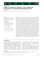
TE = 331 ms, voxel-size = 1 Â 1 Â 1 mm3). The MRI showed no
alterations in the brain parenchyma signal (Fig. 1). During acquisition, the subject was asked to perform a response-inhibition paradigm (go/nogo task) in which the subject had to respond to go
stimuli inhibiting the response to infrequent nogo stimuli (the letter ‘‘X” with a frequency of 17%) [4,5]. Functional data were
acquired using T2⁄-weighted echo plannar image (EPI)
(TR = 2.20 s,
TE = 35 ms,
slice-matrix = 64 Â 64,
slice
gap = 0.28 mm, FOV = 24 cm, flip angle = 90°, slices aligned on the
anterior commissure -posterior commissure [AC-PC] line). After
scanning, the patient was asked to provide an estimate on the
number of errors made in the experimental session. Considering
the analyses, the authors selected a volume of interest encompassing the midcingulate zone, which has been shown to be specifically
activated during tasks that require response selection and willful
generation of motor behavior. This subregion of the ACC is located
posterior to the genu of the ACC, anterior to the vertical plane passing through the anterior commissure [13,14].
In line with the authors’ hypothesis on the role of responseinhibition disabilities and ACC hypofunctionality in the arising of
715
impulsivity in this PD subject (see the introduction section), the
patient completely failed the nogo task. He also showed difficulties
in action-monitoring (in terms of number of detected nogo errors).
In particular, despite errors in the nogo condition have reached
100% [40/40], the number of reported errors was 0. Moreover,
the fMRI acquisition revealed unexpectedly total absent tasksensitive activity in the ACC and medial prefrontal cortex (MPFC)
for the contrast nogo versus go (Fig. 1).
Discussion
The primary aim of the current work was to analyze the link
between ICD, reduce response-inhibition and brain dysfunction
in a 51-year-old man with PD that was admitted to the hospital
for the ascertainment of requirements for STN-DBS surgery. This
type of investigation could be very useful, since STN has been associated with neuropsychiatric changes, including ICD [3]. Indeed,
the STN acts as a relay station for processing associative and limbic
information before they are retransmitted to other brain regions,
thus influencing behavioral changes [3]. In the limbic system, the
amygdala acts as the integrative center for emotions, emotional
behavior, and motivation [15]. Although amygdala is hypofunctioning in PD, dopamine replacement treatment induces amygdala
hyperactivity [16]. Its dysfunction contributes to metacognitiveexecutive impairment, while ICD, hallucinations, anxiety, and
panic attacks may appear in predisposed individuals [16]. Once
considered these elements, it was decided to submit the patient
to an overall cognitive test battery and behavioral assessment of
mood changes.
Integration of reward/punishment contingencies in individual
choices have been related with ICD. Significant activations during
punishment behavior have been previously found in ventral
tegmental area, right and left anterior insula, ACC, and the ventromedial prefrontal cortex [17]. ICD can also be detected by
response-inhibition, which neural substrate is linked with ACC.
In line with the neurocognitive approach, cingulate functionality
was assessed with fMRI while the patient performed a go/nogo
task that represents a classic paradigm in which the differing frequency of event types may result in response-related processing
Fig. 1. Structural MRI image (T1-weighted sequence) and fMRI results for the contrast NOGO vs GO conditions were shown. Maps were thresholded at p < 0.05 cluster-level
corrected using a small volume correction [SVC] with a sphere of 10 mm radius centered on ACC according to the coordinates reported in Palermo et al. [18] and Amanzio
et al. [19].
716
S. Palermo et al. / Journal of Advanced Research 8 (2017) 713–716
conflict. The task involves visual discrimination and a simple
choice: to respond (go) or not respond (nogo) depending on the
current stimulus. Response conflict arises from competition
between the execution and the inhibition of a single response
(response-inhibition conflict), rather than from competition
between two alternative responses (response-selection conflict).
The experimental section revealed impairment of two basic executive functions: response-inhibition and action-monitoring (i.e.,
impaired error awareness) [13,14]. These alterations have been
previously associated with a lack of recruitment of the medial prefrontal regions of the brain [13,14]. In particular, we have previously observed a relationship between action-monitoring and
lower functionality in these brain regions in Bipolar Disorder
patients unaware of their symptomatology when assessed with
the same response-inhibition test [14]. These results are also in
line with our previously published study on patients with AD
and underline a reduced functional recruitment of the cingulofrontal and parieto-temporal regions in patients with reduced
awareness [13]. Action-monitoring disabilities could be explained
by the nature of the executive deficits observed in this case report
and involving fronto-striatal dysfunctions. Indeed, previous findings demonstrated that a specific executive dysfunction - related
to action-monitoring, response-inhibition, and disinhibition derives from ACC and MPFC hypo-functionality [13,14]. This finding has been found across pathologies and could have an underlying common etiopathogenetic mechanism.
Within this fronto-striatal circuitry, ACC and its connections
could be considered part of an evaluative-affective network
involved in behavioral inhibition [13]. Moreover, previous and
current results consider the role of dopaminergic treatment on
executive functions and metacognitive abilities in the medial
prefrontal-ventral striatal non-depleted circuit [18,19]. Those
results underline how the unawareness of distinct pathologies
may exhibit overlapping symptoms in the context of overlapping
circuit-specific dysfunction [14]. fMRI data advise that in this PD
patient a functional alteration of the same cerebral network
(involved in motor and behavioral disinhibition) could be possibly
associated with ICD.
The evidences reported in this work suggest also that the execution of inappropriate motor responses reflects OFC, ACC, and MPFC
hypo-functionality, and poor impulse control. Indeed, responseinhibition could be one of the motor/behavioral aspect of impulse
control. Response-inhibition tasks may be useful in PD for better
characterizing the clinical profile evaluating treatment options. It
is relevant to note that ICD is a detrimental and underreported side
effect of dopaminergic medication. Such an assessment is
supposed to be particularly useful in the post-diagnostic phase,
to better identify individuals at risk of developing ICD with
dopaminergic medication.
Conclusions
ICD was associated with depressed mood, disinhibition, irritability, and appetite disturbance [20]. Moreover, many PD
patients have difficulties with mental processing speed,
response-inhibition, and shifting between different conceptual
sets, suggesting frontal-executive dysfunction. The authors’
hypothesis pointed out how ‘‘behavioral addiction” (i.e. ‘‘motor
impulsivity”) is related with ‘‘disinhibition of prepotent
responses”. Indeed, executive dysfunction in terms of responseinhibition could be a predisposing factor able to define the progression of ICD.
With this case report, a new suggestion for the comprehension
of the neuropsychological and neural abnormalities involved in ICD
was added. However, the relationship between ICD in Parkinson’s
disease and executive dysfunction is an intriguing question that
has yet to be resolved. Future studies are needed to verify if the risk
of ICD may best be determined through the integration of functional MRI and neuropsychological data involving responseinhibition measures.
Conflict of interest
The authors have declared no conflict of interest.
Compliance with Ethics Requirements
All procedures followed were in accordance with the ethical standards of the responsible committee on human experimentation (institutional and national) and with the Helsinki Declaration of 1975, as
revised in 2008. Informed consent was obtained from patient
described in the report.
References
[1] Zgaljardic DJ, Borod DJ, Foldi NS, Mattis PJ, Gordon MF, Feigin A, et al. An
examination of executive dysfunction associated with frontostriatal circuitry
in Parkinson’s disease. J Clin Exp Neuropsychol 2006;28:1127–44.
[2] Alexander GE, Delong MR, Strick PL. Parallel organisation of functionally
segregated circuits linking basal ganglia and cortex. Annu Rev Neurosci
1986;9:357–81.
[3] Rossi PJ, Gunduz A, Okun MS. The subthalamic nucleus, limbic function, and
impulse control. Neuropsychol Rev 2015;25:398–410.
[4] Ray N, Strafella AP. Dopamine, reward, and frontostriatal circuitry in impulse
control disorders in Parkinson’s disease: insights from functional imaging. Clin
EEG Neurosci 2010;41:87–93.
[5] Voon V, Mehta AR, Harlett M. Impulse control disorders in Parkinson’s disease:
recent advances. Curr Opin Neurol 2011;24(4):324–30.
[6] Bugalho P, Oliveira-Maia AJ. Impulse control disorders in Parkinson disease:
crossroads between neurology, psychiatry and neuroscience. Behav Neurol.
2013;547–557.
[7] Antonini A, Barone P, Bonuccelli U, Annoni K, Asgharnejad M, Stanzione P.
ICARUS study: prevalence and clinical features of impulse control disorders in
Parkinson’s disease. J Neurol Neurosurg Psychiatry 2017;88(4):317–24.
[8] Ceravolo R, Frosini D, Rossi C, Bonuccelli U. Spectrum of addictions in
Parkinson’s disease: from dopamine dysregulation syndrome to impulse
control disorders. J Neurol 2010;257(Suppl. 2):S276–83.
[9] Schreiber L, Odlaug BL, Grant JE. Impulse control disorders: updated review of
clinical characteristics and pharmacological management. Front Psychiatry
2011;2:1.
[10] Courtney KE, Ghahremani DG, Ray LA. Fronto-striatal functional connectivity
during response inhibition in alcohol dependence. Addict Biol
2013;18:593–604.
[11] Marsh R, Horga G, Parashar N, Wang Z, Peterson BS, Simpson HB. Altered
activationin fronto-striatal circuits during sequential processing of conflict in
unmedicated adults with obsessive-compulsive disorder. Biol Psychiat
2014;75:615–22.
[12] Li CR, Huang C, Yan P, Bhagwagar P, Milivojevic V, Sinha R. Neural correlates of
impulse control during stop signal inhibition in cocaine dependent men.
Neuropsychopharmacology 2008;33:1798–806.
[13] Amanzio M, Torta DM, Sacco K, Cauda F, D’Agata F, Duca S, et al. Unawareness
of deficits in Alzheimer’s disease: role of the cingulate cortex. Brain
2011;134:1061–76.
[14] Palermo S, Cauda F, Costa T, Duca S, Gallino G, Geminiani G, et al. Unawareness
of bipolar disorder: the role of the cingulate cortex. Neurocase
2015;21:438–47.
[15] Janek HJ, Tye MT. From circuits to behavior in the amygdala. Nature
2015;517:284–92.
[16] Diederich NJ, Goldman JG, Stebbins GT, Goetz CG. Failing as doorman and disc
jockey at the same time; amygdala dysfunction in parkinson’s disease. Mov
Disord 2016;31(1):11–22.
[17] Morese R, Rabellino D, Sambataro F, Perussia F, Valentini MC, Bara BG, et al.
Group membership modulates the neural circuitry underlying third party
punishment. PLoS One 2016;11(11):e0166357.
[18] Palermo S, Lopiano L, Zibetti M, Rosato R, Leotta D, Amanzio M. A novel
framework for understanding reduced awareness of dyskinesias in Parkinson’s
Disease. Parkinsonism Relat Disord 2017;39:58–63.
[19] Amanzio M, Palermo S, Zibetti M, Leotta D, Rosato R, Geminiani G, et al. Selfunawareness of levodopa induced dyskinesias in patients with Parkinson’s
disease. Brain Cogn 2014;90:135–41.
[20] Pontone G, Williams JR, Bassett SS, Marsh L. Clinical features associated with
impulse control disorders in Parkinson disease. Neurology 2006;67
(7):1258–61.