Effects of the geometrical structure of a honeycomb TCP on relationship between bone/cartilage formation and angiogenesis
Bạn đang xem bản rút gọn của tài liệu. Xem và tải ngay bản đầy đủ của tài liệu tại đây (1.78 MB, 9 trang )
Int. J. Med. Sci. 2018, Vol. 15
Ivyspring
International Publisher
1582
International Journal of Medical Sciences
Research Paper
2018; 15(14): 1582-1590. doi: 10.7150/ijms.28452
Effects of the Geometrical Structure of a Honeycomb
TCP on Relationship between Bone / Cartilage
Formation and Angiogenesis
Hiroyuki Matsuda1, Kiyofumi Takabatake1, Hidetsugu Tsujigiwa2, Satoko Watanabe3, Satoshi Ito1,
Hotaka Kawai1, Mei Hamada1, Saori Yoshida1, Keisuke Nakano1, Hitoshi Nagatsuka1
1.
2.
3.
Department of Oral Pathology and Medicine, Graduate School of Medicine, Dentistry and Pharmaceutical Science, Okayama University, Okayama, Japan
Department of life science, Faculty of Science, Okayama University of Science, Okayama, Japan
Department of Plastic and Reconstructive Surgery, Graduate School of Medicine, Dentistry and Pharmaceutical Science, Okayama University, Okayama,
Japan
Corresponding author: Hitoshi Nagatsuka, Department of Oral Pathology and Medicine, Graduate School of Medicine, Dentistry and Pharmaceutical
Sciences, Okayama University. 2-5-1 Shikata-Cho, Okayama 700-8558, Japan. Phone: (+81) 86-2351-6651, Fax: (+81) 86-235-6654 E-mail:
Kiyofumi Takabatake, Department of Oral Pathology and Medicine, Graduate School of Medicine, Dentistry and Pharmaceutical Sciences, Okayama University.
2-5-1 Shikata-Cho, Okayama 700-8558, Japan. Phone: (+81) 86-2351-6651, Fax: (+81) 86-235-6654 E-mail:
© Ivyspring International Publisher. This is an open access article distributed under the terms of the Creative Commons Attribution (CC BY-NC) license
( See for full terms and conditions.
Received: 2018.07.11; Accepted: 2018.09.13; Published: 2018.10.20
Abstract
A number of biomaterials have been developed, some of which already enjoy widespread clinic use.
We have devised a new honeycomb tricalcium phosphate (TCP) containing through-and-through
holes of various diameters to control cartilage and bone formation. However, the way in which the
geometric structure of the honeycomb TCP controls cartilage and bone tissue formation separately
remains unknown. In addition, an association has been reported between bone formation and
angiogenesis. Therefore, in the present study, we investigated the relationship between angiogenesis
and various hole diameters in our honeycomb TCP over time in a rat ectopic hard tissue formation
model. Honeycomb TCPs with hole diameters of 75, 300, and 500 µm were implanted into rat
femoral muscle. Next, ectopic hard tissue formation in the holes of the honeycomb TCP was
assessed histologically at postoperative weeks 1, 2, and 3, and CD34 immunostaining was performed
to evaluate angiogenesis. The results showed that cartilage formation accompanied by thin and poor
blood vessel formation, bone marrow-like tissue with a branching network of vessels, and vigorous
bone formation with thick linear blood vessels occurred in the TCPs with 75-µm, 300-µm, and
500-µm hole diameters, respectively. These results indicated that the geometrical structure of the
honeycomb TCP affected cartilage and bone tissue formation separately owing to the induced
angiogenesis and altered oxygen partial pressure within the holes.
Key words: Angiogenesis, Bone formation, Cartilage formation, Geometrical structure, Honeycomb TCP
Introduction
In recent years, the progression of regenerative
medicine has led to the development of various
materials. During this process, stem cells, growth
factors, and the extracellular matrix (ECM) constitute
the elements needed for cell growth and
differentiation1–5. In addition, it has recently come to
be believed that trophic resources (e.g., vessels) and
dynamic elements (e.g., mechanical stress) play an
important role in cell growth and differentiation6,7.
Mesenchymal or induced pluripotent stem cells are
often used as the stem cell element, and bone
morphogenetic protein-2 (BMP-2), among others, is
widely used as a growth factor. Furthermore, in
research focusing on the ECM, artificial biomaterials
composed of various materials have been researched
and developed to reproduce the extracellular
microenvironment and induce tissue formation and
cell growth and differentiation8–12.
Int. J. Med. Sci. 2018, Vol. 15
Recently, several studies have focused on the
geometrical structure of biomaterials because not only
the composition, but also the optimum geometrical
structure of artificial biomaterials, is considered
important for inducing cell differentiation and tissue
formation7,13. Regarding hard tissue regeneration in
the clinical setting, ceramic biomaterials with high
biocompatibility, such as hydroxyapatite and
tricalcium phosphate (TCP), have been developed and
already enjoy widespread use. Therefore, many
researchers have attempted to identify the optimal
geometrical structure of artificial biomaterials for
inducing hard tissue formation7,13–15.
Focusing on the importance of the geometrical
structure of artificial biomaterials for inducing cell
differentiation and hard tissue formation, we have
already succeeded in developing a new honeycomb
TCP structure containing through-holes of various
diameters. In our previous study, we reported that the
difference in surface properties resulting from the
sintering temperature affects the biocompatibility and
osteoinductivity of TCP16. Furthermore, changing the
geometrical structure of honeycomb TCP holes has
successfully controlled cartilage and bone formation17.
In that study, we investigated histologically how
differences in the hole diameters (75, 300, 500, and
1600 µm) of a honeycomb TCP structure with various
final contained amount. of BMP-2 (0, 125, 250, 500,
and 1000 ng) influenced bone tissue regeneration.
Cartilage formation was observed in the honeycomb
TCP with a 75-µm pore size and a low contained
amount of BMP-2 (125 ng) at 3 weeks after
implantation into rat femoral muscle. In addition, a
bone marrow-like structure was found in the
honeycomb TCP with a 300-µm pore size and a high
contained amount of BMP-2 (1000 ng), and vigorous
bone formation was observed in the honeycomb TCP
with a 500-µm pore size at 3 weeks after implantation
into rat femoral muscle. On the other hand, no bone
formation was observed in the honeycomb TCP with a
1600-µm pore size regardless of BMP-2 concentration
and TCP without BMP-2 did not show hard tissue
formation at any pore size. These findings suggest
that cartilage and bone formation can be controlled by
altering the geometric structure of artificial
biomaterials; however, the details underlying this
mechanism remain unclear.
In recent years, angiogenesis has been found to
be important for appropriate bone formation as it
supplies cells, oxygen, nutrients, and cytokines to
osteoblast progenitor cells; thus, bone formation is
thought to occur in conjunction with angiogenesis.18.
With this background, in the present study, we
analyzed the relationship between hard tissue
formation and angiogenesis in TCP holes over time to
1583
examine how the geometrical structure of a
honeycomb TCP structure affects the differentiation
mechanism of bone and cartilage formation. In this
experiment, we used the honeycomb TCP with a
75-µm pore size and a low contained amount of
BMP-2 (125 ng), which specifically recognizes
cartilage tissue formation, and the honeycomb TCP
with a 300-µm or 500-µm pore size and a high
contained amount of BMP-2 (1000 ng), which
specifically identified bone tissue formation.
Materials and Methods
Animals and implantation procedure
A total of 14 4-week-old healthy male Wister rats
were used in this experiment. All experiments were
performed in accordance with Okayama University’s
Policy on the Care and Use of the Laboratory Animals
and approved by the Animal Care and Use
Committee. All surgical procedures were performed
under general anesthesia in a pain-free state.
Preparation of honeycomb TCP containing
BMP-2
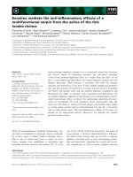
Honeycomb TCP was pressed in a cylindrical
mold with a depth of 5 mm containing
through-and-through holes with diameters of 75 µm
(75TCP), 300 µm (300TCP), and 500 µm (500TCP).
Each TCP was calcinated by heating to 1200 °C (Fig.
1). Details of TCP manufacturing method have been
described previously16.
Each TCP structure was sterilized by autoclave
and loaded with BMP-2. 75TCP was loaded with
BMP-2 diluted to a final contained amount of 125 ng
in Matrigel (BD Biosciences, Inc., NJ, USA), and
300TCP and 500TCP were loaded with BMP-2 diluted
to a final contained amount of 1000 ng in Matrigel
(BD Bioscience). Next, these TCPs were implanted
into rat femoral muscle.
Histological procedure
For histological observations, the implanted
TCPs were removed after 1, 2, and 3 weeks and fixed
in neutral buffered formalin. Next, the specimens
were decalcified in 10% ethylenediaminetetraacetic
acid for 2 weeks and then embedded in paraffin.
Finally, sections were stained with hematoxylin–eosin
(HE) and observed histologically.
CD34 immunostaining
In the present study, rabbit polyclonal anti-CD34
antibody (Abcam, Tokyo, Japan) was used as a
vascular endothelial marker. CD34 is a cell-surface
glycoprotein known to have the ability to differentiate
into all blood and endothelial cells. For that reason,
the presence of CD34 was investigated.
Int. J. Med. Sci. 2018, Vol. 15
1584
Figure 1. The honeycomb TCP structure used in this experiment.
CD34 immunostaining was carried out as
follows. The sections were deparaffinized in a series
of xylene solutions for 15 min and then rehydrated in
graded ethanol solutions. Endogenous peroxidase
activity was blocked by incubating the sections in
0.3% H2O2 in methanol for 30 min. Antigen retrieval
was performed in 0.01 mol/l citrate buffer (pH 6.0) for
1 min. After incubation with normal serum, the
sections were incubated overnight with primary
antibodies at 4 °C. Tagging of primary antibodies was
achieved by the subsequent application of anti-rabbit
IgG (ABC kit; Vector Laboratories, Inc., Burlingame,
CA, USA). Immunoreactivity was visualized using a
diaminobenzidine (DAB)/H2O2 solution (Histofine
DAB substrate; Nichirei, Tokyo, Japan), and slides
were counterstained with Mayer’s HE (Merck KGaA,
Darmstadt, Germany).
Hard tissue and vessel formation evaluation by
area measurement
To quantify the hard tissue area, hard tissue
formation was measured in five areas chosen from
randomly selected regions in HE-stained specimens
(200× magnification, n=4) using Image J software
(NIH, Bethesda, MD, USA). In each field, we
measured the total area of bone or cartilage formation
in TCP holes and the area of TCP holes and we
calculated the ratio of area of bone or cartilage area in
TCP holes to determine the average of the 5 fields. The
obtained average value was compared in each group,
the rate of bone formation and cartilage formation
were compared for different pore size. To evaluate
angiogenesis in the TCP holes of various diameters,
vessel number counts per a TCP hole, vessels area
measurements in TCP holes in a manner similar to
that of the hard tissue, and average vessel thicknesses
were evaluated in a TCP hole.
Statistical analysis
Statistical analysis was performed using
one-way analysis of variance and Fisher’s exact tests.
A P value <0.05 was considered statistically
significant. All calculations were performed using
PASW Statistics 18 (SPSS Inc., Chicago, IL, USA).
Results
Hard tissue formation in TCP holes over time
Histological findings in 75TCP over time
At 1 week, cell penetration was seen at the
entrance of the 75TCP holes, and small blood vessels
were observed penetrating into the pores (Fig. 2a). At
2 weeks, fibrous connective tissue was observed to be
filling the holes to the center; however, no hard tissue
formation was seen (Fig. 2b). At 3 weeks,
chondrogenesis which was positive of Toluidine blue
staining was observed to be filling the TCP holes, and
angiogenesis was poor compared with that seen at 2
weeks (Fig. 2c).
Histological findings in 300TCP over time
At 1 week, in the 300TCP holes, invasion and
proliferation of fibrous connective tissue were
observed in about one-third of the area from the
entrance of the TCP holes. In addition, microvessel
invasion was observed around the entrance and a
cartilage matrix with clear vesicles had formed on the
inner walls. Basic staining showed the cartilage matrix
to be homogeneous and nonstructural, and Toluidine
blue staining positive image was observed to fit the
cartilage-like tissue (Fig. 2d). At 2 weeks, cell
infiltration and microvessel invasion were observed
Int. J. Med. Sci. 2018, Vol. 15
up to the center of the holes, and the formation of
woven bone surrounded by osteoblasts was observed
around the inner walls (Fig. 2e). At 3 weeks, formation
of bone tissue was found around the inner walls. In
addition, new bone formation was observed to be
covering the inner walls, and a small amount of
cancellous trabecular bone formation was observed in
the pore cavities. A large number of polygonal
osteoblasts were arranged around the bone matrix,
suggesting that 300TCP had the highest osteogenic
activity. Moreover, the formation of numerous
vascular lumens was observed penetrating the lumen
surrounded by bone tissue; vascular lumens were
even observed in the central part of the TCP. In
addition, bone marrow-like tissue with many blood
cells was observed in the vascular lumen-rich part of
the TCP (Fig. 2f).
Histological findings in 500TCP over time
At 1 week, in 500TCP holes, as in the case of
1585
300TCP, infiltration of fibrous connective tissue and
cells was observed in about one-third of the area from
the entrance of the TCP holes. In the vicinity of the
entrance, island-shaped cartilage tissue and cartilage
formation attached to the walls of the TCP were
observed; woven bone formation was also observed in
the part following the cartilage matrix which was
Toluidine blue staining positive. Moreover, thick
straight blood vessel invasion compared with 300TCP
was observed (Fig. 2g). At 2 weeks, the proliferation
of fibrous connective tissue and the invasion of thick
linear blood vessels were observed around the central
part of the TCP, and bone formation was found
around the thick blood vessels (Fig. 2h). At 3 weeks,
500TCP showed a similar pattern of bone formation as
300TCP. In addition, numerous newly cancellous
bone tissues were found; however, no bone
marrow-like tissues were found in the area
surrounded by these bone tissues (Fig. 2i).
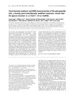
Figure 2. Histological findings. (a) Cell penetration was observed at the entrance of the TCP holes. (b) Fibrous connective tissue was observed filling the TCP holes
up to the center, but no hard tissue formation was observed. (c) Chondrogenesis was observed filling the TCP holes (white arrowheads). Inset: Toluidine blue staining
positive images. (d) Invasion and proliferation of fibrous connective tissue and cartilage formation (white arrowheads) were observed in about one-third of the area
from the entrance of the TCP holes. Inset: Toluidine blue staining positive images. (e) Cell infiltration was observed up to the center of the TCP holes, and bone
formation was observed on the inner walls (arrowheads). (f) Formation of bone and bone marrow-like tissue (asterisks) was found in the TCP holes. (g) Infiltration
of fibrous connective tissue and cartilage formation (white arrowheads) were observed in about one-third of the area from the entrance of the TCP holes. Inset:
Toluidine blue staining positive images. (h) Bone formation (arrowheads) was found around the thick blood vessels. (i) Numerous newly cancellous bone tissues were
observed (arrowheads).
Int. J. Med. Sci. 2018, Vol. 15
1586
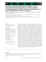
Figure 3. CD34 immunostaining. (a) Small blood vessels were observed in about one-third of the area from the entrance of the TCP holes. (b) Small blood vessels
were observed up to the center of the TCP holes. (c) Angiogenesis was poor at 3 weeks compared with that at 2 weeks. (d) Microvessel invasion was also observed
around the entrance of the TCP holes. (e) Vascular invasion was observed up to the center of the TCP holes, along with a branching network of blood vessel
formation. (f) Blood vessels similar to sinusoidal vessels were observed in the bone marrow-like structures formed within the TCP holes. (g) A thick formation of
vascular invasion was observed in the vicinity of the TCP hole entrances. (h) A plurality of thick rectilinear blood vessels penetrating into the central part of the TCP
holes was observed. (i) At 3 weeks, a pattern of blood vessel invasion similar to that at 2 weeks was seen.
Dynamics of blood vessels entering TCP over
time
We investigated the dynamics of blood vessels
entering the TCP over time using CD34
immunostaining.
At 1 week, small blood vessel invasion was
observed in the vicinity of the 75TCP hole entrances.
The amount of vascular invasion was about one per
hole (Fig. 3a). At 2 weeks, blood vessel invasion was
observed even in the central part of the TCP holes;
however, similar to the findings at 1 week, the
formation pattern was thin and the amount of
vascular invasion was about one per hole (Fig. 3b). At
3 weeks, the insides of the TCP holes were filled with
hard tissue composed mainly of cartilage, so the blood
vessel invasion was poorer than that seen at 2 weeks
(Fig. 3c).
At 1 week, multiple vascular invasions were
observed in the vicinity of the 300TCP hole entrances
(Fig. 3d). At 2 weeks, vascular invasion was observed
around the central part, along with a branching
network of blood vessel formation (Fig. 3e). At 3
weeks, blood vessels similar to sinusoidal vessels
were observed in the bone marrow-like structures
formed within the TCP holes (Fig. 3f).
At 1 week, a thick formation of vascular invasion
was observed in the vicinity of the 500TCP hole
entrances (Fig. 3g). At 2 weeks, a plurality of thick
rectilinear blood vessels penetrating into the central
part of the TCP holes was observed (Fig. 3h), and at 3
weeks, a pattern of blood vessel invasion similar to
that at 2 weeks was seen (Fig. 3i).
Quantitative examination of hard tissue and
vessels. formation in TCP
To investigate the correlation between hard
tissue formation and angiogenesis, we quantitatively
examined the area of hard tissue formation and blood
vessels invading into the TCP holes over time.
Therefore, we analyzed the area, number, and
thickness of blood vessels entering into the TCP holes.
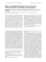
The area of bone formation tended to increase in
the TCP holes over time, regardless of diameter. The
Int. J. Med. Sci. 2018, Vol. 15
amount of bone formation amount tended to increase
with increases in hole size from 75 to 500 µm (Fig. 4a).
By contrast, cartilage formation tended to decrease in
300TCP and 500TCP over time, whereas marked
chondrogenesis was observed in 75TCP at 3 weeks
(Fig. 4b).
The area of blood vessel invasion tended to
increase with increasing hole size over time; however,
invasion at 3 weeks decreased compared with that at 2
weeks in 75TCP (Fig. 5a). The number of blood vessels
invading into the TCP holes also tended to increase
over time and with increasing pore size. In 75TCP, the
number of invasive blood vessels was two or fewer
1587
per hole, and in each TCP hole, the number of blood
vessels was almost about one. In addition, the number
of blood vessels decreased at 3 weeks (Fig. 5b). The
thickness of the blood vessels entering the TCP holes
tended to increase over time and with increasing hole
diameters (Fig. 5c).
In 300TCP and 500TCP, the invasive blood vessel
and bone tissue areas tended to increase over time,
whereas in 75TCP, although hard tissue formation,
especially cartilage formation, tended to increase over
time, the blood vessel area and thickness decreased at
3 weeks.
Figure 4. Quantitative analysis of the hard tissue area. (a) Quantification of the neonatal bone tissue area in the TCP holes. (b) Quantification of the neonatal cartilage
tissue area in the TCP holes. ✴: p<0.05
Int. J. Med. Sci. 2018, Vol. 15
1588
Figure 5. Quantitative analysis of angiogenesis. (a) Quantification of the angiogenesis area in the TCP holes over time. (b) Quantification of the amount of
angiogenesis in the TCP holes. (c) Quantification of the diameter of neonatal vessels in the TCP holes. *: p<0.05
Int. J. Med. Sci. 2018, Vol. 15
Discussion
Stem cells, scaffolds, and growth factors are
important for tissue regeneration, and normal tissue
regeneration does not occur when any of these
elements are missing1–5. Among these elements,
artificial biomaterials (scaffolds) provide the
environment for cell growth and differentiation. In
addition, ideal artificial biomaterials must have an
affinity for living tissue, a structure that cells are likely
to invade, and tissue solubility19.
As a result of CD34 immunostaining, the
formation of vascular lumens penetrating the TCP
holes was observed in 300TCP and 500TCP. Blood
vessel formation is important for not only tissue
regeneration, but also bone tissue formation20,21. The
results of the present experiment also suggested that
angiogenesis exerts a substantial influence on bone
tissue formation. The invasive blood vessel formation
pattern in 300TCP was different from that in 500TCP,
which showed the same linear angiogenesis. The
reason for this was thought to be the geometric
structure of TCP, which influenced the shape of the
invasive blood vessels and affected bone formation.
Therefore, the results of the present study suggest that
the formation of invasive blood vessels was controlled
by the pore size of the honeycomb TCP. In addition,
the differences in hard tissues formation were thought
to be the result of differences in oxygen partial
pressure caused by differences in the shape of blood
vessels influenced by the geometrical structure of the
TCP. Bassett et al.22 reported that when mesenchymal
stem cells were cultured under various conditions, the
precursor cells differentiated into osteoblasts when
cultured under high oxygen partial pressure, and into
chondrocytes otherwise. The differentiation of the
precursor cells was therefore considered to be
determined by the environment in which the
progenitor cells were placed, particularly in terms of
oxygen partial pressure. It has long been known that
bone marrow is in a hypoxic environment, and
changes in oxygen concentration have been shown to
affect the hematopoietic mechanism23,24.
In the present study, 500TCP induced vigorous
osteogenesis with linear thick blood vessel invasion
and 300TCP induced bone marrow formation with
fine reticulated vessel invasion, respectively. On the
other hand, cartilage formation with narrow blood
vessel invasion was observed in 75TCP. Therefore,
cartilage, bone marrow, and bone formation occurred
in relation to angiogenesis in the TCP holes (in the
order of 75TCP, 300TCP, 500TCP), that is, in
accordance with decreased oxygen partial pressure.
These findings suggest that controlling blood vessel
invasion into TCP structures may control the pattern
1589
of hard tissue formation. The geometrical structure of
the TCP holes with various pore sizes reflected the
oxygen partial pressure of the bone, bone marrow,
and cartilage tissue environments in the living body,
and it appears that these kinds of environments were
reproduced by the honeycomb TCP. By changing the
pore sizes of the honeycomb TCP, it was possible to
reproduce the optimal environment for the desired
regeneration of hard tissue.
Our results indicated that angiogenesis
decreased at 3 weeks in 75TCP. Chondromodulin
produced from chondrocytes contributes to normal
cartilage formation by blocking blood vessel
invasion25. Therefore, chondromodulin secreted from
the cartilage tissue filling the holes in 75TCP inhibited
angiogenesis and preserved cartilage tissue in 75TCP.
In addition, since BMP-2 has been reported to be
involved in cell aggregation and angiogenesis26,27, if
the concentration of BMP-2 is low, so is the capacity
for bone formation induced by angiogenesis.
Therefore, 75TCP with a low concentration of BMP-2
is advantageous for cartilage formation.
In conclusion, the results of the present study
indicated that the linear geometry of our honeycomb
TCP structure promoted angiogenesis and hard tissue
formation. Therefore, by altering the pore size and
controlling blood vessel invasion, our honeycomb
TCP structure may allow the selective and efficient
formation of cartilage tissue and bone.
Acknowledgments
This study was funded by the Japan Society for
Promotion of Science (JSPS) KAKENHI Grant-in-Aid
for Scientific Research (No. 16K20577) and (No.
18K17224).
Conflict of Interest
The authors declare that they have no conflict of
interest.
References
1.
2.
3.
4.
5.
6.
7.
8.
Reddi AH, Huggins CB. Infuence of geometry of transplanted tooth and bone
on transformation of broblasts. Proc Soc Exp Biol Med. 1973; 143: 634-637.
Karageorgiou V, Kaplan D. Porosity of 3D biomaterial scaffolds and
osteogenesis. Biomaterials. 2005; 26(27): 5474–5491.
Burg KJ, Porter S, Kellam JF. Biomaterial developments for bone tissue
engineering. Biomaterials. 2000; 21(23): 2347–2359.
Stevens M. Biomaterials for bone tissue engineering. Materials Today. 2008;
11(5): 18–25.
Langer R, Vacanti JP. Tissue engineering. Science. 1993; 260(5110): 920–926.
Zhang L, Liu W, Zhao J, Ma X, Shen L, Zhang Y, Jin F, Jin Y. Mechanical stress
regulates osteogenic differentiation and RANKL/OPG ratio in periodontal
ligament stem cells by the Wnt/β-catenin pathway. Biochim Biophys Acta.
2016; 1860(10): 2211-2219.
Kuboki Y, Jin Q, Takita H. Geometry of carriers controlling phenotypic in
BMP-induced osteogenesis and condrogenesis. J Bone Joint Surg. 2001; 83A:
S1-105-114.
Kawakubo A, Matsunaga T, Ishizaki H, Yamada S, Hayashi Y. Zinc as an
essential trace element in the acceleration of matrix vesicles-mediated mineral
deposition. Microsc Res Tech 2011;74:1161–1165.
Int. J. Med. Sci. 2018, Vol. 15
9.
10.
11.
12.
13.
14.
15.
16.
17.
18.
19.
20.
21.
22.
23.
24.
25.
26.
27.
1590
Porter JR, Henson A, Popat KC. Biodegradable poly(e-caprolactone)nanowires for bone tissue engineering applications. Biomateri-als 2009;
30:780–788.
Arpornmaeklong P, Pripatnanont P, Suwatwirote N. Properties of
chitosan-collagen sponges and osteogenic differentiation of rat-bone-marrow
stromal cells. Int J Oral Maxillofac Surg 2008; 37:357–366.
Abarrategi A, Moreno-Vicente C, Ramos V, Aranaz I, Sanz Casado JV,
Lopez-Lacomba JL. Improvement of porous b-TCP scaffolds with rhBMP-2
chitosan carrier film for bone tissue application. Tissue Eng A 2008;
14:1305–1319.
Wiria FE, Chua CK, Leong KF, Quah ZY, Chandrasekaran M, Lee MW.
Improved biocomposite development of poly(vinyl alcohol)-and
hydroxyapatite for tissue engineering scaffold fabrication using selective laser
sintering. J Mater Sci Mater Med 2008; 19:989–996.
Jin QM, Takita H, Kohgo T, Atsumi K, Itoh H, Kuboki Y. Effect of geometry of
hydroxyapatite as a cell substratum in BMP-induced ectopic bone formation. J
Biomed Mater Res. 2000; 51: 491–499.
Tsuruga E. Pore size of porous hydroxyapatite as the cellsubstratum controls
BMP-induced osteogenesis. J Biochem. 1997; 121: 317–324.
Kiba H, Kuboyama N, Uchida R, Ishizaki T, Nishiyama N. Bone ingrowth into
the parallel cylindrical tubes with different sizes of porous hydroxyapatite
implanted into the rabbits. J Hard Tissue Biol. 2012; 21: 307–314.
Takabatake K, Yamachika E, Tsujigiwa H, Takeda Y, Kimura M, Takagi S,
Nagatsuka H, Iida S. Effect of geometry and microstructure of honeycomb
TCP scaffolds on bone regeneration. J Biomed Mater Res A. 2013; 102(9):
2952-2960.
Watanabe S, Takabatake K, Tsujigiwa H, Watanabe T, Tokuyama E, Ito S,
Nagatsuka H, Kimata Y. Efficacy of Honeycomb TCP-induced
Microenvironment on Bone Tissue Regeneration in Craniofacial Area. Int J
Med Sci. 2016; 13(6): 466-476.
Kusumbe AP, Ramasamy SK, Adams RH. Coupling of angiogenesis and
osteogenesis by a specific vessel subtype in bone. Nature. 2014; 507: 323–328.
Yang HY, Thompson I, Yang SF, Chi XP, Evans JR, Cook RJ. Dis-solution
characteristics of extrusion free formed hydroxyapatite-tricaicium phosphate
scaffolds. J Mater Sci Mater Med 2008; 19:3345–3353
Petersen W, Tillmann B. Structure and vascularization of the cruciate
ligaments of the human knee joint. Anat Embryol. 1999; 200: 325-334.
Hossler FE, Douglas JE. Vascular Corrosion Casting: Review of Advantages
and Limitations in the Application of Some Simple Quantitative Methods.
Microsc Microanal. 2001; 7: 253-264.
Bassett C, Herrmann I. Influence of oxygen concentration and mechanical
factors on differentiation of connective tissues in vitro. Nature. 1961; 190:
460-461.
Guitart AV, Hammoud M, Dello Sbarba P, Ivanovic Z, Praloran V.
Slow-cycling/quiescence balance of hematopoietic stem cells is related to
physiological gradient of oxygen. Exp Hematol. 2010; 38: 847–851.
Harrison JS, Rameshwar P, Chang V, Bandari P. Oxygen saturation in the bone
marrow of healthy volunteers. Blood. 2001; 99(1): 394.
Hiraki Y, Inoue H, Iyama K, Kamizono A, Ochiai M, Shukunami C, Iijima S,
Suzuki F, Kondo J. Identification of Chondromodulin I as a Novel Endothelial
Cell Growth Inhibitor. J.Biol. Chem. 1997; 272: 32419-32426.
Chen WC, Chung CH, Lu YC, Wu MH, Chou PH, Yen JY, Lai YW, Wang GS,
Liu SC, Cheng JK, Wu YJ, Yeh HI, Wang LY, Wang SW. BMP-2 induces
angiogenesis by provoking integrin α6 expression in human endothelial
progenitor cells. Biochem Pharmacol. 2018; 150: 256-266.
Pan Y, Chen J, Yu Y, Dai K, Wang J, Liu C. Biomater Sci. Enhancement of
BMP-2-mediated angiogenesis and osteogenesis by 2-N,6-O-sulfated chitosan
in bone regeneration. Biomater Sci. 2018; 6(2): 431-439.