The modulation of erythrocyte Na+/K+-ATPase activity by curcumin
Bạn đang xem bản rút gọn của tài liệu. Xem và tải ngay bản đầy đủ của tài liệu tại đây (2.13 MB, 8 trang )
Journal of Advanced Research (2015) 6, 1023–1030
Cairo University
Journal of Advanced Research
ORIGINAL ARTICLE
The modulation of erythrocyte Na+/K+-ATPase
activity by curcumin
Prabhakar Singh a, Rajesh Kumar Kesharwani b, Krishna Misra b,
Syed Ibrahim Rizvi a,*
a
Department of Biochemistry, University of Allahabad, Allahabad 211002, India
Division of Applied Science & Indo-Russian Center For Biotechnology [IRCB], Indian Institute of Information Technology,
Allahabad 211012, India
b
A R T I C L E
I N F O
Article history:
Received 4 October 2014
Received in revised form 20 December
2014
Accepted 23 December 2014
Available online 22 January 2015
Keywords:
Curcumin
Erythrocytes
Na+/K+ ATPase
In silico
A B S T R A C T
Curcumin, an active biphenolic molecule present in turmeric (Curcuma longa), has been
reported to elicit plethora of health protective effects. The present study was carried out
in vitro, in vivo and in silico to investigate the modulatory effects of curcumin on erythrocyte
membrane Na+/K+-ATPase activity. In vitro curcumin (10À5 M to 10À8 M) was incubated with
human erythrocytes membrane. In vivo curcumin (340 mg/kg b.w. and 170 mg/kg b.w.) was supplemented to wistar rats for 21 days. In silico, catalytic unit a of Na+/K+-ATPase (3b8e.pdb)
protein was used as a receptor for the natural ligand ATP to study curcumin-mediated docking
simulation using AutoDock4. The in vitro effect of curcumin on the Na+/K+-ATPase activity
in human erythrocytes was biphasic. An inhibitory response was observed at 10À5 M
(p < 0.001). An activation of the Na+/K+-ATPase activity was observed at 10À7 and 10À8 M
(p < 0.001 and p < 0.01). In vivo, curcumin supplementation to rats increased the Na+/K+ATPase activity at doses 340 mg/kg b.w. (p < 0.001) as well as at 170 mg/kg b.w., (p < 0.01).
AutoDock4 docking simulation study showed that both ligands curcumin and ATP actively
interacted with amino acids Glu214, Ser215, Glu216, Thr371, Asn377, Arg378, Met379,
Arg438, Val440, Ala444, Lys451 and Asp586 at the catalytic cavity of Na+/K+-ATPase.
ATP had more H bonding and hydrophobic interaction with active site amino acid residues
compared to curcumin. These finding may explain some of the health beneficial properties of
curcumin associated with deregulated Na+/K+-ATPase activity or ions homeostasis.
ª 2015 Production and hosting by Elsevier B.V. on behalf of Cairo University.
Abbreviations: CUR, curcumin; ATP, Adenosine Tri-Phosphate;
RBCs, Red Blood Cells; RMS, root mean square.
* Corresponding author. Tel.: +91 9415305910; fax: +91
5322242116.
E-mail address: (S.I. Rizvi).
Peer review under responsibility of Cairo University.
Production and hosting by Elsevier
Introduction
Curcumin (1E,6E)-1,7-Bis(4-hydroxy-3-methoxyphenyl)-1,6heptadiene-3,5-dione, a natural biphenolic compound isolated
from turmeric (Curcuma longa) has been reported to elicit a
plethora of health protective effects [1]. Curcumin is lipophilic
and therefore has the ability to localize between polar head
and non-polar tail of lipid molecule in plasma membrane. This
interaction influences the fluidity and thickness of the
/>2090-1232 ª 2015 Production and hosting by Elsevier B.V. on behalf of Cairo University.
1024
membrane. The localization between the membrane lipid bilayers significantly weakens the elasticity moduli of the bilayers
[2,3]. Insertion of curcumin into the membrane modifies the
activity of several functionally unrelated membrane-bound
proteins and related signalling cascade systems [4,5].
Na+/K+-ATPase (EC 3.6.3.9) is a heterodimeric, transmembrane, ubiquitously present protein that regulates neuronal signalling, ion homeostasis, substrate transportation and
muscle contraction [6]. Besides its inotropic effects, Na+/
K+-ATPase also acts as a signal transducer regulating many
cellular events including those associated with tumour cell
growth [7]. In view of its important cellular role, there is an
increasing interest in the characterization of chemicals which
may modulate this enzyme.
Na+/K+-ATPase (P-type ATPases) is made up of an
active a unit (110 kDa) with 10 Trans membrane segments
(TMS; aM1–aM10), sugar rich auxiliary b unit (55 kDa)
and a hydrophobic single membrane crossing protein c unit
(12 kDa) for regulating ionic gradient across the cell membrane [8]. There are several isoforms of the binding units
in the primary catalytic unit present in different tissues: a1
in nerves, kidney and lung, a2 in heart and skeletal muscle,
a3 in the brain and a4 in testis and specifically in spermatozoa [9].
The membrane’s physical and biochemical properties are
strongly regulated by lipid composition and redox status of
the environment. Changes in membrane fluidity have been
shown to modulate the activity of membrane bound receptors,
enzymes and ion-exchangers [10,11]. Na+/K+ ATPase activity
is modulated by the surrounding microenvironment of lipids;
thus, modifications in the membrane fluidity translate into
effects on Na+/K+ ATPase activity. An altered Na+/K+ATPase activity has been reported during late complications
of diabetes mellitus such as nephropathy, neuropathy, retinopathy and in the development of diabetic vascular complications
[12–14]. Elevation of intracellular sodium and potassium value
was associated with reduced activity of erythrocyte Na+/K+ATPase pump [15]. The present study was carried out in vitro,
in vivo and in silico to investigate the modulatory effects of curcumin on ouabain-sensitive Na+/K+-ATPase from erythrocyte membrane of humans and rats. In addition, catalytic
unit a of Na+/K+-ATPase (3b8e.pdb) protein has been used
as a receptor for natural ligand adenosine triphosphate
(ATP) and curcumin-mediated docking simulation using
AutoDock4. The suitable docked conformation between
receptor and ligands was predicted on the basis of cluster
analysis.
Material and methods
P. Singh et al.
Experimental study
Human erythrocyte
Human venous blood was collected in heparin from 26 healthy
volunteers of both sexes between the 24 and 45 years of age by
venipuncture. The human subjects were screened for diabetes
mellitus, asthma, tuberculosis, or other major illness. None
of the subjects were smokers or were taking any medication.
All 26 selected subjects gave informed consent for the use of their
blood samples for the research study. The protocol of study was
in conformity with the guidelines of the Institutional Ethical
Committee, University of Allahabad. The heparinized blood
was centrifuged at 800g at 4 °C for 10 min. After the removal
of plasma, buffy coat and top layer comprising approximately
15–20% of the packed cells, the remaining packed erythrocytes
were washed twice with 10 mM phosphate buffered saline
pH 7.4.
Animal
Male albino rats (Wistar strain) of 5–6 months weighing
between 150 and 200 g were purchased from CDRI, Lucknow,
India. Animals were housed in polypropylene cages at
24 ± 2 °C (6 rats per cage) and 12 h light:12 h dark cycles.
Animals were fed with standard pellet diet obtained from
Dayal Industries Limited, Lucknow, India, and had free access
to drinking water. Rats were acclimated for one week before
treatment. The protocol of study was in conformity with the
guidelines of the Institutional Ethical Committee of University
of Allahabad.
Twenty-four male Wistar rats were randomly divided into
four groups (six rats/group). Group [I]: Control, receiving no
treatment/supplementation. Group [II]: Experimental control,
rats were supplemented with 0.9% NaCl solution through oral
route. Group [III]: Curcumin-treated group (340 mg/kg b.w.,
saline) [16]. Group [IV]: Curcumin-treated group (170 mg/kg
b.w., saline).
Curcumin and saline treatments were given through oral
route for 21 days at fixed time 11.00 am to 12.00 pm to avoid
circadian disturbance. At the end of treatment, rats were sacrificed under light anaesthesia. The blood was collected in heparinized tubes by heart puncture. The heparinized blood was
centrifuged at 800g at 4 °C for 10 min. After the removal of
plasma, buffy coat and upper 15–20% of the packed RBCs
cells, the remaining RBCs were washed twice with 10 mM
phosphate buffered saline pH 7.4.
Preparation of erythrocytes membrane
Chemicals and instrument
Curcumin ((1E,6E)-1,7-bis(4-hydroxy-3-methoxyphenyl)-1,
6-heptadiene-3,5-dione) was purchased from Bio Basic
Inc., Ontario, Canada (cat. # CB0346), and Imidazole,
Ouabain, ATP, Bovine Serum Albumin (BSA) were
purchased from Sigma Aldrich, India. Other chemicals of
highest purity were purchased from Merck, India, and
HIMEDIA Labs, India. Spectrophotometric measurements
were performed on Shimadzu-UV-1800 (Japan) UV–VIS
spectrophotometer.
The erythrocyte membrane was isolated according to the
method of Marchesi and Palade [17]. The erythrocyte membrane proteins were quantified according to the method of
Lowry et al. [18].
Measurement of Na+/K+-ATPase activity
Na+/K+-ATPase activity was measured according to the
method of Suhail and Rizvi [13]. The final assay mixture contained 0.5–1.2 mg membrane protein/mL, 20 mmol/L KCl,
Na+/K+-ATPase and curcumin
140 mmol/L NaCl, 3 mmol/L MgCl2, 30 mmol/L imidazole
(pH 7.24), with or without 5 · 10–4 mol/L ouabain and
6 mmol/L ATP. Assay mixture was incubated for 30 min at
37 °C and the reaction was stopped by the addition of
3.5 mL of a solution-A (0.5% ammonium molybdate,
0.5 mol/L H2SO4, and 2% sodium dodecyl sulphate). The
amount of liberated phosphate (Pi) was estimated according
to the method of Fiske and Subbarow [19]. In vitro experiment
was carried out by adding curcumin (final concentration
10À5 M to 10À8 M) to the assay mixture and incubated for
30 min at 37 °C prior to enzyme assay. Na+/K+-ATPase
activity was expressed as nmol pi released/mg protein per hour
at 37 °C.
Statistical analysis was performed by the software GraphPad
Prism 5 version 5.01. One way analysis of variance (ANOVA)
was performed for multiple comparisons. P-values were evaluated by two tailed method. Statistical differences shown in
Fig. 1 represent in vitro study (n = 26) on human erythrocytes,
and Fig. 2 represents in vivo study (n = 6) on rat erythrocytes.
p < 0.05 was considered to be significant. Values are represented as ±SD in graphs. Significance between experimental
and control is represented by star (*) in graph.
Computational study
Selection of protein and ligand
A three-dimensional X-ray crystallized structure of Na+/K+ATPase subunit a-1 protein complexed with 2 PC1 (2-diacylsn-glycero-3-phosphocholine) as a ligand and 6 Rb+ and
2 Mg++ together with 2 F4MgÀÅÀ as a cofactor (PDB ID:
3B8E, Resolution = 3.50 A˚, Chains: A, B, C, D, G, H) were
downloaded from the Protein Data Bank [20,21]. Pubchem
compound database was used to retrieve the 3 dimensional
Na+/K+ ATPase Activity
[nmol pi released/h/mg membrane protein]
700
In vivo: Rat Erythrocytes
In vitro: Human Erythrocytes
***
***
600
***
500
400
300
200
100
0
1
Control
(Group-I)
Statistical analysis
100
Na+/K+ ATPase Activity
[nmol pi released/h/mg membrane protein]
1025
Ex. Control
(Group-II)
CUR (170)
(Group-IV)
CUR (340)
(Group-III)
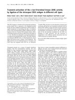
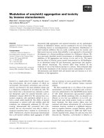
Fig. 2 In vivo effect of curcumin (340 mg/kg b.w. and 170 mg/
kg b.w. oral) on wistar albino rat’s erythrocyte membrane Na+/
K+-ATPase activity (Ouabain-sensitive). Na+/K+-ATPase activity was expressed as nmol pi released/h/mg membrane protein at
37 °C. Values (n = 6) are means ± S.D.
structures of selected ligands (ATP and curcumin) and energy
minimization was done using UCSF Chimera software (Developed by the Resource for Biocomputing, Visualization, and
Informatics and can be downloaded from .
ucsf.edu/chimera/).
Proteins and ligands structure preparation
The selected target protein (PDB ID: 3B8E) and ligands
(curcumin and ATP) were prepared as an input file for
docking by using MGL (Molecular Graphics Laboratory)
Tool 1.5.6 developed by The Scripps Research Institute for
visualization and analysis of molecular structures [22,23].
The co-crystallized heteroatoms and ligands were removed
and final .pdbqs format file of receptor protein was used
during docking simulation. On the basis of X-ray crystallized bound ligand MF4 (magnesium tetrafluoride), the
active site was chosen for current docking study with curcumin and ATP [22].
80
Grid design and docking simulation
***
60
***
40
20
0
Control
Ex.
Control
10 -8 M
1
10-7 M
10 -6 M
10 -5 M
Curcumin [Molar]
À5
Fig. 1 In vitro effect of curcumin (10 M to 10À8 M) on human
erythrocyte’s membrane Na+/K+-ATPase activity (Ouabain-sensitive). Na+/K+-ATPase activity was expressed as nmol pi
released/h/mg membrane protein at 37 °C. Values (n = 26) are
means ± S.D.
AutoGrid 4 was used to obtain the grid maps required prior to
docking and AutoDock 4 for docking study [24]. AutoDock4
and AutoGrid4 tool have been developed by The Scripps
Research Institute, U.S.A. which can be downloaded from
The user defined three
dimensional grid covered the region of active or binding site of
interest in the receptor and selected ligands and were limited to
this search space during docking. A grid box size set on the
basis of requirement (100 · 100 · 100 points with spacing of
0.375 A˚) was used for the study. The binding constant or inhibition constant (kI) was directly calculated by AutoDock4
during the simulation for each conformation [22,24]. The
docked conformation of selected ligands with the receptor
has been demonstrated in 2-dimensional page by using LIGPLOT version 4.5.3 (Developed by Wallace et al., 1995 and
can be downloaded from www.ebi.ac.uk/thornton-srv/software/LIGPLOT/) [25].
1026
P. Singh et al.
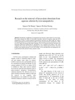
formed with amino acids at active site by ATP. However,
curcumin formed only three hydrogen bonds with different
amino acids at active cavity. Table 1 shows that the binding
energy of curcumin was relatively higher (À7.4 kcal) than
ATP (À11.55 kcal).
During docking simulation with cluster root mean
square (RMS) = 2.0, different clusters were formed and
each cluster except a single cluster (having 17 conformation) had 1–6 conformation of curcumin based cluster
RMS value. ATP docking simulation showed several
clusters with average conformation of 1–3. Fig. 5a and b
shows all the conformations for clusters ranked according
to binding energy.
Discussion
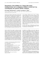
Fig. 3 Secondary structure (cartoon) representation at the active
site of Na+/K+-ATPase subunit a-1 together with docked
conformation of ligand ATP and curcumin.
Results
Fig. 1 shows that the effect of curcumin on the Na+/K+-ATPase activity in human erythrocytes was biphasic. An inhibitory
response was observed at 10À5 M (P < 0.001) while an activation of the activity was observed at 10À7 M (P < 0.001) and
10À8 M (p < 0.001).
Fig. 2 shows that in vivo, curcumin (340 mg/kg b.w, oral)
supplementation to rats significantly (P < 0.001) increased
the Na+/K+-ATPase activity in erythrocyte membrane as
compared with control and experimental control rats. Lower
dose of curcumin supplementation (170 mg/kg b.w., oral) also
significantly (P < 0.001) increased the Na+/K+-ATPase
activity.
Fig. 3 shows the secondary structure (cartoon representation) of Na+/K+-ATPase subunit a-1 together with docked
conformation of ligand ATP and curcumin. The AutoDock4
docking simulation results presented in Fig. 4(a) show that
the active site residues at catalytic unit a of Na+/K+-ATPase (3b8e.pdb) Glu214, Ser215, Glu216, Thr371, Asn377,
Arg378, Met379, Leu436, Lys437, Arg438, Ala439, Val440,
Ala444, Lys451, Asp586 were actively involved in hydrogen
bonding and hydrophobic interaction with natural ligand
ATP. On the other hand Fig. 4(b) shows that curcumin
interacted actively with Glu214, Ser215, Glu216, Lys370,
Thr371, Asn377, Arg378, Met379, Arg438, Val440, Ala444,
Ser447, Lys451, Arg544, Asp586, Asp612, Thr616 amino
acids at the catalytic centre. Eight hydrogen bonds were
Na+/K+-ATPase regulates different physiological and metabolic processes through regulating ion gradient across the
cell membrane of excitable tissues [9,26]. Several drugs
modulate the Na+/K+-ATPase activity by binding at the
active site, allosteric site, lipid microenvironment of the
membrane or through a synergistic mechanism. Na+/K+ATPase activity was reported to strongly synchronize with
the fluidity of membrane and by drugs affecting fluidity
[5,27]. Na+/K+-ATPase activity inhibitory effect in the
presence of high concentration of curcumin was reported
by Mahmmoud (2005). This report showed that in shark
rectal glands and pig kidney the EC50 of curcumin for
Na+/K+-ATPase activity was 15.8 ± 1.12 lM and
5.24 ± 1.0 lM respectively [28]. Sharks contain higher cholesterol than pig kidney membranes; hence, the higher
EC50 value of curcumin for Na+/K+-ATPase activity
may be due to differences in membrane fluidity [29]. It
was observed that curcumin interacted with trans-membrane domain(s) of the a-subunit of Na+/K+ ATPase protein and reduced the oligomycin inhibitory effects [16].
Oligomycin and curcumin have similar modulatory effects
on the kinetic properties of the Na+/K+-ATPase [28].
We propose that higher concentrations of curcumin
(10À5 M) caused down-regulation of Na+/K+-ATPase
activity in human erythrocyte membrane by directly interacting at the active catalytic centre of the enzyme. However, lower concentrations of curcumin (10À7 M)
modulated the membrane fluidity resulting in an increased
activity of the enzyme.
Curcumin reduced the Na+/K+-ATPase activity in human
blood mononuclear cells during initial days (up to 3 days of
7 days) of incubation, but prolonged incubation increased
the Na+/K+-ATPase activity [30]. Curcumin supplementation
to retinol deficient rats resulted in increased activity of brain
microsomal membrane Na+/K+-ATPase activity mediated
through improved cholesterol:phospholipid ratio [31]. In vitro
we observed that curcumin at 10À7 M concentration increased
the Na+/K+-ATPase activity maximally which decreased further on decreasing the curcumin concentration till 10À8 M. In
vivo curcumin supplementation (340 mg/kg b.w. and 170 mg/
kg b.w.) to rats increased the Na+/K+-ATPase activity in
erythrocyte membrane thus corroborating the in vitro curcumin effects on Na+/K+-ATPase activity.
Minimum energy docked conformation after cluster
analysis in MGL Tools and AutoDock4 docking simulation
Na+/K+-ATPase and curcumin
1027
(a) ATP
Fig. 4 Docked conformation of hydrogen bonding view and hydrophobic interaction of (a) ATP, (b) curcumin with amino acids of
human Na+/K+-ATPase subunit a-1 protein (3b8e.pdb) at the active site cavity (hydrogen bonds as green dashed lines between the atoms
involved and hydrophobic contacts as an arc with spokes radiating towards the ligand atoms).
suggested that curcumin actively interacted to form hydrogen
bond with amino acids viz., Lys370, Lys451 and Asp612 at
the active site cavity of Na+/K+-ATPase a unit. Comparative
binding analysis of curcumin and ATP shows that both ligands
have common amino acids Glu214, Ser215, Glu216, Thr371,
Asn377, Arg378, Met379, Arg438, Val440, Ala444, Lys451,
Asp586 and among these Lys451 is the key residue for hydrogen bonding. Table 1 shows that ATP had more H bonding
interactions compared to curcumin at the active site cavity of
receptor protein.
1028
P. Singh et al.
(b) Curcumin
Fig. 4. (continued)
Table 1 Comparative docking simulation result of ligands
(ATP and curcumin) with Na+/K+-ATPase protein
(3b8e.pdb).
1.
2.
3.
4.
5.
6.
Properties
ATP
Curcumin
Binding energy (kcal/mol)
Ligand efficiency
Inhibition constant (kI)
Intermole energy (kcal/mol)
Torsional energy (kcal/mol)
No. of H-bond interactions
À11.55
À0.37
3.34eÀ009
À16.034
4.47
8
À7.4
À0.27
3.75eÀ006
À10.39
2.98
3
In silico results show that curcumin has lower binding affinity due to less hydrogen bonding and hydrophobic interaction
at the active site of Na+/K+ ATPase protein in comparison
with natural ligand ATP. Down-regulating Na+/K+ ATPase
activity effects of curcumin at higher concentration >10À6 M
were hypothesized to be due to the interaction of curcumin
with amino acids involved in active catalysis at a-subunit of
Na+/K+ ATPase. However, increased Na+/K+ ATPase
activity at lower concentration <10À7 M of curcumin is
thought to be due to altering the membrane fluidity in favour
of increased Na+/K+ ATPase activity in erythrocyte
membrane.
Na+/K+-ATPase and curcumin
1029
(a) ATP
(b) Curcumin
Fig. 5 Possible cluster between conformations and binding energy within range of root mean square (RMS) tolerance (a) ATP, (b)
curcumin.
Conclusions
The ion gradient and active membrane potential derived from
Na+/K+-ATPase activity form the basis for a range of necessary cellular processes, especially Na+ and H+-dependent secondary transport systems. Curcumin binds and interacts at the
active site cavity of Na+/K+-ATPase to down-regulate the
enzyme activity at higher concentration (>10À6 M). However,
curcumin surprisingly increased the enzyme activity at lower
concentration (<10À7 M). In vivo oral supplementation of curcumin also increased Na+/K+-ATPase activity. The study concludes that curcumin has significant potential to modulate Na+/
K+-ATPase activity dose dependently and the present findings
may help to explain some of the biological effects of curcumin.
Conflict of Interest
The authors have declared no conflict of interest.
Acknowledgements
Prabhakar Singh acknowledges the support of Council of
Scientific and Industrial Research (CSIR), New Delhi, India,
for providing Senior Research Fellowship. Rajesh Kumar
Kesharwani acknowledges the Indian Council of Medical
Research (ICMR), New Delhi, India, for providing financial
support as Senior Research Fellowship (SRF). Department
of Biochemistry is also a recipient of FIST grant from Department of Science and Technology, Govt. of India.
References
[1] Hung WC, Chen FY, Lee CC, Sun Y, Lee MT, Huang HW.
Membrane-thinning effect of curcumin. Biophys. J.
2008;94:4331–8.
[2] Kesharwani KR, Misra K. Prediction of binding site for
curcuminoids at human toposomerase II a protein; an in silico
approach. Curr. Sci. 2011;101:1060–5.
[3] Ingolfsson HI, Koeppe 2nd RE, Andersen OS. Curcumin is a
modulator of bilayer material properties. Biochemistry
2007;46:10384–91.
[4] Singh P, Rizvi SI. Curcumin activates erythrocyte
membrane acetylcholinesterase. Lett. Drug Des. Discov.
2013;10:550–6.
[5] Mahmmoud YA. Curcumin is a lipid dependent inhibitor of the
Na, K-ATPase that likely interacts at the protein-lipid interface.
Biochim. Biophys. Acta 2011;1808:466–73.
[6] Wang HY, O’Doherty GA. Modulators of Na/K-ATPase: a
patent review. Exp. Opin. Ther. Pat. 2012;22:587–605.
1030
[7] Rocafull MA, Thomas LE, del Castillo JR. The second sodium
pump: from the function to the gene. Pflugers Arch.
2012;463:755–77.
[8] Suhail M. Na+, K+-ATPase: ubiquitous multifunctional
transmembrane protein and its relevance to various
pathophysiological conditions. J. Clin. Med. Res. 2010;2:1–17.
[9] Kaplan JH. Biochemistry of Na, K-ATPase. Annu. Rev.
Biochem. 2002;71:511–35.
[10] Halliwell B, Gutteridge JMC. Oxygen free radicals and iron in
relation to biology and medicine: some problems and concept.
Arch. Biochem. Biophys. 1986;246:501–14.
[11] Maridonneau I, Barquet P, Garay RP. Na+ and K+ transport
damage induced by oxygen free radicals in human red cell
membranes. J. Biol. Chem. 1983;258:3107–13.
[12] Jain SK, Lim G. Lipoic acid decreases lipid peroxidation and
protein glycosylation and increases (Na(+) + K(+)) and Ca(++)ATPase activities in high glucose-treated human erythrocytes.
Free Radic. Biol. Med. 2000;29:1122–8.
[13] Suhail M, Rizvi SI. Red cell membrane (Na+ + K+)-ATPase in
diabetes mellitus. Biochem. Biophys. Res. Commun.
1987;146:179–86.
[14] Jeffcoate SL. Diabetes control and complications: the role of
glycated haemoglobin, 25 years on. Diabet. Med.
2004;21:657–65.
[15] Garay R, Adragna N, Canessa M, Tosteson D. Outward sodium
and potassium co-transport in human red cells. J. Membr. Biol.
1981;62:169–74.
[16] Marczylo TH, Verschoyle RD, Cooke DN, Morazzoni P,
Steward WP, Gescher AJ. Comparison of systemic availability
of curcumin with that of curcumin formulated with
phosphatidylcholine.
Cancer
Chemother.
Pharmacol.
2007;60:171–7.
[17] Marchesi VT, Palade GE. The localization of Mg–Na–Kactivated adenosine triphosphatase on red cell ghost
membranes. J. Cell. Biol. 1967;35:385–404.
[18] Lowry OH, Rosebrough NJ, Farr AL, Randall RJ. Protein
measurement with the Folin phenol reagent. J. Biol. Chem.
1951;193:265–75.
[19] Fiske C, Subbarow Y. The colourimetric determination of
phosphorus. J. Biol. Chem. 1925;66:375–400.
P. Singh et al.
[20] Morth JP, Pedersen BP, Toustrup-Jensen MS, Sørensen TLM,
Petersen J, Andersen JP, et al. Crystal structure of the sodium
potassium pump. Nature 2007;450:1043–50.
[21] Berman HM, Westbrook J, Feng Z, Gilliland G, Bhat TN,
Weissig H, et al. The protein data bank. Nucl. Acids Res.
2000;28:235–42.
[22] Morris GM, Goodsell DS, Halliday RS, Huey R, Hart WE,
Belew RK, et al. Automated docking using a Lamarckian
genetic algorithm and empirical binding free energy function. J.
Comput. Chem. 1998;19:1639–62.
[23] Sanner MF. Python: a programming language for software
integration and development. J. Mol. Graph. Model.
1999;17:57–61.
[24] Morris GM, Goodsell DS, Huey R, Olson AJ. Distributed
automated docking of flexible ligands to proteins: parallel
applications of AutoDock 2.4. J. Comput. Aided Mol. Des.
1996;10:293–304.
[25] Wallace AC, Laskowski RA, Thornton JM. LIGPLOT: a
program to generate schematic diagrams of protein–ligand
interactions. Prot. Eng. 1995;8:127–34.
[26] Takeuchi A, Reyes N, Artigas P, Gadsby DC. The ion pathway
through the opened Na(+), K(+)-ATPase pump. Nature
2008;456:413–6.
[27] Lingwood D, Simons K. Lipid rafts as a membrane-organizing
principle. Science 2010;327:46–50.
[28] Mahmmoud YA. Curcumin modulation of Na+, K+-ATPase:
phosphoenzyme accumulation, decreased K+ occlusion, and
inhibition of hydrolytic activity. Br. J. Pharmacol. 2005;145:
236–45.
[29] Cornelius F, Turner N, Christensen HR. Modulation of Na, KATPase by phospholipids and cholesterol. II. Steady-state and
pre steady-state kinetics. Biochemistry 2003;42(28):8541–9.
[30] Hari Cohly HP, Rao MR, Kanji VK, Patlolla B, Taylor A,
Wilson MT, et al. Effect of turmeric, turmerin and curcumin on
Ca2+, Na+/K+ ATPases in concanavalin a-stimulated human
blood mononuclear cells. Int. J. Mol. Sci. 2003;4:34–44.
[31] Kaul S, Krishnakanth TP. Effect of retinol deficiency and
curcumin or turmeric feeding on brain Na(+)–K(+) adenosine
triphosphatase activity. Mol. Cell. Biochem. 1994;137:101–7.